DEPARTAMENTO DE CIÊNCIAS DA VIDA ... - Estudo Geral
DEPARTAMENTO DE CIÊNCIAS DA VIDA ... - Estudo Geral
DEPARTAMENTO DE CIÊNCIAS DA VIDA ... - Estudo Geral

Create successful ePaper yourself
Turn your PDF publications into a flip-book with our unique Google optimized e-Paper software.
18<br />
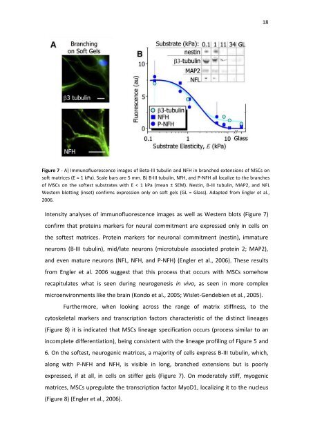
Figure 7 - A) Immunofluorescence images of Beta-III tubulin and NFH in branched extensions of MSCs on<br />
soft matrices (E ≈ 1 kPa). Scale bars are 5 mm. B) B-III tubulin, NFH, and P-NFH all localize to the branches<br />
of MSCs on the softest substrates with E < 1 kPa (mean ± SEM). Nestin, B-III tubulin, MAP2, and NFL<br />
Western blotting (inset) confirms expression only on soft gels (GL = Glass). Adapted from Engler et al.,<br />
2006.<br />
Intensity analyses of immunofluorescence images as well as Western blots (Figure 7)<br />
confirm that proteins markers for neural commitment are expressed only in cells on<br />
the softest matrices. Protein markers for neuronal commitment (nestin), immature<br />
neurons (B-III tubulin), mid/late neurons (microtubule associated protein 2; MAP2),<br />
and even mature neurons (NFL, NFH, and P-NFH) (Engler et al., 2006). These results<br />
from Engler et al. 2006 suggest that this process that occurs with MSCs somehow<br />
recapitulates what is seen during neurogenesis in vivo, as seen in more complex<br />
microenvironments like the brain (Kondo et al., 2005; Wislet-Gendebien et al., 2005).<br />
Furthermore, when looking across the range of matrix stiffness, to the<br />
cytoskeletal markers and transcription factors characteristic of the distinct lineages<br />
(Figure 8) it is indicated that MSCs lineage specification occurs (process similar to an<br />
incomplete differentiation), being consistent with the lineage profiling of Figure 5 and<br />
6. On the softest, neurogenic matrices, a majority of cells express B-III tubulin, which,<br />
along with P-NFH and NFH, is visible in long, branched extensions but is poorly<br />
expressed, if at all, in cells on stiffer gels (Figure 7). On moderately stiff, myogenic<br />
matrices, MSCs upregulate the transcription factor MyoD1, localizing it to the nucleus<br />
(Figure 8) (Engler et al., 2006).