DEPARTAMENTO DE CIÊNCIAS DA VIDA ... - Estudo Geral
DEPARTAMENTO DE CIÊNCIAS DA VIDA ... - Estudo Geral
DEPARTAMENTO DE CIÊNCIAS DA VIDA ... - Estudo Geral

Create successful ePaper yourself
Turn your PDF publications into a flip-book with our unique Google optimized e-Paper software.
25<br />
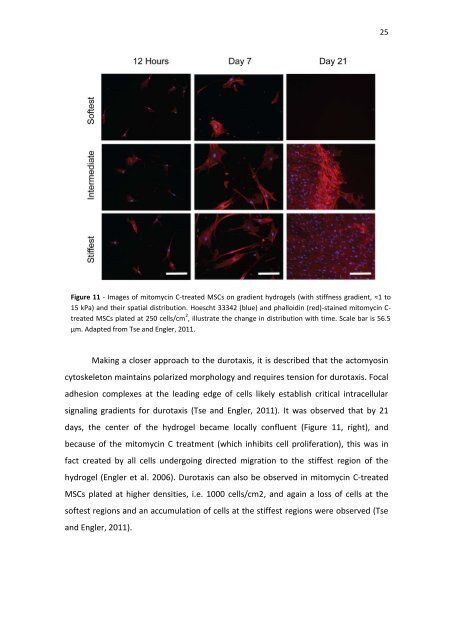
Figure 11 - Images of mitomycin C-treated MSCs on gradient hydrogels (with stiffness gradient, ≈1 to<br />
15 kPa) and their spatial distribution. Hoescht 33342 (blue) and phalloidin (red)-stained mitomycin C-<br />
treated MSCs plated at 250 cells/cm 2 , illustrate the change in distribution with time. Scale bar is 56.5<br />
µm. Adapted from Tse and Engler, 2011.<br />
Making a closer approach to the durotaxis, it is described that the actomyosin<br />
cytoskeleton maintains polarized morphology and requires tension for durotaxis. Focal<br />
adhesion complexes at the leading edge of cells likely establish critical intracellular<br />
signaling gradients for durotaxis (Tse and Engler, 2011). It was observed that by 21<br />
days, the center of the hydrogel became locally confluent (Figure 11, right), and<br />
because of the mitomycin C treatment (which inhibits cell proliferation), this was in<br />
fact created by all cells undergoing directed migration to the stiffest region of the<br />
hydrogel (Engler et al. 2006). Durotaxis can also be observed in mitomycin C-treated<br />
MSCs plated at higher densities, i.e. 1000 cells/cm2, and again a loss of cells at the<br />
softest regions and an accumulation of cells at the stiffest regions were observed (Tse<br />
and Engler, 2011).