DEPARTAMENTO DE CIÊNCIAS DA VIDA ... - Estudo Geral
DEPARTAMENTO DE CIÊNCIAS DA VIDA ... - Estudo Geral
DEPARTAMENTO DE CIÊNCIAS DA VIDA ... - Estudo Geral

Create successful ePaper yourself
Turn your PDF publications into a flip-book with our unique Google optimized e-Paper software.
12.5% Ac PBS<br />
Fold increase of hMSCs<br />
46<br />
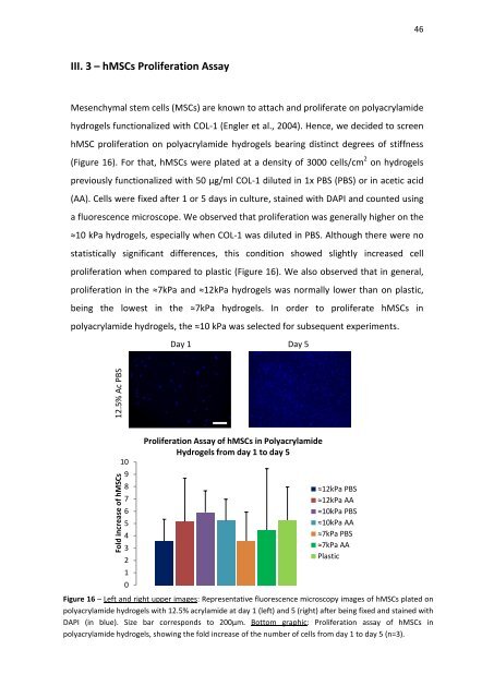
III. 3 – hMSCs Proliferation Assay<br />
Mesenchymal stem cells (MSCs) are known to attach and proliferate on polyacrylamide<br />
hydrogels functionalized with COL-1 (Engler et al., 2004). Hence, we decided to screen<br />
hMSC proliferation on polyacrylamide hydrogels bearing distinct degrees of stiffness<br />
(Figure 16). For that, hMSCs were plated at a density of 3000 cells/cm 2 on hydrogels<br />
previously functionalized with 50 µg/ml COL-1 diluted in 1x PBS (PBS) or in acetic acid<br />
(AA). Cells were fixed after 1 or 5 days in culture, stained with <strong>DA</strong>PI and counted using<br />
a fluorescence microscope. We observed that proliferation was generally higher on the<br />
≈10 kPa hydrogels, especially when COL-1 was diluted in PBS. Although there were no<br />
statistically significant differences, this condition showed slightly increased cell<br />
proliferation when compared to plastic (Figure 16). We also observed that in general,<br />
proliferation in the ≈7kPa and ≈12kPa hydrogels was normally lower than on plastic,<br />
being the lowest in the ≈7kPa hydrogels. In order to proliferate hMSCs in<br />
polyacrylamide hydrogels, the ≈10 kPa was selected for subsequent experiments.<br />
Day 1 Day 5<br />
10<br />
9<br />
8<br />
7<br />
6<br />
5<br />
4<br />
3<br />
2<br />
1<br />
0<br />
Proliferation Assay of hMSCs in Polyacrylamide<br />
Hydrogels from day 1 to day 5<br />
≈12kPa PBS<br />
≈12kPa AA<br />
≈10kPa PBS<br />
≈10kPa AA<br />
≈7kPa PBS<br />
≈7kPa AA<br />
Plastic<br />
Figure 16 – Left and right upper images: Representative fluorescence microscopy images of hMSCs plated on<br />
polyacrylamide hydrogels with 12.5% acrylamide at day 1 (left) and 5 (right) after being fixed and stained with<br />
<strong>DA</strong>PI (in blue). Size bar corresponds to 200µm. Bottom graphic: Proliferation assay of hMSCs in<br />
polyacrylamide hydrogels, showing the fold increase of the number of cells from day 1 to day 5 (n=3).