DEPARTAMENTO DE CIÊNCIAS DA VIDA ... - Estudo Geral
DEPARTAMENTO DE CIÊNCIAS DA VIDA ... - Estudo Geral
DEPARTAMENTO DE CIÊNCIAS DA VIDA ... - Estudo Geral

You also want an ePaper? Increase the reach of your titles
YUMPU automatically turns print PDFs into web optimized ePapers that Google loves.
26<br />
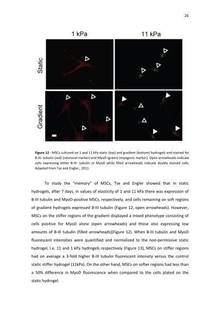
Figure 12 - MSCs cultured on 1 and 11 kPa static (top) and gradient (bottom) hydrogels and stained for<br />
B-III- tubulin (red) (neuronal marker) and MyoD (green) (myogenic marker). Open arrowheads indicate<br />
cells expressing either B-III- tubulin or MyoD while filled arrowheads indicate doubly stained cells.<br />
Adapted from Tse and Engler,, 2011.<br />
To study the “memory” of MSCs, Tse and Engler showed that in static<br />
hydrogels, after 7 days, in values of elasticity of 1 and 11 kPa there was expression of<br />
B-III tubulin and MyoD-positive MSCs, respectively, and cells remaining on soft regions<br />
of gradient hydrogels expressed B-III tubulin (Figure 12, open arrowheads). However,<br />
MSCs on the stiffer regions of the gradient displayed a mixed phenotype consisting of<br />
cells positive for MyoD alone (open arrowheads) and those also expressing low<br />
amounts of B-III tubulin (filled arrowheads)(Figure 12). When B-III tubulin and MyoD<br />
fluorescent intensities were quantified and normalized to the non-permissive static<br />
hydrogel, i.e. 11 and 1 kPa hydrogels respectively (Figure 13), MSCs on stiffer regions<br />
had on average a 3-fold higher B-III tubulin fluorescent intensity versus the control<br />
static stiffer hydrogel (11kPa). On the other hand, MSCs on softer regions had less than<br />
a 50% difference in MyoD fluorescence when compared to the cells plated on the<br />
static hydrogel.