Spectroscopy in a Suitcase - Royal Society of Chemistry
Spectroscopy in a Suitcase - Royal Society of Chemistry
Spectroscopy in a Suitcase - Royal Society of Chemistry

Create successful ePaper yourself
Turn your PDF publications into a flip-book with our unique Google optimized e-Paper software.
SPECTROSCOPY<br />
INTRODUCTION 2<br />
When matter absorbs electromagnetic radiation the<br />
change which occurs depends on the type <strong>of</strong> radiation,<br />
and therefore the amount <strong>of</strong> energy, be<strong>in</strong>g absorbed.<br />
Absorption <strong>of</strong> energy causes an electron or molecule to go<br />
from an <strong>in</strong>itial energy state (ground state) to a high energy<br />
state (excited state) which could take the form <strong>of</strong> the<br />
<strong>in</strong>creased rotation, vibration or electronic excitation.<br />
By study<strong>in</strong>g this change <strong>in</strong> energy state scientists are able<br />
to learn more about the physical and chemical properties<br />
<strong>of</strong> the molecules.<br />
• Radio waves can cause nuclei <strong>in</strong> some atoms to<br />
change magnetic orientation and this forms the basis<br />
<strong>of</strong> a technique called nuclear magnetic resonance<br />
(NMR) spectroscopy.<br />
• Molecular rotations are excited by microwaves.<br />
• Electrons are promoted to higher orbitals by<br />
ultraviolet or visible light.<br />
• Vibrations <strong>of</strong> bonds are excited by <strong>in</strong>frared radiation.<br />
The energy states are said to be quantised because a<br />
photon <strong>of</strong> precise energy and frequency (or wavelength)<br />
must be absorbed to excite an electron or molecule from<br />
the ground state to a particular excited state.<br />
S<strong>in</strong>ce molecules have a unique set <strong>of</strong> energy states that<br />
depend on their structure, IR, UV-visible and NMR<br />
spectroscopy will provide valuable <strong>in</strong>formation about the<br />
structure <strong>of</strong> the molecule.<br />
To ‘see’ a molecule we need to use light hav<strong>in</strong>g a<br />
wavelength smaller than the molecule itself (approximately<br />
10 –10 m). Such radiation is found <strong>in</strong> the X-ray region <strong>of</strong> the<br />
electromagnetic spectrum and is used <strong>in</strong> the field <strong>of</strong><br />
X-ray crystallography. This technique yields very detailed<br />
three-dimensional pictures <strong>of</strong> molecular structures –<br />
the only drawback be<strong>in</strong>g that it requires high quality<br />
crystals <strong>of</strong> the compound be<strong>in</strong>g studied. Although other<br />
spectroscopic techniques do not yield a three-dimensional<br />
picture <strong>of</strong> a molecule they do provide <strong>in</strong>formation about its<br />
characteristic features and are therefore used rout<strong>in</strong>ely <strong>in</strong><br />
structural analysis.<br />
Mass spectrometry is another useful technique used by<br />
chemists to help them determ<strong>in</strong>e the structure <strong>of</strong><br />
molecules. Although sometimes referred to as mass<br />
spectroscopy it is, by def<strong>in</strong>ition, not a spectroscopic<br />
technique as it does not make use <strong>of</strong> electromagnetic<br />
radiation. Instead the molecules are ionised us<strong>in</strong>g high<br />
energy electrons and these molecular ions subsequently<br />
undergo fragmentation. The result<strong>in</strong>g mass spectrum<br />
conta<strong>in</strong>s the mass <strong>of</strong> the molecule and its fragments<br />
which allows chemists to piece together its structure.<br />
In all spectroscopic techniques only very small quantities<br />
(milligrams or less) <strong>of</strong> sample are required, however, <strong>in</strong><br />
mass spectrometry the sample is destroyed <strong>in</strong> the<br />
fragmentation process whereas the sample can be<br />
recovered after us<strong>in</strong>g IR, UV-visible and NMR<br />
spectroscopy.<br />
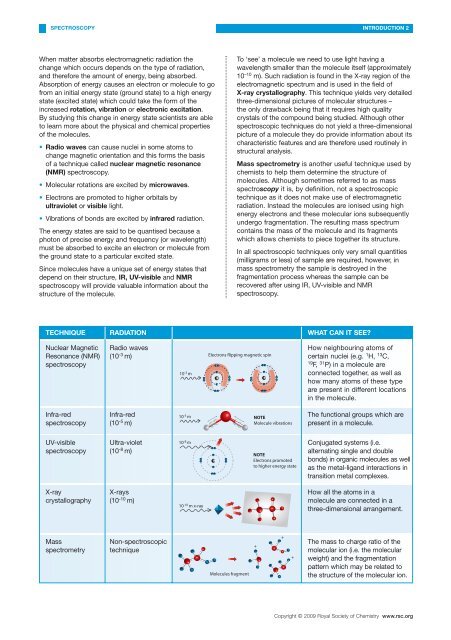
TECHNIQUE RADIATION WHAT CAN IT SEE?<br />
Nuclear Magnetic<br />
Resonance (NMR)<br />
spectroscopy<br />
Radio waves<br />
(10 -3 m)<br />
10 -3 m<br />
Electrons flipp<strong>in</strong>g magnetic sp<strong>in</strong><br />
How neighbour<strong>in</strong>g atoms <strong>of</strong><br />
certa<strong>in</strong> nuclei (e.g. 1 H, 13 C,<br />
19 F, 31 P) <strong>in</strong> a molecule are<br />
connected together, as well as<br />
how many atoms <strong>of</strong> these type<br />
are present <strong>in</strong> different locations<br />
<strong>in</strong> the molecule.<br />
Infra-red<br />
spectroscopy<br />
Infra-red<br />
(10 -5 m)<br />
10 -5 m NOTE<br />
Molecule vibrations<br />
The functional groups which are<br />
present <strong>in</strong> a molecule.<br />
UV-visible<br />
spectroscopy<br />
Ultra-violet<br />
(10 -8 m)<br />
10 -8 m<br />
NOTE<br />
Electrons promoted<br />
to higher energy state<br />
Conjugated systems (i.e.<br />
alternat<strong>in</strong>g s<strong>in</strong>gle and double<br />
bonds) <strong>in</strong> organic molecules as well<br />
as the metal-ligand <strong>in</strong>teractions <strong>in</strong><br />
transition metal complexes.<br />
X-ray<br />
crystallography<br />
X-rays<br />
(10 -10 m)<br />
10 -10 m x-ray<br />
How all the atoms <strong>in</strong> a<br />
molecule are connected <strong>in</strong> a<br />
three-dimensional arrangement.<br />
Mass<br />
spectrometry<br />
Non-spectroscopic<br />
technique<br />
Molecules fragment<br />
+<br />
+<br />
+<br />
The mass to charge ratio <strong>of</strong> the<br />
molecular ion (i.e. the molecular<br />
weight) and the fragmentation<br />
pattern which may be related to<br />
the structure <strong>of</strong> the molecular ion.<br />
Copyright © 2009 <strong>Royal</strong> <strong>Society</strong> <strong>of</strong> <strong>Chemistry</strong> www.rsc.org