Real time PCR - European Pharmaceutical Review
Real time PCR - European Pharmaceutical Review
Real time PCR - European Pharmaceutical Review

You also want an ePaper? Increase the reach of your titles
YUMPU automatically turns print PDFs into web optimized ePapers that Google loves.
SUBSCRIBE<br />
ISSUE<br />
2009 PAT<br />
a bioprocess further emphasises the<br />
need of a thorough analysis of testing<br />
and optimisation possibilities with<br />
modern PAT methods.<br />
The variety of procedural<br />
alternatives for propagation,<br />
expansion, differentiation,<br />
preservation and characterisation of<br />
hESC are indeed multi-parametrical<br />
and need to be adapted, refined and<br />
optimised to satisfy the demands<br />
of the regulators, including the<br />
expectations inherent in guidelines<br />
of QbD and PAT.<br />
Stem cell technology<br />
The first step in the preparation of<br />
hESC and hESC derived products is to<br />
enzymatically digest the inner cell<br />
mass of embryonic blastocytes 11-12 .<br />
The isolated cell mass is placed on a<br />
layer of growth-inhibited mouse<br />
fibroblast feeder cells in tissue culture<br />
dishes. After one or two weeks the<br />
outgrown cells from the inner cell<br />
mass are isolated, by dissection or<br />
enzymatic treatment, and transferred<br />
to new culture dishes for further<br />
growth in an un-dissociated cellular<br />
state. The feeder cells provide<br />
differentiation inhibitors which hinder<br />
the stem cells to differentiate.<br />
The stability of the cell culture is<br />
monitored through characterising<br />
specific biomarker molecules,<br />
typically cell surface proteins<br />
(e.g. SSEA-3/4, Oct3/4 and Nanog)<br />
which indicate that the cell mass<br />
remains in the undifferentiated state 13 .<br />
Additional characterisation and<br />
control methods are appropriate to<br />
use, such as gene array analyses, flow<br />
cytometry other methods 14,15 .<br />
If the production goal is to<br />
produce undifferentiated stem cells,<br />
the characterised cells are isolated<br />
and further expanded under<br />
controlled conditions (see Figure 1 on<br />
page 34). Up to 100 passages have<br />
been reported provided efficient<br />
inhibition factors are supplied 10 .<br />
If the goal of the production is a<br />
particular cell type, a well defined<br />
differentiation protocol ensues in a<br />
stepwise procedure of propagation<br />
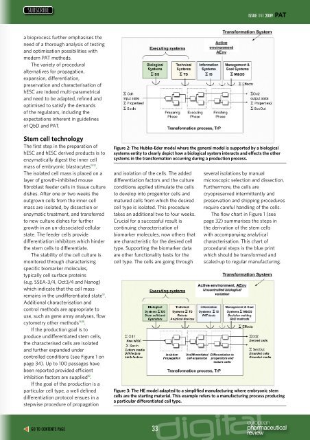
Figure 2: The Hubka-Eder model where the general model is supported by a biological<br />
systems entity to clearly depict how a biological system interacts and effects the other<br />
systems in the transformation occurring during a production process.<br />
and isolation of the cells. The added<br />
differentiation factors and the culture<br />
conditions applied stimulate the cells<br />
to develop into progenitor cells and<br />
matured cells from which the desired<br />
cell type is isolated. This procedure<br />
takes an additional two to four weeks.<br />
Crucial for a successful result is<br />
continuing characterisation of<br />
biomarker molecules, now others that<br />
are characteristic for the desired cell<br />
type. Supporting the biomarker data<br />
are other functionality tests for the<br />
cell type. The cells are going through<br />
several isolations by manual<br />
microscopic selection and dissection.<br />
Furthermore, the cells are<br />
cryopreserved intermittently and<br />
preservation and shipping procedures<br />
require careful handling of the cells.<br />
The flow chart in Figure 1 (see<br />
page 32) summarises the steps in<br />
the derivation of the stem cells<br />
with accompanying analytical<br />
characterisation. This chart of<br />
procedural steps is the blue print<br />
which should be transformed and<br />
scaled-up to regular manufacturing.<br />
Figure 3: The HE model adapted to a simplified manufacturing where embryonic stem<br />
cells are the starting material. This example refers to a manufacturing process producing<br />
a particular differentiated cell type.<br />
GO TO CONTENTS PAGE 33