Abstracts of the Academy of Dental Materials Annual ... - IsiRed
Abstracts of the Academy of Dental Materials Annual ... - IsiRed
Abstracts of the Academy of Dental Materials Annual ... - IsiRed

You also want an ePaper? Increase the reach of your titles
YUMPU automatically turns print PDFs into web optimized ePapers that Google loves.
e20 dental materials 26S (2010) e1–e84<br />
and each wafer specimen were analyzed with a FTIR spectrometer<br />
(BRUKER TENSOR 27, Germany) operating 16 scans at<br />
4cm −1 resolution. Two and three way ANOVA were performed<br />
on data <strong>of</strong> depth <strong>of</strong> cure and DC, respectively. Also, Tukey HSD<br />
were performed at <strong>the</strong> 95% significance level.<br />
Results: Statistical analysis revealed that <strong>the</strong> depth <strong>of</strong> cure<br />
was affected by temperature and that <strong>the</strong> specimens cured at<br />
37 ◦ C had statistically significant higher depth <strong>of</strong> cure compared<br />
to <strong>the</strong> specimens cured at 5 ◦ C in both QTH and LED<br />
groups (p < 0.01). Also, lower depth <strong>of</strong> cure was obtained with<br />
QTH light curing system. Temperature increase also gave<br />
higher DC. The results were statistically significant at three<br />
different temperature groups (p < 0.05) regardless <strong>of</strong> <strong>the</strong> curing<br />
unit. Also, a significant difference found between top and bottom<br />
surfaces <strong>of</strong> specimens polymerized at room temperature.<br />
However, no significant differences were found between top<br />
and 2 mm depth on materials cured at 37 ◦ C and 5 ◦ C(p > 0.05).<br />
Conclusions: Increasing <strong>the</strong> temperature <strong>of</strong> resin composites<br />
has an important influence on its degree <strong>of</strong> conversion<br />
and depth <strong>of</strong> cure. Thus, it may affect <strong>the</strong> physical properties.<br />
Thus, increasing <strong>the</strong> shelf life <strong>of</strong> composites by storing<br />
<strong>the</strong>m in refrigerator may show a negative-influence on<br />
polymerization.<br />
doi:10.1016/j.dental.2010.08.049<br />
42<br />
Time-dependent fracture toughness <strong>of</strong> conventional glassionomer<br />
cements<br />
R. Belli, A. Petschelt, U. Lohbauer<br />
<strong>Dental</strong> Clinic 1, University <strong>of</strong> Erlangen-Nuernberg, Germany<br />
Objectives: To investigate <strong>the</strong> fracture toughness KIc <strong>of</strong><br />
three commercial dental glassionomer (GI) cements at different<br />
storage intervals.<br />
<strong>Materials</strong> and methods: Bars with dimensions <strong>of</strong><br />
25 mm × 2mm× 2 mm were produced out <strong>of</strong> Fuji IX GP<br />
(GC Europe, Belgium), Ketac Molar (3M ESPE, Germany) and<br />
Chemfil Rock (Dentsply, Germany) and stored for 3 h, 24 h, 7<br />
days, and 21 days in water at 37 ◦C. After <strong>the</strong> storage periods,<br />
sharp notches were cut on <strong>the</strong> lower side <strong>of</strong> <strong>the</strong> bars using<br />
a diamond wheel saw blade and a razor blade. Fracture<br />
toughness (n = 15) was measured in bending using <strong>the</strong> SENB<br />
(single edge notched beam) method. The actual notch depth<br />
for each specimen was measured after fracture under a<br />
light microscope (40×). KIc values were analysed by two-way<br />
ANOVA and mod. LSD test at p < 0.05.<br />
Results: The major increase in KIc was measured between<br />
3 h and 24 h for all <strong>the</strong> GI cements (p < 0.05). Chemfil<br />
Rock presented <strong>the</strong> fastest initial toughness, reaching<br />
0.44 ± 0.06 MPam0.5 after 3 h, whereas KIc for Fuji IX GP and<br />
Ketac Molar were 0.36 ± 0.04 MPam0.5 and 0.34 ± 0.04 MPam0.5 ,<br />
respectively at <strong>the</strong> same period. Final KIc at 21 days was<br />
not statistically different from that <strong>of</strong> 24 h for Chemfil<br />
Rock and Ketac Molar, while Fuji IX GP showed continuous<br />
KIc increase over <strong>the</strong> test periods. However, final<br />
(21 days) KIc were not statistically different for <strong>the</strong> three<br />
GI systems (0.57 ± 0.05 MPam0.5 , 0.57 ± 0.08 MPam0.5 and<br />
0.53 ± 0.09 MPam0.5 Molar, respectively).<br />
for Chemfil Rock, Fuji IX GP and Ketac<br />
Conclusions: Although fracture toughness development<br />
has shown to be material-dependent, GI cements are<br />
more prone to fracture before 24 h due to <strong>the</strong> early stage <strong>of</strong><br />
poly-acid/glass reaction kinetics. Since GI restorations are subjected<br />
to normal chewing forces from first hours <strong>of</strong> placement,<br />
fast fracture toughness development is <strong>of</strong> clinical relevance to<br />
prevent early fracture. In this sense, Chemfil Rock has shown<br />
to reach a statistically higher KIc at <strong>the</strong> earliest test period.<br />
This study was supported by Dentsply, Germany.<br />
doi:10.1016/j.dental.2010.08.050<br />
43<br />
Bond strength to enamel and flexural strength <strong>of</strong> fiberreinforced<br />
composites<br />
M. Beloica 1 , C. Goracci 2 , N. Chieffi 2 , A. Vichi 2 , Z.R.<br />
Vulicevic 1 , M. Ferrari 2<br />
1 University <strong>of</strong> Belgrade, Serbia<br />
2 University <strong>of</strong> Siena, Italy<br />
Objectives: To assess shear bond strength to enamel and<br />
flexural strength <strong>of</strong> reinforcing fibers used in combination<br />
with a flowable resin composite as splinting materials.<br />
<strong>Materials</strong> and methods: Quartz Splint (RTD, QS), Triaxial<br />
(Ribbond, TX), THM (Ribbond, THM), Construct (Kerr, CNS),<br />
Connect (Kerr, CNN) and everStick PERIO (StickTech, ES) were<br />
tested. QS, CNS, and CNN were impregnated with <strong>the</strong> proprietary<br />
resin prior to application. Premise Flow (Kerr) was<br />
used in combination. For shear bond strength testing 10 sound<br />
extracted molars per group were selected. On <strong>the</strong> buccal surface<br />
<strong>of</strong> each tooth enamel was etched with 37% phosphoric<br />
acid and Optibond FL adhesive (Kerr) was applied. A 1 mmthick<br />
increment <strong>of</strong> resin composite was layered in a cylindrical<br />
mould and an adequately sized segment <strong>of</strong> reinforcing fiber<br />
was placed on top. The fiber was <strong>the</strong>n overlaid by ano<strong>the</strong>r 1mm<br />
thick layer <strong>of</strong> resin composite. Twenty-four hours after<br />
preparation, <strong>the</strong> bonded cylinder <strong>of</strong> fiber-reinforced resin composite<br />
was sheared <strong>of</strong>f using a universal testing machine.<br />
Failure modes were noted. For flexural strength assessment<br />
10 bar-shaped specimens <strong>of</strong> fiber-reinforced composite per<br />
group were prepared and tested according to ISO Standard<br />
4049/2000. Shear bond strength and flexural strength<br />
<strong>of</strong> Premise Flow without fiber reinforcement were measured<br />
with <strong>the</strong> same methods. The ‘No fiber’ group served as control.<br />
Between-group comparisons in shear bond strength,<br />
failure modes distribution and flexural strength were statistically<br />
evaluated (statistical tests: one-way ANOVA, Tukey;<br />
Pearson’s chi-square; Kruskall–Wallis ANOVA, Dunn’s multiple<br />
range, p < 0.05). Scanning electron microscope observations<br />
were taken to visualize <strong>the</strong> structure <strong>of</strong> <strong>the</strong> reinforcing fibers.<br />
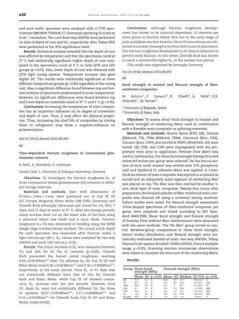
Results:<br />
Group Shear bond<br />
strength (MPa)<br />
Flexural strength (MPa)<br />
Mean SD p < 0.05 Mean SD Median 25–75% p < 0.05<br />
QS 15.4 5.2 A 24.9 4.5 13.7 10.6–14.3 A<br />
TX 16.7 6.2 A 19.3 8.6 18.6 12.5–26.7 ABC<br />
THM 18.6 5.6 A 12.4 3 24.7 21.2–25.9 CD<br />
CNS 16 5.5 A 17.5 5.3 13.6 11–18.6 ABC<br />
CNN 14.4 4.1 A 14.3 5.2 19.7 11.6–21.7 BCD<br />
ES 4.6 3.2 B 18.5 2.6 19.3 18.4–19.5 ABC<br />
No fiber 12.8 4.7 A 5.8 1.4 6.3 4.8–6.9 D