Abstracts of the Academy of Dental Materials Annual ... - IsiRed
Abstracts of the Academy of Dental Materials Annual ... - IsiRed
Abstracts of the Academy of Dental Materials Annual ... - IsiRed

Create successful ePaper yourself
Turn your PDF publications into a flip-book with our unique Google optimized e-Paper software.
for each ceramic system, maintaining <strong>the</strong> overall total thickness<br />
at 1.3 mm. The thicknesses <strong>of</strong> <strong>the</strong> layers were: metal<br />
base disc 0.3 mm, opaque 0.15 mm, dentin opaque 0.35 mm<br />
and 0.45 mm, dentin 0.5 mm and 0.7 mm, enamel 0.1 mm and<br />
0.2 mm. The thickness <strong>of</strong> each layer was verified after each firing.<br />
A clinical spectrophotometer (Vita Easyshade) was used<br />
for color comparison in “Restoration” mode, which allows<br />
comparing <strong>the</strong> instrument set color data with <strong>the</strong> measured<br />
ones. The obtained �E values (differences between intended<br />
and obtained shade) were statistically analyzed with a 3-way<br />
ANOVA and <strong>the</strong> Tukey HSD test (˛ = 0.05).<br />
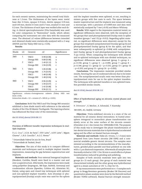
Results:<br />
Shade LS Ceramic �E Significance<br />
2M3 1 VM13 0.98 (0.08) a<br />
Omega 900 0.88 (0.08) a<br />
2 VM13 1.40 (0.12) b<br />
Omega 900 1.94 (0.09) c<br />
3M2 1 VM13 1.06 (0.08) a<br />
Omega 900 1.76 (0.05) c<br />
2 VM13 1.48 (0.08) b<br />
Omega 900 1.48 (0.04) b<br />
4M2 1 VM13 0.96 (0.09) a<br />
Omega 900 1.90 (0.07) c<br />
2 VM13 1.42 (0.08) b<br />
Omega 900 0.96 (0.11) a<br />
Significance column = homogeneous subsets (Tukey HSD test<br />
level = 0.05).<br />
Interaction shade × LS × ceramic (F = 189.62, p < 0.001).<br />
Conclusions: Both Vita VM13 and Vita Omega 900 ceramics<br />
exhibited a close shade match with reference to <strong>the</strong> selected<br />
shades <strong>of</strong> <strong>the</strong> Vita 3D Master Toothguide. The layering scheme<br />
has influenced <strong>the</strong> final color <strong>of</strong> <strong>the</strong> restorations.<br />
doi:10.1016/j.dental.2010.08.111<br />
104<br />
Precision <strong>of</strong> different transfer impression techniques in multiple<br />
implants<br />
F.L.G. Cruz 1 , J.C.B. de Faria 2 , F.P.P. Leite 1 , A.P.P. Leite 1 ,Mgam<br />
Chaves 1 , L.R.S. Concílio 2 , A.C.C. Neves 2<br />
1 Universidade Federal De Juiz De Fora, Brazil<br />
2 Universidade de Taubaté, Brazil<br />
Objectives: The aim <strong>of</strong> this study is to compare different<br />
materials and techniques used in multiple implant transfer<br />
impressions, measuring <strong>the</strong> gap between superstructure and<br />
implants.<br />
<strong>Materials</strong> and methods: Four external hexagonal implants<br />
(Neodent, Curitiba, Brazil) were fixed in a master cast and<br />
over a superstructure. Afterwards, <strong>the</strong> impressions were made<br />
using polye<strong>the</strong>r (Impregum, 3M Espe, Seefeld, Germany) and<br />
condensation silicone (Speedex, Coltène AG, Altstatten SG,<br />
Swiss), using open and closed tray techniques with splinted<br />
and non-splinted implant transfers. Auto (Duralay) or photopolymerized<br />
(Duolay) acrylic resin was used for <strong>the</strong> splints<br />
dental materials 26S (2010) e1–e84 e49<br />
and <strong>the</strong> implant transfers were splinted, or not, resulting in<br />
sixteen groups with five casts in each. The space between<br />
master superstructure and <strong>the</strong> implants was measured using<br />
a microscope, with a precision <strong>of</strong> 0.0005 mm and data were<br />
statistically analyzed by T test (=0.05) and Mann–Whitney.<br />
Results: With regard to <strong>the</strong> impression material tested, no<br />
significant differences were observed, with <strong>the</strong> exception <strong>of</strong><br />
<strong>the</strong> groups that used photopolymerized Duolay resin to splint<br />
transfers (groups 7 and 15) (p = 0.012). Regarding splint material,<br />
no significant differences were observed, except for <strong>the</strong><br />
groups that employed autopolymerized Duralay (group 4) and<br />
photopolymerized Duolay (group 8) for <strong>the</strong> splint, and that<br />
were subsequently re-splinted (p = 0.004) with autopolymerized<br />
Duolay (group 5) and photopolymerized Duolay (group<br />
7) (p = 0.012). When comparing <strong>the</strong> techniques with divided<br />
and re-splinted techniques with non-divided techniques, no<br />
significant differences were observed (group 3 × group 4 –<br />
p = 0.158; group 5 × group 6 – p = 0.094; group 7 × group 8 –<br />
p = 0.144; group 11 × group 12 – p = 0.331; group 13 × group 14<br />
– p = 0.203; and group 15 × group 16 – p = 0.092).<br />
Conclusions: The impression materials provided similar<br />
results, favoring <strong>the</strong> use <strong>of</strong> condensed silicone due to its lower<br />
cost. The autopolymerized acrylic resin was better than photopolymerized<br />
resin for use in <strong>the</strong> splint implant transfers.<br />
The techniques with splinted transfers or non-splinted transfers<br />
provided similar results.<br />
doi:10.1016/j.dental.2010.08.112<br />
105<br />
Effect <strong>of</strong> hydro<strong>the</strong>rmal aging on zirconia crystal phases and<br />
strength<br />
R. Dittmann ∗ , E. Mecher, A. Schmalzl, T. Kuretzky<br />
3M ESPE, AG, Seefeld, Germany<br />
Objectives: Yttria-stabilized zirconia is a state <strong>of</strong> <strong>the</strong> art<br />
material for all ceramic dental restorations. In humid atmosphere<br />
tetragonal to monoclinic phase transformation can<br />
slowly occur at <strong>the</strong> outer surface <strong>of</strong> <strong>the</strong> zirconia ceramic<br />
(Chevalier et al. J Am Ceram Soc 1999;82(8):2150). The purpose<br />
<strong>of</strong> this study was to investigate <strong>the</strong> phase transformation <strong>of</strong><br />
two dental zirconia materials due to hydro<strong>the</strong>rmal accelerated<br />
aging and <strong>the</strong> effect on biaxial fracture strength.<br />
<strong>Materials</strong> and methods: Disks (d =14mm, t = 1.5 mm) were<br />
prepared from Lava Frame zirconia (3M ESPE) and a commercial<br />
available generic dental zirconia. The samples were<br />
sintered according to <strong>the</strong> recommendations <strong>of</strong> <strong>the</strong> manufacturers,<br />
grinded plane-parallel and final polished on one side<br />
with a 1 �m diamond suspension. Afterwards all samples were<br />
<strong>the</strong>rmally etched at 1350 ◦ C for 30 min to avoid effects <strong>of</strong> phase<br />
transformation induced by grinding and polishing.<br />
Samples <strong>of</strong> both zirconia materials were split into two<br />
groups (n = 15 each group). One group <strong>of</strong> each material was<br />
characterized initially and <strong>the</strong> o<strong>the</strong>r group was hydro<strong>the</strong>rmal<br />
aged in a steam autoclave at 135 ◦ C, 2 bar for 5 h.<br />
X-ray measurements were performed on n = 5 samples each<br />
group in Bragg-Brentano geometry (Bruker D8 Discover) and<br />
quantitative phase analysis were done by <strong>the</strong> Rietveld method<br />
(Bruker TOPAS s<strong>of</strong>tware). Biaxial fracture strength measure-