Abstracts of the Academy of Dental Materials Annual ... - IsiRed
Abstracts of the Academy of Dental Materials Annual ... - IsiRed
Abstracts of the Academy of Dental Materials Annual ... - IsiRed

You also want an ePaper? Increase the reach of your titles
YUMPU automatically turns print PDFs into web optimized ePapers that Google loves.
Conclusions: In conclusion, HA coating seemed to improve<br />
differentiation potential <strong>of</strong> osteoblasts but also signaling for<br />
clasts differentiation and bone remodeling.<br />
doi:10.1016/j.dental.2010.08.150<br />
143<br />
Effects <strong>of</strong> external bleaching agents on dental tissues and<br />
cement–enamel junction<br />
M.G.A.M. Chaves, M. Oliveira, M.N. Pereira, M.R.S.P. Soares,<br />
H.D.M. Chaves Filho, F.P.P. Leite<br />
Universidade Federal De Juiz De Fora, Brazil<br />
Objectives: Evaluate with a scanning electron microscope<br />
(SEM) <strong>the</strong> alterations occurring in enamel, cementum and<br />
cementum–enamel junction after <strong>the</strong> use <strong>of</strong> different external<br />
bleaching agents.<br />
<strong>Materials</strong> and methods: Fifteen healthy pre-molars were<br />
sectioned in <strong>the</strong> middle with <strong>the</strong> aid <strong>of</strong> a cutting machine<br />
(Labcut 1010), in <strong>the</strong> longitudinal direction along <strong>the</strong> axis<br />
<strong>of</strong> <strong>the</strong> teeth, obtaining mesial and distal surfaces <strong>of</strong> each<br />
tooth. The mesial surfaces were divided into three groups<br />
in accord with <strong>the</strong> material used (AI – carbamide peroxide<br />
10% – Whiteness Standard 10 FGM ® , AII – carbamide<br />
peroxide 16% Whiteness Standard 16 FGM ® , e AIII – hydrogen<br />
peroxide 35% Whiteness HP Maxx FGM ® ), applied<br />
according to <strong>the</strong> instructions <strong>of</strong> <strong>the</strong> manufacturers. The<br />
distal surfaces <strong>of</strong> each tooth were maintained in artificial<br />
saliva, in a bacteriological sterilizer at 37 ◦ C. After whitening<br />
<strong>the</strong> specimens were dehydrated in increasing solutions<br />
<strong>of</strong> acetone and metalized for evaluation using a scanning<br />
electron microscope. The results were submitted to statistical<br />
evaluation using <strong>the</strong> Kruskal–Wallis and Mann–Whitey<br />
tests.<br />
Results: Statistically significant differences were observed<br />
between <strong>the</strong> whitened and unwhitened specimens (p < 0.001).<br />
In enamel, insignificant alterations were observed, and no differences<br />
between <strong>the</strong> groups were found. Loss <strong>of</strong> structure,<br />
with formation <strong>of</strong> gaps and dental exposure were observed in<br />
<strong>the</strong> cemento-enamel junction <strong>of</strong> <strong>the</strong> three whitened groups.<br />
Conclusions: Tooth whitening should be done with<br />
caution principally in patients who possess gingival recession.<br />
doi:10.1016/j.dental.2010.08.151<br />
144<br />
Solubility in water or DMEM <strong>of</strong> F-doped MTA cements with<br />
increasing F-content<br />
A. Colin, C. Prati, G.A. Pelliccioni, M.G. Gandolfi<br />
Laboratory <strong>of</strong> Biomaterials and Oral Pathology, Department <strong>of</strong><br />
Odontostomatological Sciences, University <strong>of</strong> Bologna, Bologna,<br />
Italy<br />
Objectives: Fluoride increases <strong>the</strong> expansion and retards<br />
<strong>the</strong> setting time <strong>of</strong> F-doped calcium-silicate MTA cements.<br />
These cements may be successfully used as endodontic sealer.<br />
The assessment <strong>of</strong> <strong>the</strong> solubility <strong>of</strong> <strong>the</strong> endodontic cements<br />
dental materials 26S (2010) e1–e84 e67<br />
allows to predict <strong>the</strong> loss <strong>of</strong> material from <strong>the</strong> surgical site,<br />
where <strong>the</strong>y are in contact with physiological fluids (blood,<br />
tubular fluid). The aim <strong>of</strong> <strong>the</strong> study was to evaluate <strong>the</strong> solubility<br />
in deionized water (ISO 6876) or in simulated body fluid<br />
(DMEM) <strong>of</strong> designed F-doped calcium-silicate MTA cements<br />
with increasing amount <strong>of</strong> fluoride.<br />
<strong>Materials</strong> and methods: A calcium-silicate powder containing<br />
di- and tricalcium-silicate, calcium sulphate and barium<br />
sulphate was prepared. An increasing percentage <strong>of</strong> fluoride<br />
(F0%, F1%, F5% and F10%) was added to <strong>the</strong> powder<br />
to obtain 4 cements. The cements were mixed with DPBS<br />
(liquid/powder ratio 0.22), placed into a mould (10 mm diameter;<br />
2.21 mm height) and stored at 37 ◦ C and 98% relative<br />
humidity for <strong>the</strong> 75% <strong>of</strong> <strong>the</strong>ir own setting time. Each sample<br />
(n = 5 for each cement) was weighed (initial weight, IW)<br />
and immersed in 10 mL <strong>of</strong> DMEM or deionized water keeping<br />
a vertical position. After 1 or 21 days <strong>the</strong> samples were<br />
dehydrated at 37 ◦ C and weighed (final weight, FW), <strong>the</strong>n<br />
demoulded and <strong>the</strong> moulds weighed (mould weight, MW).<br />
The percentage <strong>of</strong> weight loss (solubility) was calculated as<br />
[(IW − FW)/(IW − MW)] × 100.<br />
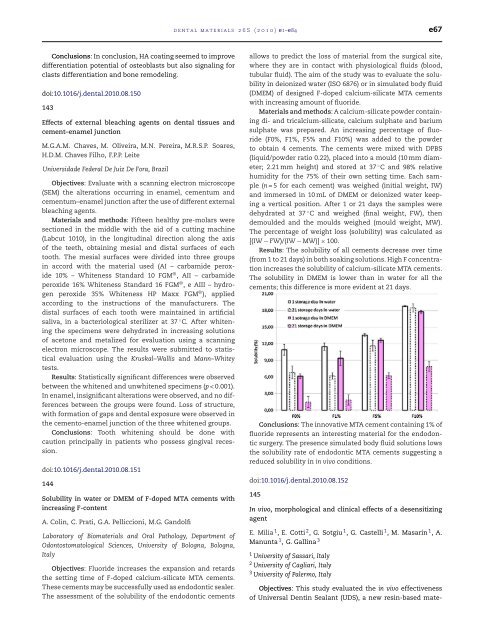
Results: The solubility <strong>of</strong> all cements decrease over time<br />
(from 1 to 21 days) in both soaking solutions. High F concentration<br />
increases <strong>the</strong> solubility <strong>of</strong> calcium-silicate MTA cements.<br />
The solubility in DMEM is lower than in water for all <strong>the</strong><br />
cements; this difference is more evident at 21 days.<br />
Conclusions: The innovative MTA cement containing 1% <strong>of</strong><br />
fluoride represents an interesting material for <strong>the</strong> endodontic<br />
surgery. The presence simulated body fluid solutions lows<br />
<strong>the</strong> solubility rate <strong>of</strong> endodontic MTA cements suggesting a<br />
reduced solubility in in vivo conditions.<br />
doi:10.1016/j.dental.2010.08.152<br />
145<br />
In vivo, morphological and clinical effects <strong>of</strong> a desensitizing<br />
agent<br />
E. Milia 1 , E. Cotti 2 , G. Sotgiu 1 , G. Castelli 1 , M. Masarin 1 ,A.<br />
Manunta 1 , G. Gallina 3<br />
1 University <strong>of</strong> Sassari, Italy<br />
2 University <strong>of</strong> Cagliari, Italy<br />
3 University <strong>of</strong> Palermo, Italy<br />
Objectives: This study evaluated <strong>the</strong> in vivo effectiveness<br />
<strong>of</strong> Universal Dentin Sealant (UDS), a new resin-based mate-