Abstracts of the Academy of Dental Materials Annual ... - IsiRed
Abstracts of the Academy of Dental Materials Annual ... - IsiRed
Abstracts of the Academy of Dental Materials Annual ... - IsiRed
Create successful ePaper yourself
Turn your PDF publications into a flip-book with our unique Google optimized e-Paper software.
152<br />
Structural analysis <strong>of</strong> <strong>the</strong> enamel subjected to pre- and postbleaching<br />
agents<br />
C. Francci 1,2 , M.N. Gomes 1,2 , A.G. Devito-Moraes 1,2 , F.P.<br />
Rodrigues 1,2 , L.C. Yamazaki 1,2 , L.M. Silva 1,2 , N.R.G. Fróes-<br />
Salgado 1,2 , A.C. Nishida 1,2<br />
1 University <strong>of</strong> São Paulo-USP, Brazil<br />
2 Bandeirante University <strong>of</strong> São Paulo -UNIBAN, Brazil<br />
Objective: To evaluate <strong>the</strong> effects <strong>of</strong> pre and post-bleaching<br />
agents as NaOCl and ACP-CPP on <strong>the</strong> structure <strong>of</strong> <strong>the</strong> enamel<br />
surface.<br />
Material and methods: Six human premolars were buccallingual<br />
and mesio-distal sectioned and <strong>the</strong>n subjected to <strong>the</strong><br />
following experimental conditions (n = 24): G1: 35% H2O2 (Pola<br />
Office, Australia, SDI) – 4 sessions <strong>of</strong> 8 min; G2: 5.25% NaOCl<br />
during 1 min previously to <strong>the</strong> application <strong>of</strong> 35% H2O2 and<br />
G3: similar to G1 + ACP-CPP (MI-Paste, Japão, GC America), during<br />
5 min daily repeated during 7 days. The samples were<br />
covered with platinum to be evaluated with Scanning Electron<br />
Microscopy (Stereoscan 440, LEO) and with High Vacuum<br />
Field Emission Microscopy (Quanta 600 FEG, FEI). The images<br />
were obtained using secondary electrons detector (Everhart-<br />
Thornley), with a work distance <strong>of</strong> 10 mm, and a high voltage<br />
<strong>of</strong> 10 kV, probe <strong>of</strong> 500 A with magnifications from 3000 through<br />
100,000.<br />
Results: In group G1, erosions on enamel prisms were<br />
noticed. In G2, <strong>the</strong> application <strong>of</strong> <strong>the</strong> NaOCl previously to<br />
<strong>the</strong> bleaching generated a smoo<strong>the</strong>r surface. There was a<br />
covering mantle in G3 around <strong>the</strong> enamel prisms and lower<br />
spaces among <strong>the</strong> hydroxyapatite crystals. The use <strong>of</strong> NaOCl<br />
associated to <strong>the</strong> bleaching with hydrogen peroxide modifies<br />
<strong>the</strong> enamel morphology.<br />
Conclusion: The application <strong>of</strong> <strong>the</strong> ACP-CPP post-bleaching<br />
agent recovers <strong>the</strong> enamel surface when applied during a<br />
week after in-<strong>of</strong>fice bleaching sessions.<br />
doi:10.1016/j.dental.2010.08.160<br />
dental materials 26S (2010) e1–e84 e71<br />
153<br />
Biomimetic dentine remineralization by a novel bioactive<br />
light-curing calcium-silicate composite<br />
M.G. Gandolfi 1 , F. Siboni 1 , P. Taddei 2 , E. Modena 2 , C. Prati 1<br />
1 Department <strong>of</strong> Odontostomatological Sciences, University <strong>of</strong><br />
Bologna, Bologna, Italy<br />
2 Department <strong>of</strong> Chemistry, University <strong>of</strong> Bologna, Bologna, Italy<br />
Objectives: A light-curing resin-modified calcium-silicate<br />
cement was designed to obtain a bioactive calciumreleasing<br />
remineralizing material (Ic-MTA) for rehabilitation<br />
<strong>of</strong> deciduous teeth and remineralization <strong>of</strong> dentine<br />
lesions.<br />
<strong>Materials</strong> and methods: Ic-MTA was prepared by mixing<br />
a tailored calcium-silicate bioactive filler with an anphiphilic<br />
HEMA/TEGDMA-based resin. Calcium release and alkalinizing<br />
activity (i.e. pH <strong>of</strong> soaking water) <strong>of</strong> <strong>the</strong> cement<br />
were tested. The dentine remineralization capability and <strong>the</strong><br />
bioactivity (i.e. apatite-forming ability) were investigated by<br />
ATR-FTIR and ESEM/EDX analyses after immersion in DPBS<br />
at 37 ◦ C for 7 days. Dentine disks 0.8 ± 0.2 mm thick were<br />
obtained from coronal side <strong>of</strong> healthy third molars and<br />
demineralized in 15 mL <strong>of</strong> sterile 17% EDTA for 2 h. EDX<br />
element distribution maps confirmed <strong>the</strong> complete absence<br />
<strong>of</strong> apatite on dentine disks from <strong>the</strong> surface to a 10 �m<br />
depth.<br />
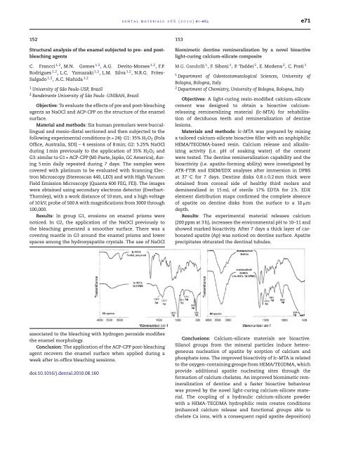
Results: The experimental material releases calcium<br />
(200 ppm at 3 h), increases <strong>the</strong> environmental pH to 10–11 and<br />
showed marked bioactivity. After 7 days a thick layer <strong>of</strong> carbonated<br />
apatite (Ap) was noticed on dentine surface. Apatite<br />
precipitates obturated <strong>the</strong> dentinal tubules.<br />
Conclusions: Calcium-silicate materials are bioactive.<br />
Silanol groups from <strong>the</strong> mineral particles induce heterogeneous<br />
nucleation <strong>of</strong> apatite by sorption <strong>of</strong> calcium and<br />
phosphate ions. The improved bioactivity <strong>of</strong> Ic-MTA is related<br />
to <strong>the</strong> oxygen-containing groups from HEMA/TEGDMA, which<br />
provide additional apatite nucleating sites through <strong>the</strong><br />
formation <strong>of</strong> calcium chelates. An improved biomimetic remineralization<br />
<strong>of</strong> dentine and a faster bioactive behaviour<br />
was proved by <strong>the</strong> novel light-curing calcium-silicate material.<br />
The coupling <strong>of</strong> a hydraulic calcium-silicate powder<br />
with a HEMA-TEGDMA hydrophilic resin creates conditions<br />
(enhanced calcium release and functional groups able to<br />
chelate Ca ions, with a consequent rapid apatite deposition)


