Abstracts of the Academy of Dental Materials Annual ... - IsiRed
Abstracts of the Academy of Dental Materials Annual ... - IsiRed
Abstracts of the Academy of Dental Materials Annual ... - IsiRed
Create successful ePaper yourself
Turn your PDF publications into a flip-book with our unique Google optimized e-Paper software.
e76 dental materials 26S (2010) e1–e84<br />
<strong>the</strong> parameters to compute surface tension by using image<br />
analysis s<strong>of</strong>tware (Sigma Scan Pro V). ANOVA was used to<br />
investigate differences <strong>of</strong> <strong>the</strong> surface tension properties <strong>of</strong> various<br />
root canal sealers at <strong>the</strong> significance level <strong>of</strong> 0.05, using<br />
SPSS s<strong>of</strong>tware.<br />
Results: All sealers exhibited varied degrees <strong>of</strong> surface tension<br />
regardless <strong>of</strong> <strong>the</strong> type <strong>of</strong> <strong>the</strong> sealers. <strong>Dental</strong>is ® exhibited<br />
<strong>the</strong> highest (59.28 mN/m) while EndoREZ ® showed <strong>the</strong> lowest<br />
(27.93 mN/m). Statistically significant differences were found<br />
between <strong>the</strong> sealers (P < 0.05).<br />
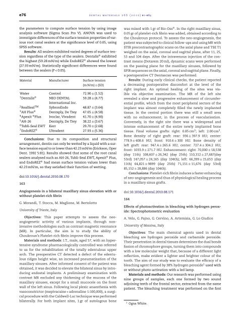
Material Manufacturer Surface tension<br />
(m N/m) ± (SD)<br />
Water Control 72.90 ± (1.52)<br />
1<strong>Dental</strong>is ® NEO DENTAL<br />
International Inc.<br />
59.28 ± (4.77)<br />
2RealSealTM SybronEndo 48.87 ± (3.64)<br />
3AH Plus ® Dentsply, De Trey 47.65 ± (4.58)<br />
4Apexit ® Plus Ivoclar, Vivadent 41.70 ± (4.90)<br />
5AH-26 Dentsply, De Trey 38.22 ± (3.67)<br />
6Tubli-Seal EWT Kerr 29.70 ± (2.68)<br />
7EndoREZ ® Ultradent 27.93 ± (5.36)<br />
Conclusions: Due to its composition and structural<br />
arrangement, dentin can only be wetted by a liquid with a surface<br />
tension equal to or lower than 42.23 mN/m (Erickson, Oper<br />
Dent. 1992 5:81). Results showed that some <strong>of</strong> <strong>the</strong> root canal<br />
sealers analysed such as AH-26, Tubli-Seal EWT, Apexit ® Plus,<br />
and EndoREZ ® had mean surface tension values lower than<br />
42.23 mN/m, so <strong>the</strong>y satisfied <strong>the</strong>ir function <strong>of</strong> wetting.<br />
doi:10.1016/j.dental.2010.08.170<br />
163<br />
Angiogenesis in a bilateral maxillary sinus elevation with or<br />
without platelet-rich fibrin<br />
G. Morandi, T. Stocca, M. Maglione, M. Bertolotto<br />
University <strong>of</strong> Trieste, Italy<br />
Objectives: This paper attempts to assess <strong>the</strong> neoangiogenetic<br />
activity <strong>of</strong> various implants, through noninvasive<br />
methodologies such as contrast magnetic resonance<br />
(MR). In particular, <strong>the</strong> aim is to study <strong>the</strong> ability <strong>of</strong><br />
Choukroun’s Platelet-rich fibrin improve this process.<br />
<strong>Materials</strong> and methods: I.T., male, aged 57, with an hypertensive<br />
syndrome pharmacologically controlled was referred<br />
to us for <strong>the</strong> rehabilitation <strong>of</strong> <strong>the</strong> totally edentulous upper<br />
arch. The preoperative CT detected a defect <strong>of</strong> <strong>the</strong> edentulous<br />
ridges height wise, an increased pneumatisation <strong>of</strong> <strong>the</strong><br />
maxillary sinuses. After informed consent <strong>of</strong> <strong>the</strong> patient was<br />
obtained, it was decided to elevate <strong>the</strong> bilateral sinus by introducing<br />
endostal implants. A preliminary examination with<br />
contrast MR excluded any thickening <strong>of</strong> <strong>the</strong> mucosa <strong>of</strong> <strong>the</strong><br />
maxillary sinuses, except for a small mucocele on <strong>the</strong> front<br />
wall <strong>of</strong> <strong>the</strong> left sinus. Following local plexic anaes<strong>the</strong>sia with<br />
vasoconstrictor (mepivacaine + adrenaline 1:100,000), a surgical<br />
procedure with <strong>the</strong> Caldwell-Luc technique was performed<br />
bilaterally. For both implant sites, 1 gr <strong>of</strong> autologous bone<br />
was mixed with 1 gr <strong>of</strong> Bio-Oss ® . In <strong>the</strong> right maxillary sinus,<br />
0.05 gr <strong>of</strong> platelet-rich fibrin was added, obtained according to<br />
<strong>the</strong> Choukroun protocol. To assess <strong>the</strong> neo-angiogenesis, <strong>the</strong><br />
patient was subjected to clinical follow-ups and using MR with<br />
STIR precontrastographic scans on <strong>the</strong> axial plane and TSE T1<br />
weighed on <strong>the</strong> axial, coronal and sagittal plane, after 11, 25,<br />
53 and 104 days. After <strong>the</strong> intravenous injection <strong>of</strong> <strong>the</strong> contrast<br />
means (Dotarem 20 ml), dynamic scans were performed<br />
on <strong>the</strong> passing plane for <strong>the</strong> maxillary sinuses, followed by<br />
SPIR sequences on <strong>the</strong> axial, coronal and sagittal plane. Finally,<br />
a postoperative CT Dentascan was performed.<br />
Results: During early clinical checks, <strong>the</strong> patient reported<br />
a decreasing postoperative discomfort at <strong>the</strong> level <strong>of</strong> <strong>the</strong><br />
right implant. An optimal healing <strong>of</strong> <strong>the</strong> sites was visible<br />
via objective examination. The MR <strong>of</strong> <strong>the</strong> left site<br />
revealed a slow and progressive enhancement <strong>of</strong> circumferential<br />
pr<strong>of</strong>ile, which from <strong>the</strong> most peripheral sectors <strong>of</strong> <strong>the</strong><br />
implant was almost completely filled <strong>the</strong> newly implanted<br />
tissue. In <strong>the</strong> central portion <strong>the</strong>re was still a small area<br />
with no enhancement, in <strong>the</strong> process <strong>of</strong> vascularisation.<br />
Conversely, in <strong>the</strong> right site <strong>the</strong>re was a widespread and<br />
intense enhancement <strong>of</strong> <strong>the</strong> entire newly implanted bone<br />
tissue. Final volume grafts: right: 0.85 cm 3 ; left: 2.00 cm 3 .<br />
Bone density <strong>of</strong> right graft: rear: 936 ± 397.9 HU; center:<br />
778.9 ± 408.6 HU; front: 910.6 ± 300 HU. Bone density <strong>of</strong><br />
left graft: rear: 947.4 ± 265.6 HU; center: 727.4 ± 304.2 HU;<br />
front: 1033.9 ± 271.7 HU. Enhancement: right: 70,000 ± 18,538<br />
(day 11th) 108,607 ± 26,342 (day 25th) 110,312 ± 27,687(day<br />
53rd) 147,057 ± 24,165 (day 104th); left: 66,399 ± 15,653 (day<br />
11th) 64,821 ± 9899 (day 25th) 71,151 ± 11,676 (day 53rd)<br />
85,152 ± 28,886 (day 104th).<br />
Conclusions: Platelet-rich fibrin induces a faster enhancing<br />
<strong>of</strong> neo-angiogenesis and thus <strong>of</strong> physiological healing process<br />
in a maxillary sinus grafts.<br />
doi:10.1016/j.dental.2010.08.171<br />
164<br />
Effects <strong>of</strong> photoactivation in bleaching with hydrogen peroxide:<br />
Spectrophotometric evaluation<br />
A. Velo, G. Pajno, G. Cervino, A. Artemisia, G. Lo Giudice<br />
University <strong>of</strong> Messina, Italy<br />
Objectives: The main chemical agents used in dental<br />
bleaching are hydrogen peroxide and carbamide peroxide.<br />
Their penetration in dental tissues determines <strong>the</strong> dual bonds<br />
fission <strong>of</strong> chromophore groups, turning <strong>the</strong>m into compounds<br />
with a low molecular weight that, because <strong>of</strong> a different light<br />
reflection, make evident a lighter and brighter colour <strong>of</strong> <strong>the</strong><br />
tooth. The aim <strong>of</strong> our study was to evaluate <strong>the</strong> efficacy <strong>of</strong> a<br />
bleaching agent formed by 38% hydrogen peroxide 1 used with<br />
or without photo-activation with a led lamp.<br />
<strong>Materials</strong> and methods: Our research was performed using<br />
nine groups <strong>of</strong> samples, each one formed by two sound<br />
adjoining teeth <strong>of</strong> <strong>the</strong> frontal sector, extracted from <strong>the</strong> same<br />
patient. The bleaching treatment was performed on <strong>the</strong> first<br />
1 Ogna White.


