Raman Spectroscopy of nanomaterials - institut de chimie et des ...
Raman Spectroscopy of nanomaterials - institut de chimie et des ...
Raman Spectroscopy of nanomaterials - institut de chimie et des ...

You also want an ePaper? Increase the reach of your titles
YUMPU automatically turns print PDFs into web optimized ePapers that Google loves.
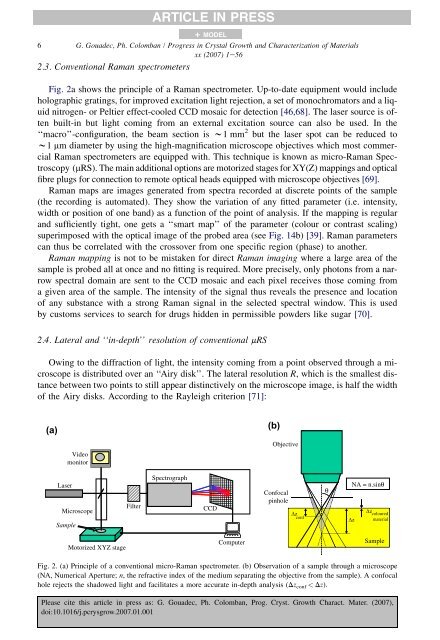
ARTICLE IN PRESS+ MODEL6 G. Goua<strong>de</strong>c, Ph. Colomban / Progress in Crystal Growth and Characterization <strong>of</strong> Materialsxx (2007) 1e562.3. Conventional <strong>Raman</strong> spectrom<strong>et</strong>ersFig. 2a shows the principle <strong>of</strong> a <strong>Raman</strong> spectrom<strong>et</strong>er. Up-to-date equipment would inclu<strong>de</strong>holographic gratings, for improved excitation light rejection, a s<strong>et</strong> <strong>of</strong> monochromators and a liquidnitrogen- or Peltier effect-cooled CCD mosaic for <strong>de</strong>tection [46,68]. The laser source is <strong>of</strong>tenbuilt-in but light coming from an external excitation source can also be used. In the‘‘macro’’-configuration, the beam section is w1 mm 2 but the laser spot can be reduced tow1 mm diam<strong>et</strong>er by using the high-magnification microscope objectives which most commercial<strong>Raman</strong> spectrom<strong>et</strong>ers are equipped with. This technique is known as micro-<strong>Raman</strong> <strong>Spectroscopy</strong>(mRS). The main additional options are motorized stages for XY(Z) mappings and opticalfibre plugs for connection to remote optical heads equipped with microscope objectives [69].<strong>Raman</strong> maps are images generated from spectra recor<strong>de</strong>d at discr<strong>et</strong>e points <strong>of</strong> the sample(the recording is automated). They show the variation <strong>of</strong> any fitted param<strong>et</strong>er (i.e. intensity,width or position <strong>of</strong> one band) as a function <strong>of</strong> the point <strong>of</strong> analysis. If the mapping is regularand sufficiently tight, one g<strong>et</strong>s a ‘‘smart map’’ <strong>of</strong> the param<strong>et</strong>er (colour or contrast scaling)superimposed with the optical image <strong>of</strong> the probed area (see Fig. 14b) [39]. <strong>Raman</strong> param<strong>et</strong>erscan thus be correlated with the crossover from one specific region (phase) to another.<strong>Raman</strong> mapping is not to be mistaken for direct <strong>Raman</strong> imaging where a large area <strong>of</strong> thesample is probed all at once and no fitting is required. More precisely, only photons from a narrowspectral domain are sent to the CCD mosaic and each pixel receives those coming froma given area <strong>of</strong> the sample. The intensity <strong>of</strong> the signal thus reveals the presence and location<strong>of</strong> any substance with a strong <strong>Raman</strong> signal in the selected spectral window. This is usedby customs services to search for drugs hid<strong>de</strong>n in permissible pow<strong>de</strong>rs like sugar [70].2.4. Lateral and ‘‘in-<strong>de</strong>pth’’ resolution <strong>of</strong> conventional mRSOwing to the diffraction <strong>of</strong> light, the intensity coming from a point observed through a microscopeis distributed over an ‘‘Airy disk’’. The lateral resolution R, which is the smallest distanceb<strong>et</strong>ween two points to still appear distinctively on the microscope image, is half the width<strong>of</strong> the Airy disks. According to the Rayleigh criterion [71]:(a)Vi<strong>de</strong>omonitor(b)ObjectiveLaserMicroscopeSampleFilterSpectrographCCDConfocalpinholez confθNA = n.sinθzz colouredmaterialMotorized XYZ stageComputerSampleFig. 2. (a) Principle <strong>of</strong> a conventional micro-<strong>Raman</strong> spectrom<strong>et</strong>er. (b) Observation <strong>of</strong> a sample through a microscope(NA, Numerical Aperture; n, the refractive in<strong>de</strong>x <strong>of</strong> the medium separating the objective from the sample). A confocalhole rejects the shadowed light and facilitates a more accurate in-<strong>de</strong>pth analysis (Dz conf < Dz).Please cite this article in press as: G. Goua<strong>de</strong>c, Ph. Colomban, Prog. Cryst. Growth Charact. Mater. (2007),doi:10.1016/j.pcrysgrow.2007.01.001