- Page 1:
COUNCIL ON DENTAL EDUCATION AND LIC
- Page 4 and 5:
A. General InformationThe American
- Page 7 and 8:
The American College of Prosthodont
- Page 9 and 10:
The American College of Prosthodont
- Page 11 and 12:
The American College of Prosthodont
- Page 13 and 14:
The American College of Prosthodont
- Page 15 and 16:
The American College of Prosthodont
- Page 17 and 18:
Changes in Scope of PracticeThe Ame
- Page 19 and 20:
The American College of Prosthodont
- Page 22 and 23:
The American College of Prosthodont
- Page 24 and 25:
The American College of Prosthodont
- Page 26 and 27:
ReferencesThe American College of P
- Page 28 and 29:
The American College of Prosthodont
- Page 30 and 31:
COUNCIL ON DENTAL EDUCATION AND LIC
- Page 33 and 34:
ENVISIONED FUTURE~ 10-30 YEAR HORIZ
- Page 35 and 36:
Prosthodontists will provide prosth
- Page 37 and 38:
GOALS AND OBJECTIVES~ 3-5 YEAR PLAN
- Page 39 and 40:
E4. Create effective linkages to fu
- Page 41 and 42:
Journal of ProsthodonticsImplant, E
- Page 43 and 44:
December 2005, Supplement, Volume 1
- Page 45 and 46:
December 2005, Supplement, Volume 1
- Page 47 and 48:
December 2005, Supplement, Volume 1
- Page 49 and 50:
December 2005, Supplement, Volume 1
- Page 51 and 52:
December 2005, Supplement, Volume 1
- Page 53 and 54:
December 2005, Supplement, Volume 1
- Page 55 and 56:
December 2005, Supplement, Volume 1
- Page 57 and 58:
December 2005, Supplement, Volume 1
- Page 59 and 60:
December 2005, Supplement, Volume 1
- Page 61 and 62:
December 2005, Supplement, Volume 1
- Page 63 and 64:
December 2005, Supplement, Volume 1
- Page 65 and 66:
December 2005, Supplement, Volume 1
- Page 67 and 68:
December 2005, Supplement, Volume 1
- Page 69 and 70:
December 2005, Supplement, Volume 1
- Page 71 and 72:
December 2005, Supplement, Volume 1
- Page 73 and 74:
December 2005, Supplement, Volume 1
- Page 75 and 76:
December 2005, Supplement, Volume 1
- Page 77 and 78:
December 2005, Supplement, Volume 1
- Page 79 and 80:
December 2005, Supplement, Volume 1
- Page 81 and 82:
December 2005, Supplement, Volume 1
- Page 83 and 84:
December 2005, Supplement, Volume 1
- Page 85 and 86:
December 2005, Supplement, Volume 1
- Page 87 and 88:
December 2005, Supplement, Volume 1
- Page 89 and 90:
December 2005, Supplement, Volume 1
- Page 91 and 92:
December 2005, Supplement, Volume 1
- Page 93 and 94:
December 2005, Supplement, Volume 1
- Page 95 and 96:
December 2005, Supplement, Volume 1
- Page 97 and 98: December 2005, Supplement, Volume 1
- Page 99 and 100: December 2005, Supplement, Volume 1
- Page 101 and 102: December 2005, Supplement, Volume 1
- Page 103 and 104: December 2005, Supplement, Volume 1
- Page 105 and 106: December 2005, Supplement, Volume 1
- Page 107 and 108: December 2005, Supplement, Volume 1
- Page 109 and 110: December 2005, Supplement, Volume 1
- Page 111 and 112: December 2005, Supplement, Volume 1
- Page 113 and 114: December 2005, Supplement, Volume 1
- Page 115 and 116: December 2005, Supplement, Volume 1
- Page 117 and 118: December 2005, Supplement, Volume 1
- Page 119 and 120: December 2005, Supplement, Volume 1
- Page 121 and 122: December 2005, Supplement, Volume 1
- Page 123 and 124: December 2005, Supplement, Volume 1
- Page 125 and 126: December 2005, Supplement, Volume 1
- Page 127 and 128: December 2005, Supplement, Volume 1
- Page 129 and 130: December 2005, Supplement, Volume 1
- Page 131 and 132: December 2005, Supplement, Volume 1
- Page 133 and 134: December 2005, Supplement, Volume 1
- Page 135 and 136: December 2005, Supplement, Volume 1
- Page 137 and 138: December 2005, Supplement, Volume 1
- Page 139 and 140: December 2005, Supplement, Volume 1
- Page 141 and 142: December 2005, Supplement, Volume 1
- Page 143 and 144: December 2005, Supplement, Volume 1
- Page 145 and 146: COUNCIL ON DENTAL EDUCATION AND LIC
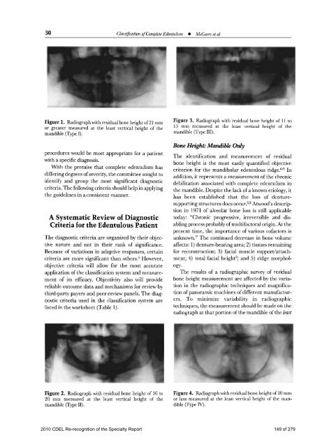
- Page 147: 28 Cllas$caizon of Complete Edentul
- Page 151 and 152: 32 Clarszjicatwn ofcomplete Edeiitu
- Page 153 and 154: Figure 14. Class Ipatient. (A) Pano
- Page 155 and 156: Figure 16. Class KU patient. (A) Pa
- Page 157 and 158: 38 Clm$cation of Cumllete EdClass 1
- Page 159 and 160: COUNCIL ON DENTAL EDUCATION AND LIC
- Page 161 and 162: 182 Classification System for Parti
- Page 163 and 164: 184 Classification System Jor Parti
- Page 165 and 166: 186 Classification Systemfor Partia
- Page 167 and 168: 188 Classification System jor Parti
- Page 169 and 170: Figure 4. Class IV palienl. Edenlul
- Page 171 and 172: I192 Classification Sy stemfor Part
- Page 173 and 174: COUNCIL ON DENTAL EDUCATION AND LIC
- Page 175 and 176: 74 Classification System for the Co
- Page 177 and 178: 76 Classification System for the Co
- Page 179 and 180: 78 Classification System for the Co
- Page 181 and 182: 80 Classification System for the Co
- Page 183 and 184: 82 Classification System for the Co
- Page 185 and 186: ORALCANCERSCREENING,EXAMINATIONANDB
- Page 187 and 188: ChrisBornITProjectManagerLomaLindaU
- Page 189 and 190: THOSEATRISKFORORALCANCERWhoisatrisk
- Page 191 and 192: Inaprospectivestudy(NIH‐AARPDieta
- Page 193 and 194: AgedataWhenexaminingdatafrom19agegr
- Page 195 and 196: Whatisthestatusofcommunityscreening
- Page 197 and 198: ReferencesArbesSJ,OlshanAF.CaplanDJ
- Page 199 and 200:
THEPROCESSOFONCOGENESISIntroduction
- Page 201 and 202:
tightregulationoftranscriptionaleve
- Page 203 and 204:
withalloftheotherhallmarksinplace,a
- Page 205 and 206:
THERADIOGRAPHICEXAMINATIONCanradiog
- Page 207 and 208:
thinningorperforationofthecortices,
- Page 209 and 210:
lymphoma)andprimarycarcinomas,areen
- Page 211 and 212:
Figure4EffectsonadjacentstructuresW
- Page 213 and 214:
fromaprimarylesioninboneorthesoftti
- Page 215 and 216:
THEORALCANCERSCREENINGEXAMINATIONTh
- Page 217 and 218:
Society’sABCDERule:lookforAsymmet
- Page 219 and 220:
Function:Paraesthesia,paralysis,pai
- Page 221 and 222:
Examinetheentireheadandneckincludin
- Page 223 and 224:
Figure11IntraoralexaminationTheintr
- Page 225 and 226:
Figure13Thesoftpalate,uvula,andtons
- Page 227 and 228:
Figure15Examinationofthetongueisthe
- Page 229 and 230:
Figure18Finally,thefloorofthemouthn
- Page 231 and 232:
MANAGEMENTOFSUSPICIOUSORALLESIONSIn
- Page 233 and 234:
Tables1through10ThoseatriskTable1 4
- Page 235 and 236:
Table3 512010 CDEL Re-recognition o
- Page 237 and 238:
Table6 532010 CDEL Re-recognition o
- Page 239 and 240:
Table9Table10 552010 CDEL Re-recogn
- Page 241 and 242:
CPE Course History From 2000-2010Da
- Page 243 and 244:
COUNCIL ON DENTAL EDUCATION AND LIC
- Page 245 and 246:
Accreditation Standards forAdvanced
- Page 247 and 248:
Document Revision History (continue
- Page 249 and 250:
Mission Statement of theCommission
- Page 251 and 252:
PrefaceMaintaining and improving th
- Page 253 and 254:
Definitions of Terms Used in Prosth
- Page 255 and 256:
Educationally Qualified: An individ
- Page 257 and 258:
Intent: Major changes have a direct
- Page 259 and 260:
STANDARD 2 - PROGRAM DIRECTOR AND T
- Page 261 and 262:
STANDARD 3 - FACILITIES AND RESOURC
- Page 263 and 264:
STANDARD 4 - CURRICULUM AND PROGRAM
- Page 265 and 266:
Intent: Students will have in depth
- Page 267 and 268:
4-18 Students/Residents must be exp
- Page 269 and 270:
STANDARD 5 - ADVANCED EDUCATION STU
- Page 271 and 272:
STANDARD 6 - RESEARCHAdvanced speci
- Page 273 and 274:
2010 CDEL Re-recognition of the Spe
- Page 275 and 276:
2010 CDEL Re-recognition of the Spe
- Page 277 and 278:
2010 CDEL Re-recognition of the Spe
- Page 279:
2010 CDEL Re-recognition of the Spe