Dental Asia May/June 2018
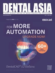
For more than two decades, Dental Asia is the premium journal in linking dental innovators and manufacturers to its rightful audience. We devote ourselves in showcasing the latest dental technology and share evidence-based clinical philosophies to serve as an educational platform to dental professionals. Our combined portfolio of print and digital media also allows us to reach a wider market and secure our position as the leading dental media in the Asia Pacific region while facilitating global interactions among our readers.
For more than two decades, Dental Asia is the premium journal in linking dental innovators
and manufacturers to its rightful audience. We devote ourselves in showcasing the latest dental technology and share evidence-based clinical philosophies to serve as an educational platform to dental professionals. Our combined portfolio of print and digital media also allows us to reach a wider market and secure our position as the leading dental media in the Asia Pacific region while facilitating global interactions among our readers.
- No tags were found...
You also want an ePaper? Increase the reach of your titles
YUMPU automatically turns print PDFs into web optimized ePapers that Google loves.
Behind the Scenes<br />
increase the accuracy of the restoration and<br />
to decrease the diculty of implementation<br />
and the necessary time of processing.<br />
Clinical case<br />
A 54-year-old, male patient, non-smoker<br />
with no major clinical diseases, came to<br />
the clinician with an upper edentulous<br />
arch with some lower teeth present (from<br />
34 to 45). The upper total prosthesis was<br />
incongruous, and caused some diculties<br />
in chewing and phonation. The patient<br />
manifested a psychological discomfort<br />
due the prosthesis condition in relation<br />
to his age, which hindered his speech<br />
when socialising with his co-workers. He<br />
also expressed the wish to replace the<br />
removable upper dentures with implants,<br />
quoting his own words, “something that<br />
will stay xed in the mouth and without<br />
palate.”<br />
The clinician started constructing the<br />
upper jaw with a new provisional but still<br />
entirely removable prosthesis for diagnostic<br />
purposes, and with the periodontal<br />
treatment of the lower arch, with a<br />
provisional and removable partial denture.<br />
Once the aesthetics and correct occlusal<br />
plane was restored, it is fundamental to<br />
maintain the buccal flange to support<br />
the upper lip. Some diagnostic tests were<br />
performed to study the placement of<br />
implants with a panoramic radiography and<br />
a computed tomography. In accordance<br />
to the patient’s approval, the clinician<br />
proceeds with the following treatment plan:<br />
insertion of four implants, in 14, 12, 22 and 24<br />
areas and the construction of an implant -<br />
mucosal supported prosthesis with a milled<br />
bar on the upper arch; and the lower jaw<br />
was maintained healthy and restored with<br />
a removable partial denture.<br />
Once the implants were placed, the<br />
position was established according to the<br />
availability of the bone and the prosthetic<br />
requirements, the patient waited for the<br />
successful osseointegration with the total<br />
temporary prosthesis, suitably modied.<br />
In this period, the patient was subjected<br />
to periodontal maintenance therapy. The<br />
same prosthesis was used as a base for the<br />
nal restoration.<br />
Implementation of the transparent<br />
acrylic resin replica<br />
The provisional prosthesis was positioned<br />
with a precision silicone with a hardness of<br />
70 Shore-A.<br />
The prosthesis and model silicone<br />
obtained were placed in a ask and then<br />
an insulator (insulating silicone spray,<br />
Transformer) was applied. Another silicone<br />
was placed between the replica and the<br />
cover of the ask, which was closed and<br />
held in place until the full curing of the<br />
silicone. The prosthesis was removed<br />
from the ask and two holes through the<br />
upper silicon (a 0.5 cm diameter for the<br />
input channel along with a 0.3 cm for the<br />
output channel) to allow the injection of<br />
the transparent acrylic resin. The resin was<br />
mixed and injected inside the ask, which<br />
was maintained at 50°C for 25 minutes at a<br />
pressure of 2.5 bars. Once cured, the ask<br />
was opened and the replica was nished<br />
with rotary instruments mounted on a<br />
laboratory handpiece and delivered to the<br />
clinician.<br />
Fig. 1: Impression obtained with<br />
the prosthesis replica<br />
Fig. 2: Placing of the laboratory analogues and<br />
the articial gingiva<br />
Impression with the prosthesis replica<br />
The transparent resin replica was used in a<br />
single chairside appointment, as customised<br />
tray, as a reference of the teeth set-up<br />
(control of the vertical dimension, the<br />
masticatory plane and the relationship<br />
with the antagonist), and as a rst test of<br />
the aesthetics (smile line, midline, etc). The<br />
clinician proceeded with the insertion of<br />
the replica in the oral cavity, checking the<br />
occlusion and removing the wrong occlusal<br />
contacts. The precise occlusion was then<br />
recorded using an additional fast-curing<br />
silicone. The replica had been perforated<br />
in correspondence with the emergence of<br />
the implants and daubed with adhesive.<br />
For the impression an addition silicone has<br />
been used and the replica was maintained<br />
in position by the patient with his bite until<br />
the complete polymerisation occurs; before<br />
its removal, a face bow was recorded. After<br />
removing the replica from the oral cavity,<br />
the silicone inside the holes was removed<br />
with a scalpel to allow the insertion of the<br />
pick-up transfers. The replica was placed<br />
back into the oral cavity and the<br />
transfers were screwed on the<br />
implants. Keeping the prosthesis in<br />
place, the transfers were blocked<br />
on the replica using light curing<br />
resin with low shrinkage. The xing<br />
screws were removed from the<br />
transfers and the impression was<br />
delivered to the laboratory after the<br />
disinfection protocol (Fig. 1).<br />
Fabrication of the master model<br />
and aesthetic try-in<br />
A silicone reproducing the soft<br />
tissues was placed at the area<br />
around the transfers then a proper<br />
insulation was daubed (Fig. 2).<br />
The master model was poured<br />
by developing the impression<br />
obtained with the replica with a<br />
class IV plaster; according to the<br />
manufacturer’s instructions (Fig. 3-4).<br />
Once hardened, the transfers were<br />
removed and the master model was<br />
positioned on the articulator using<br />
the replica and the face bow. The<br />
antagonist model was placed on<br />
the articulator with the silicone bite.<br />
DENTAL ASIA<br />
MAY / JUNE <strong>2018</strong><br />
59


