

Thoracic Imaging 2003 - Society of Thoracic Radiology
Thoracic Imaging 2003 - Society of Thoracic Radiology
Thoracic Imaging 2003 - Society of Thoracic Radiology

You also want an ePaper? Increase the reach of your titles
YUMPU automatically turns print PDFs into web optimized ePapers that Google loves.
TUESDAY<br />
170<br />
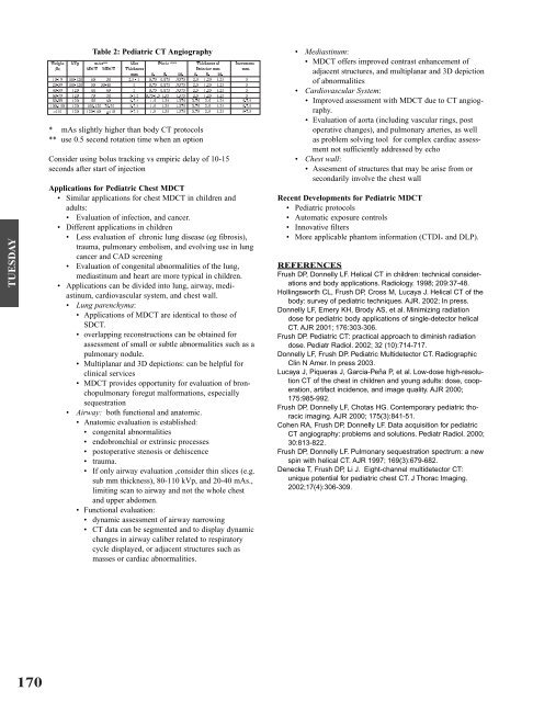
Table 2: Pediatric CT Angiography<br />
* mAs slightly higher than body CT protocols<br />
** use 0.5 second rotation time when an option<br />
Consider using bolus tracking vs empiric delay <strong>of</strong> 10-15<br />
seconds after start <strong>of</strong> injection<br />
Applications for Pediatric Chest MDCT<br />
• Similar applications for chest MDCT in children and<br />
adults:<br />
• Evaluation <strong>of</strong> infection, and cancer.<br />
• Different applications in children<br />
• Less evaluation <strong>of</strong> chronic lung disease (eg fibrosis),<br />
trauma, pulmonary embolism, and evolving use in lung<br />
cancer and CAD screening<br />
• Evaluation <strong>of</strong> congenital abnormalities <strong>of</strong> the lung,<br />
mediastinum and heart are more typical in children.<br />
• Applications can be divided into lung, airway, mediastinum,<br />
cardiovascular system, and chest wall.<br />
• Lung parenchyma:<br />
• Applications <strong>of</strong> MDCT are identical to those <strong>of</strong><br />
SDCT.<br />
• overlapping reconstructions can be obtained for<br />
assessment <strong>of</strong> small or subtle abnormalities such as a<br />
pulmonary nodule.<br />
• Multiplanar and 3D depictions: can be helpful for<br />
clinical services<br />
• MDCT provides opportunity for evaluation <strong>of</strong> bronchopulmonary<br />
foregut malformations, especially<br />
sequestration<br />
• Airway: both functional and anatomic.<br />
• Anatomic evaluation is established:<br />
• congenital abnormalities<br />
• endobronchial or extrinsic processes<br />
• postoperative stenosis or dehiscence<br />
• trauma.<br />
• If only airway evaluation ,consider thin slices (e.g.<br />
sub mm thickness), 80-110 kVp, and 20-40 mAs.,<br />
limiting scan to airway and not the whole chest<br />
and upper abdomen.<br />
• Functional evaluation:<br />
• dynamic assessment <strong>of</strong> airway narrowing<br />
• CT data can be segmented and to display dynamic<br />
changes in airway caliber related to respiratory<br />
cycle displayed, or adjacent structures such as<br />
masses or cardiac abnormalities.<br />
• Mediastinum:<br />
• MDCT <strong>of</strong>fers improved contrast enhancement <strong>of</strong><br />
adjacent structures, and multiplanar and 3D depiction<br />
<strong>of</strong> abnormalities<br />
• Cardiovascular System:<br />
• Improved assessment with MDCT due to CT angiography.<br />
• Evaluation <strong>of</strong> aorta (including vascular rings, post<br />
operative changes), and pulmonary arteries, as well<br />
as problem solving tool for complex cardiac assessment<br />
not sufficiently addressed by echo<br />
• Chest wall:<br />
• Assesment <strong>of</strong> structures that may be arise from or<br />
secondarily involve the chest wall<br />
Recent Developments for Pediatric MDCT<br />
• Pediatric protocols<br />
• Automatic exposure controls<br />
• Innovative filters<br />
• More applicable phantom information (CTDIw and DLP).<br />
REFERENCES<br />
Frush DP, Donnelly LF. Helical CT in children: technical considerations<br />
and body applications. <strong>Radiology</strong>. 1998; 209:37-48.<br />
Hollingsworth CL, Frush DP, Cross M, Lucaya J. Helical CT <strong>of</strong> the<br />
body: survey <strong>of</strong> pediatric techniques. AJR. 2002; In press.<br />
Donnelly LF, Emery KH, Brody AS, et al. Minimizing radiation<br />
dose for pediatric body applications <strong>of</strong> single-detector helical<br />
CT. AJR 2001; 176:303-306.<br />
Frush DP. Pediatric CT: practical approach to diminish radiation<br />
dose. Pediatr Radiol. 2002; 32 (10):714-717.<br />
Donnelly LF, Frush DP. Pediatric Multidetector CT. Radiographic<br />
Clin N Amer. In press <strong>2003</strong>.<br />
Lucaya J, Piqueras J, Garcia-Peña P, et al. Low-dose high-resolution<br />
CT <strong>of</strong> the chest in children and young adults: dose, cooperation,<br />
artifact incidence, and image quality. AJR 2000;<br />
175:985-992.<br />
Frush DP, Donnelly LF, Chotas HG. Contemporary pediatric thoracic<br />
imaging. AJR 2000; 175(3):841-51.<br />
Cohen RA, Frush DP, Donnelly LF. Data acquisition for pediatric<br />
CT angiography: problems and solutions. Pediatr Radiol. 2000;<br />
30:813-822.<br />
Frush DP, Donnelly LF. Pulmonary sequestration spectrum: a new<br />
spin with helical CT. AJR 1997; 169(3):679-682.<br />
Denecke T, Frush DP, Li J. Eight-channel multidetector CT:<br />
unique potential for pediatric chest CT. J Thorac <strong>Imaging</strong>.<br />
2002;17(4):306-309.


