- Page 2 and 3:
Welcome Welcome to Guangzhou and th
- Page 4 and 5:
09:00-19:00 Registration Friday, No
- Page 6 and 7:
14:20-14:40 17 HIGH EXPRESSION LEVE
- Page 8 and 9:
09:45-10:05 29 INDUCTION OF EPSTEIN
- Page 10 and 11:
Invited Presentation 4 Sunday, Nove
- Page 12 and 13:
10:50-11:10 49 BLOOD DIFFUSION AND
- Page 14 and 15:
Poster sessions Poster session 1: L
- Page 16 and 17:
74 REGULATION OF EPSTEIN-BARR VIRUS
- Page 18 and 19:
91 EPSTEIN-BARR VIRUS SM PROTEIN IN
- Page 20 and 21:
108 THE EVOLUTION OF EPSTEIN-BARR V
- Page 22 and 23:
124 EPSTEIN-BARR VIRUS ENCODED LATE
- Page 24 and 25:
143 THE EPSTEIN-BARR VIRUS (EBV)-EN
- Page 26 and 27:
159 HIGH PERFORIN EXPRESSION ON T C
- Page 28 and 29:
177 CHD5 IS A NOVEL 1P36 TUMOR SUPP
- Page 30 and 31:
191 EBV-POSITIVE NK/T-CELL LYMPHOMA
- Page 32 and 33:
206 RAPID TEST FOR DIAGNOSIS OF MON
- Page 34 and 35:
219 THE EPSTEIN-BARR VIRUS PROTEASE
- Page 36 and 37:
1 (RegID: 1068) Wolfgang Hammerschm
- Page 38 and 39:
3 Yi-xin Zeng, Tiebang Kang State K
- Page 40 and 41:
5 (RegID: 1840) Kenzo Takada Instit
- Page 42 and 43:
NOTES: EBV Conference 2008 Guangzho
- Page 44 and 45:
7 (RegID: 1268; 1269) Paul Lieberma
- Page 46 and 47:
9 (RegID: 1538) Lorand L.Kis Instit
- Page 48 and 49:
NOTES: EBV Conference 2008 Guangzho
- Page 50 and 51:
11 (RegID:1801) George Sai Wah Tsao
- Page 52 and 53:
13 (RegID: 1742) Natalie Sutkowski
- Page 54 and 55:
NOTES: EBV Conference 2008 Guangzho
- Page 56 and 57:
15 (RegID:1042; 1043; 1044) Evelyne
- Page 58 and 59:
17 (RegID: 1773) Derek Daigle Insti
- Page 60 and 61:
19 (RegID:1252; 1254; 1255) Andrew
- Page 62 and 63:
Oral speaker Abstract Session 4: GE
- Page 64 and 65:
21 (RegID: 1125) Dong-Yan Jin Insti
- Page 66 and 67:
23 (RegID:1933; 1934; 1775) Lifu Hu
- Page 68 and 69:
NOTES: EBV Conference 2008 Guangzho
- Page 70 and 71:
25 (RegID:1109) Zhenguo Wu Institut
- Page 72 and 73:
27 (RegID:1191; 1192) Arnd Kieser I
- Page 74 and 75:
29 (RegID:1213) Yao Chang Instituti
- Page 76 and 77:
NOTES: EBV Conference 2008 Guangzho
- Page 78 and 79:
31 (RegID:1114; 1115; 1116) Elena K
- Page 80 and 81:
33 (RegID: 1249; 1250) Ellen Cahir-
- Page 82 and 83:
NOTES: EBV Conference 2008 Guangzho
- Page 84 and 85:
35 (RegID:1170; 1171; 1172) Joanna
- Page 86 and 87:
37 (RegID: 1062) Maria Lung Institu
- Page 88 and 89:
39 Mu Shen zeng, Li-bing Song Insti
- Page 90 and 91:
Oral speaker Abstract Session 8: BU
- Page 92 and 93:
41 (RegID:1133; 1134) Rosemary Roch
- Page 94 and 95:
43 (RegID: 1760; 1761; 1763) Shidon
- Page 96 and 97:
Oral speaker Abstract Session 9: OT
- Page 98 and 99:
45 (RegID: 1495) Kimberley Jones In
- Page 100 and 101:
NOTES: EBV Conference 2008 Guangzho
- Page 102 and 103:
47 (RegID: 1716) Hans Wolf Institut
- Page 104 and 105:
49 (RegID: 1883; 1884; 1885) Pierre
- Page 106 and 107:
51 (RegID: 1725) Riccardo Dolcetti
- Page 108 and 109:
53 (RegID: 1276; 1277; 1278) Laura
- Page 110 and 111:
Oral speaker Abstract Session 11: D
- Page 112 and 113:
55 (RegID: 1290) Jaap Middeldorp In
- Page 114 and 115:
NOTES: EBV Conference 2008 Guangzho
- Page 116 and 117:
57 (RegID: 1087) Maha Al-Mozaini In
- Page 118 and 119:
59 (RegID: 1107 ; 1108) Tathagata C
- Page 120 and 121:
61 (RegID: 1117) Barbro Ehlin-Henri
- Page 122 and 123:
63 (RegID: 1236) Junying Jia Instit
- Page 124 and 125:
65 (RegID: 1320) Kudakwashe Mutyamb
- Page 126 and 127:
67 (RegID: 1459) Koichi Katsumura I
- Page 128 and 129:
69 (RegID: 1676; 1678; 1679) Martin
- Page 130 and 131:
71 (RegID: 1715; 1717; 1722) Richar
- Page 132 and 133:
NOTES: EBV Conference 2008 Guangzho
- Page 134 and 135:
73 (RegID: 1025; 1026) Wim Burmeist
- Page 136 and 137:
75 (RegID: 1110) Markus Kalla Insti
- Page 138 and 139:
77 (RegID: 1168; 1169; 1248) Claude
- Page 140 and 141:
79 (RegID: 1240) Haide Qin Institut
- Page 142 and 143:
81 (RegID: 1524) Stefano Gastaldell
- Page 144 and 145:
83 (RegID: 1708; 1710; 1711) Ingema
- Page 146 and 147:
85 (RegID: 1764; 1765; 1766; 1767)
- Page 148 and 149:
87 (RegID: 1989) Hongyu Deng Center
- Page 150 and 151:
89 (RegID: 1184; 1185; 1186) Alison
- Page 152 and 153:
91 (RegID: 1261) Dinesh Verma Insti
- Page 154 and 155:
93 (RegID:1449; 1450; 1451) Vicki G
- Page 156 and 157:
95 (RegID: 1752; 1754; 1755; 1756;
- Page 158 and 159:
97 (RegID: 1753) Stacy Hagemeier In
- Page 160 and 161:
NOTES: EBV Conference 2008 Guangzho
- Page 162 and 163:
99 (RegID: 1135; 1136) Liang Wu Ins
- Page 164 and 165:
101 (RegID: 1265; 1289) Sarah Leona
- Page 166 and 167:
103 (RegID: 1282) Ting Ting Yang In
- Page 168 and 169:
105 (RegID: 1294; 1296; 1297; 1298;
- Page 170 and 171: 107 (RegID: 1518) Jamie Nourse Inst
- Page 172 and 173: 109 (RegID: 1625; 1629) Shu-Jen Che
- Page 174 and 175: 111 (RegID: 1774) Alan Chiang Insti
- Page 176 and 177: 113 (RegID: 1819) Mingfang Ji Insti
- Page 178 and 179: 115 (RegID: 1944; 1943) Changzhan g
- Page 180 and 181: NOTES: EBV Conference 2008 Guangzho
- Page 182 and 183: 117 (RegID: 1013) Xuechi Lin Instit
- Page 184 and 185: 119 (RegID: 1082; 1083) Laila Canci
- Page 186 and 187: 121 (RegID: 1105; 1106) Stephanie M
- Page 188 and 189: 123 (RegID: 1118) Surya Pavan Yenam
- Page 190 and 191: 125 (RegID: 1121) Lili Li Instituti
- Page 192 and 193: 127 (RegID: 1174; 1175) Xiangning Z
- Page 194 and 195: 129 (RegID: 1198; 1200; 1223; 1224)
- Page 196 and 197: 131 (RegID: 1215; 1216; 1217; 1218;
- Page 198 and 199: 133 (RegID: 1232; 1233) Thomas Owen
- Page 200 and 201: 135 (RegID: 1246; 1247) Christopher
- Page 202 and 203: 137 (RegID: 1284; 1285) Su-Fang Lin
- Page 204 and 205: 139 (RegID: 1465; 1505) Huang Sheng
- Page 206 and 207: 141 (RegID: 1474; 1475) chia-chun c
- Page 208 and 209: 143 (RegID: 1707; 1721) Mhairi Morr
- Page 210 and 211: 145 (RegID: 1730) Che-Pei (Pat) Kun
- Page 212 and 213: 147 (RegID: 1740) Anjali Bheda Inst
- Page 214 and 215: 149 (RegID: 1874; 1875) Seiji Maruo
- Page 216 and 217: 151 (RegID: 1926) Masanao Murakami
- Page 218 and 219: Poster sessions Session 5: Immune M
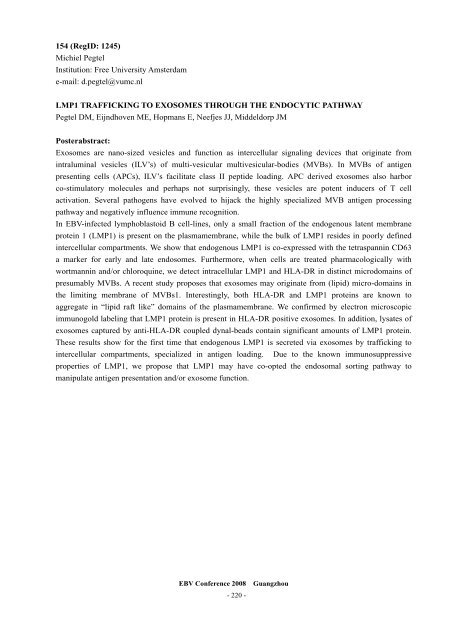
- Page 222 and 223: 155 (RegID: 1251; 1253) Akihiko Mae
- Page 224 and 225: 157 (RegID:1301; 1302、1303) Heath
- Page 226 and 227: 159 (RegID: 1355; 1383) Floor Piete
- Page 228 and 229: 161 (RegID: 1454) Hideyuki Ishii In
- Page 230 and 231: 163 (RegID: 1647) Carol Sze-Ki LEUN
- Page 232 and 233: 165 (RegID: 1741) Gretchen Bentz In
- Page 234 and 235: 167 (RegID: 1770; 1771; 1772; 1953)
- Page 236 and 237: 169 (RegID: 1784) Mark Fogg Institu
- Page 238 and 239: Poster sessions Session 6: Nasophar
- Page 240 and 241: 171 (RegID: 1027) Nicolas Tarbourie
- Page 242 and 243: 173 (RegID: 1063) King Chi Chan Ins
- Page 244 and 245: 175 (RegID: 1132) Qian Tao Institut
- Page 246 and 247: 177 (RegID: 1132) Qian Tao Institut
- Page 248 and 249: 179 (RegID: 1503; 1504; 1506; 1507;
- Page 250 and 251: 181 (RegID: 1706; 1785) Ziming Du I
- Page 252 and 253: 183 (RegID: 1931; 1932) Alan Khoo I
- Page 254 and 255: Poster sesions Session 7: Burkitt
- Page 256 and 257: 185 (RegID:1188) Qiu-liang Wu(1) In
- Page 258 and 259: 187 GAN Runliang (甘润良) Instit
- Page 260 and 261: NOTES: EBV Conference 2008 Guangzho
- Page 262 and 263: 189 (RegID: 1071; 1072) Shigetaka M
- Page 264 and 265: 191 (RegID: 1190) Qiu-liang Wu(2) I
- Page 266 and 267: 193 (RegID: 1195) Stephanie Volke I
- Page 268 and 269: 195 (RegID: 1274; 1275) Miki Takaha
- Page 270 and 271:
197 (RegID: 1723) Katerina Vrzaliko
- Page 272 and 273:
199 (RegID: 1803) Suk Kyeong Lee In
- Page 274 and 275:
201 (RegID: 1863) Julinor Bacani In
- Page 276 and 277:
Poster sessions Session 9: Diagnost
- Page 278 and 279:
203 (RegID: 1291) Jaap Middeldorp I
- Page 280 and 281:
205 (RegID: 1292) Jaap Middeldorp I
- Page 282 and 283:
207 (RegID: 1526) Patrice MORAND In
- Page 284 and 285:
NOTES: EBV Conference 2008 Guangzho
- Page 286 and 287:
209 (RegID: 1065) Corey Smith Insti
- Page 288 and 289:
211 (RegID: 1141) Graham Taylor Ins
- Page 290 and 291:
213 (RegID: 1685; 1687) Frank van S
- Page 292 and 293:
215 (RegID: 1923) Claire Gourzones
- Page 294 and 295:
Poster sessions Session 11: Drug De
- Page 296 and 297:
217 (RegID: 1181) Kwai Fung Hui Ins
- Page 298 and 299:
219 (RegID: 1227) Raphele Germi Ins
- Page 300 and 301:
221 (RegID: 1466; 1467) Lih-Chyang
- Page 302 and 303:
223 (RegID: 1744) Qiao Meng Institu
- Page 304 and 305:
NOTES: EBV Conference 2008 Guangzho
- Page 306 and 307:
NOTES: EBV Conference 2008 Guangzho


