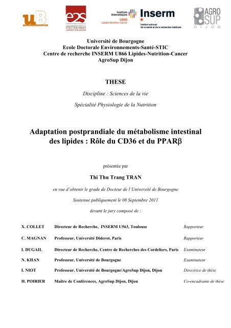
Université de Bourgogne
Université de Bourgogne
Université de Bourgogne

Create successful ePaper yourself
Turn your PDF publications into a flip-book with our unique Google optimized e-Paper software.
Adaaptation<br />
postpprandiiale<br />
du métabbolismee<br />
intest tinal<br />
<strong>de</strong>s llipi<strong>de</strong>s<br />
: Rôlee<br />
du CD D36 et du PPAARβ<br />
X. COLLETT<br />
Direccteur<br />
<strong>de</strong> Reccherche,<br />
INNSERM<br />
U56 63, Toulousee<br />
C. MAGNAAN<br />
Proffesseur,<br />
Univversité<br />
Di<strong>de</strong>rot,<br />
Paris<br />
I. DUGAILL<br />
Direccteur<br />
<strong>de</strong> Reccherche,<br />
Ceentre<br />
<strong>de</strong> Rec cherches <strong>de</strong>ss<br />
Cor<strong>de</strong>liers, , Paris Ex xaminateur<br />
N. KHAN<br />
I. NIOT<br />
Universsité<br />
<strong>de</strong> <strong>Bourgogne</strong><br />
Ecole Doctorale<br />
Environnements-Saanté-STICC<br />
Centre d<strong>de</strong><br />
rechercche<br />
INSERM<br />
U866 6 Lipi<strong>de</strong>s-Nutritionn-Cancer<br />
AggroSup<br />
Di ijon<br />
THESE E<br />
DDisciplinee<br />
: Science es <strong>de</strong> la viee<br />
Spéccialité<br />
Phyysiologie<br />
<strong>de</strong> d la Nutrrition<br />
pprésentée<br />
pa ar<br />
Thi Thhu<br />
Trang TRAN<br />
en vuue<br />
d’obtenir le gra<strong>de</strong> <strong>de</strong>e<br />
Docteur <strong>de</strong> d l’<strong>Université</strong><br />
<strong>de</strong> Bourg rgogne<br />
Soutennue<br />
publiquuement<br />
le 08 8 Septembree<br />
2011<br />
<strong>de</strong>vant lle<br />
jury comp posé <strong>de</strong> :<br />
Proffesseur,<br />
Univversité<br />
<strong>de</strong> Boourgogne<br />
Proffesseur,<br />
Univversité<br />
<strong>de</strong> Boourgogne/A<br />
AgroSup Dijoon,<br />
Dijon<br />
H. POIRIEER<br />
Maîttre<br />
<strong>de</strong> Conféérences,<br />
AgrroSup<br />
Dijon n, Dijon<br />
Ra apporteur<br />
Ra apporteur<br />
Ex xaminateur<br />
Di irectrice <strong>de</strong> thhèse<br />
Co o-encadrantee<br />
<strong>de</strong> thèse
Remerciements<br />
Quatre années <strong>de</strong> plaisir et <strong>de</strong> joie à découvrir <strong>de</strong> nouveaux résultats.<br />
Des soirées si contente car les expériences marchent bien et d’autres soirées pleines <strong>de</strong><br />
doutes : Et si notre hypothèse n’est pas bonne? Heureusement que « la nuit porte conseil!»…<br />
Quatre années passionnantes d’échanges et <strong>de</strong> moments inoubliables avec mes collègues.<br />
Je remercie toutes les personnes qui ont contribuées <strong>de</strong> près ou <strong>de</strong> loin à cette thèse.<br />
Je tiens à remercier Monsieur Philippe BESNARD, Professeur et Directeur du Laboratoire <strong>de</strong><br />
Physiologie <strong>de</strong> la Nutrition, <strong>de</strong> m’avoir accueillie au sein <strong>de</strong> son équipe, ainsi que son esprit<br />
critique et ses conseils.<br />
Toute ma gratitu<strong>de</strong> à Madame Isabelle NIOT, Professeur et Directrice <strong>de</strong> thèse, <strong>de</strong> m’avoir<br />
accordé sa gran<strong>de</strong> disponibilité, ainsi que pour la confiance qu’elle m’a témoigné et pour ses<br />
encouragements sans cesse renouvelés.<br />
Je souhaite également témoigner à Madame Hélène POIRIER, Maître <strong>de</strong> Conférences et Coencadrante<br />
<strong>de</strong> thèse, toute ma reconnaissance pour m’avoir communiqué son enthousiasme<br />
pour la recherche, pour son soutien permanent et ses remarques constructives tout au long <strong>de</strong><br />
mon stage <strong>de</strong> recherche DEA et <strong>de</strong> ma thèse. Un grand merci à toutes les <strong>de</strong>ux pour votre<br />
dévouement, votre rigueur, vos connaissances scientifiques et votre implication dans ce<br />
travail, ainsi que pour votre gentillesse et votre énergie qui furent enrichissantes aussi bien<br />
dans le travail que dans la vie.<br />
Je tiens à exprimer ma gratitu<strong>de</strong> et mon respect aux membres du jury.<br />
Je remercie tout particulièrement Monsieur Xavier COLLET, Directeur <strong>de</strong> recherche à<br />
l’INSERM U563, et Monsieur Christophe MAGNAN, Professeur à l’<strong>Université</strong> Di<strong>de</strong>rot pour<br />
m’avoir fait l’honneur d’être les rapporteurs <strong>de</strong> cette thèse.<br />
Je remercie également Madame Isabelle DUGAIL, Directeur <strong>de</strong> Recherche au Centre <strong>de</strong><br />
Recherches <strong>de</strong>s Cor<strong>de</strong>liers et Monsieur Naim KHAN, Professeur à <strong>Université</strong> <strong>de</strong> <strong>Bourgogne</strong><br />
qui ont accepté d’examiner ce travail.<br />
1
Je souhaite remercier l’ensemble <strong>de</strong>s membres du Laboratoire <strong>de</strong> physiologie <strong>de</strong> la Nutrition<br />
pour la bonne ambiance au travail.<br />
Mes plus vifs remerciements vont à Madame Marie-Clau<strong>de</strong> MONNOT, Technicienne du<br />
Laboratoire. Je la remercie pour sa gentillesse, son dévouement, sa bonne humeur et son<br />
implication dans ce travail. Sans elle, on n’aurait jamais réussi les « segments congelés ! »<br />
Je remercie Jean-François MERLIN pour son ai<strong>de</strong>, notamment pour l’étu<strong>de</strong> Echomeri <strong>de</strong>s<br />
souris KOCD36 et sa sympathie, ainsi que Madame Patricia DEGRACE pour son<br />
encouragement.<br />
Je remercie chaleureusement tous les doctorants : Valérie, Dany, Céline, Michaël, Marjorie,<br />
Véronique avec qui j’ai partagé non seulement le bureau pendant ces années <strong>de</strong> thèse mais<br />
également beaucoup <strong>de</strong> moments difficiles et joyeux (surtout toi Marjo pour la « relecture »<br />
<strong>de</strong> cette thèse !).<br />
Un grand merci à Lorène LEBRUN, stagiaire du Laboratoire pour sa contribution dans le<br />
résultat sur les souris KOPPARβ.<br />
Je remercie également mes amis: Chi, Hanh, Thoai, Hue, Quang, Phu, Huong, Truc,<br />
Floriane…<strong>de</strong>s tous les moments passés ensembles.<br />
Je voudrais remercier mes parents et ma petite sœur qui m’ont toujours encouragée et m’ont<br />
témoignée une confiance absolue.<br />
Je remercie <strong>de</strong> tout cœur mon mari, An Ninh TA, pour son amour et son soutien permanent.<br />
Merci d’être toujours à côté <strong>de</strong> moi.<br />
2
Résumé<br />
L’hypertriglycéridémie postprandiale représente un facteur <strong>de</strong> risque émergent <strong>de</strong>s maladies<br />
cardiovasculaires et est retrouvé en cas <strong>de</strong> syndrome métabolique, d’obésité et d’insulinorésistance.<br />
L’intestin grêle conditionne la triglycéridémie postprandiale puisque la taille et <strong>de</strong><br />
la quantité <strong>de</strong>s chylomicrons sécrétés modulent l’activité <strong>de</strong> la Lipoprotéine Lipase (LPL). La<br />
synthèse <strong>de</strong>s chylomicrons est un mécanisme complexe dont l’étape <strong>de</strong> lipidation <strong>de</strong><br />
l’Apolipoprotéine B48 (ApoB48) par la Microsomal Triglyceri<strong>de</strong> Transfert Protein (MTP) et<br />
celle <strong>de</strong> leur transfert du réticulum vers le Golgi dans laquelle intervient la Liver Fatty Acid<br />
binding Protein (L-FABP) sont limitantes. Des expériences menées in vivo chez <strong>de</strong>s animaux<br />
sauvages et transgéniques et ex vivo sur <strong>de</strong>s segments intestinaux, nous ont permis <strong>de</strong><br />
démontrer qu’il existe une adaptation postprandiale du métabolisme intestinal <strong>de</strong>s lipi<strong>de</strong>s.<br />
Cette adaptation postprandiale est déclenchée par la glycoprotéine CD36 qui en présence<br />
d’aci<strong>de</strong>s gras à longue chaîne (AGLC) régule la voie ERK1/2 et conduit à l’induction <strong>de</strong><br />
l’ApoB48, <strong>de</strong> la MTP et <strong>de</strong> la L-FABP. La dégradation rapi<strong>de</strong> du CD36 par la voie<br />
ubiquitine-protéasome en présence d’AGLC, qui conduit à la désactivation <strong>de</strong> la voie<br />
ERK1/2, est typique d’un récepteur. Puisque d’une part les souris invalidées pour le<br />
Peroxisome Proliferator Activated receptor β (PPARβ) présentent une altération <strong>de</strong><br />
l’adaptation et une hypertriglycéridémie postprandiale et que d’autre part les lipi<strong>de</strong>s<br />
alimentaires induisent le PPARβ via CD36, CD36 et PPARβ pourraient faire partie d’un<br />
mécanisme commun <strong>de</strong> régulation. En conclusion, CD36 et PPARβ contribuent au sensing<br />
entérocytaire <strong>de</strong>s AGLC d’origine alimentaire, responsable <strong>de</strong> l'adaptation postprandiale du<br />
métabolisme <strong>de</strong>s lipi<strong>de</strong>s qui favorise la formation <strong>de</strong> gros chylomicrons efficacement épurés<br />
<strong>de</strong> la circulation sanguine.<br />
Mots-clés : CD36, PPARβ, Récepteur, Chylomicrons, Triglycéridémie postprandiale.<br />
3
Abstract<br />
Postprandial hypertriglyceri<strong>de</strong>mia is an emerging risk factor for cardiovascular diseases and is<br />
associated with metabolic syndrome, obesity and insulin resistance. The small intestine<br />
participates in the postprandial triglyceri<strong>de</strong>mia since both the size and number of secreted<br />
chylomicrons modulate lipoprotein lipase activity (LPL). Chylomicron synthesis is a complex<br />
mechanism in which the lipidation of Apolipoprotein B48 (ApoB48) by the Microsomal<br />
Triglyceri<strong>de</strong> Transfer Protein (MTP) and the transfer between reticulum and Golgi in which<br />
the Liver Fatty Acid Binding Protein (L -FABP) is involved are limiting steps. An intestinal<br />
fat-mediated adaptation in postprandial period has been <strong>de</strong>monstrated by in vivo (transgenic<br />
and wild type mice) and ex vivo (intestinal segments) approches. This postprandial adaptation<br />
is triggered by the glycoprotein CD36 in the presence of Long chain Fatty Acids (LCFA) that<br />
regulates the ERK1/2 pathway and leads to the induction of ApoB48, MTP and L-FABP. The<br />
rapid <strong>de</strong>gradation of CD36 by the ubiquitin-proteasome pathway in the presence of LCFA,<br />
which leads to ERK1/2 <strong>de</strong>activation, has a feature of a receptor. Since firstly, Peroxisome<br />
Proliferator Activated Receptor β (PPAR β) knockout mice display an alteration of<br />
postprandial adaptation associated with a hypertriglyceri<strong>de</strong>mia and secondly, dietary fat-<br />
mediated PPARβ up-regulation is CD36 <strong>de</strong>pen<strong>de</strong>nt, CD36 and PPARβ might participate to a<br />
common regulation mechanism. In conclusion, CD36 and PPARβ contribute to the enterocyte<br />
LCFA sensing responsible for postprandial adaptation that promotes the formation of large<br />
chylomicrons efficiently cleared into the blood.<br />
Key words : CD36, PPARβ, Receptor, Chylomicrons, Postprandial triglyceri<strong>de</strong>mia.<br />
4
Table <strong>de</strong>s matières<br />
REMERCIEMENTS ....................................................................................................................... 1<br />
RESUME ...................................................................................................................................... 3<br />
ABSTRACT .................................................................................................................................. 4<br />
TABLE DES MATIERES ................................................................................................................ 5<br />
ABREVIATIONS ......................................................................................................................... 10<br />
LISTE DES FIGURES .................................................................................................................. 12<br />
LISTE DES TABLEAUX ............................................................................................................... 16<br />
INTRODUCTION ........................................................................................................................ 17<br />
SYNTHESE BIBLIOGRAPHIQUE ................................................................................................. 24<br />
LES LIPIDES ALIMENTAIRES .................................................................................................... 25<br />
1.1 Fonctions structurales et énergétiques .................................................................. 27<br />
1.2 Fonctions dans la signalisation cellulaire ............................................................. 27<br />
ABSORPTION INTESTINALE DES LIPIDES ALIMENTAIRES ........................................................ 28<br />
1 Les spécificités morphologiques et fonctionnelles <strong>de</strong> l’intestin grêle .......................... 28<br />
1.1 Anatomie complexe <strong>de</strong> l’épithélium intestinal ....................................................... 28<br />
1.2 Composition cellulaire <strong>de</strong> l’épithélium intestinal .................................................. 30<br />
1.3 Renouvellement intense <strong>de</strong> l’épithélium intestinal ................................................. 32<br />
2 Digestion <strong>de</strong>s lipi<strong>de</strong>s .................................................................................................... 34<br />
3 Absorption intestinale <strong>de</strong>s lipi<strong>de</strong>s ................................................................................ 35<br />
3.1 Captage transmembranaire <strong>de</strong>s AGLC .................................................................. 36<br />
3.1.1 FABPpm ....................................................................................................... 38<br />
3.1.2 FATP4 .......................................................................................................... 39<br />
3.1.3 CD36 ............................................................................................................ 40<br />
3.2 Le transport cytosolique <strong>de</strong>s AGLC ....................................................................... 48<br />
3.3 La ré-estérification <strong>de</strong>s AGLC en TG .................................................................... 51<br />
3.3.1 MGAT - Monoacylglycerol acyltransferase ................................................ 51<br />
5
3.3.2 DGAT - Diacylglycerol acyltransferase ....................................................... 51<br />
3.4 Formation <strong>de</strong>s chylomicrons .................................................................................. 52<br />
3.4.1 Assemblage <strong>de</strong>s préchylomicrons : caractéristique unique <strong>de</strong>s entérocytes 52<br />
3.4.2 Transport spécifique <strong>de</strong>s préchylomicrons vers le Golgi : une <strong>de</strong>uxième<br />
étape limitante .............................................................................................................. 63<br />
3.4.3 Maturation golgienne et sécrétion <strong>de</strong>s chylomicrons ................................... 65<br />
3.5 Métabolisme post-intestinal <strong>de</strong>s chylomicrons : impact sur la triglycéridémie<br />
postprandiale. ................................................................................................................... 65<br />
3.6 PPARs : régulateur potentiel <strong>de</strong> la triglycéridémie postprandiale ....................... 69<br />
PROBLEMATIQUE ..................................................................................................................... 73<br />
RESULTATS .............................................................................................................................. 75<br />
EXISTE-T-IL UNE ADAPTATION POSTPRANDIALE DU METABOLISME INTESTINAL DES<br />
LIPIDES ? .................................................................................................................................. 76<br />
1 L’ingestion <strong>de</strong> lipi<strong>de</strong>s alimentaires affecte rapi<strong>de</strong>ment le métabolisme intestinal <strong>de</strong>s<br />
lipi<strong>de</strong>s ................................................................................................................................... 77<br />
1.1 Cinétique <strong>de</strong> l’effet <strong>de</strong>s lipi<strong>de</strong>s alimentaires sur la teneur en triglycéri<strong>de</strong>s <strong>de</strong> la<br />
muqueuse intestinale et la triglycéridémie postprandiale. ............................................... 77<br />
1.2 L’ingestion <strong>de</strong> lipi<strong>de</strong>s alimentaires augmente l’expression <strong>de</strong>s gènes clefs<br />
impliqués dans l’absorption intestinale <strong>de</strong>s AGLC. ......................................................... 78<br />
1.2.1 Effet d’une charge en lipi<strong>de</strong>s sur le taux d’ARNm jéjunal <strong>de</strong>s LBPs<br />
impliquées dans le captage et le trafic intracellulaire <strong>de</strong>s AGLC ................................ 78<br />
1.2.2 Effet d’une charge en lipi<strong>de</strong>s sur le taux d’ARNm jéjunal <strong>de</strong>s protéines<br />
impliquées dans la formation et la sécrétion <strong>de</strong>s chylomicrons ................................... 79<br />
1.2.3 Effet d’une charge en lipi<strong>de</strong>s sur le taux d’ARNm jéjunal <strong>de</strong> l’ApoC-II et <strong>de</strong><br />
l’ApoC-III ..................................................................................................................... 81<br />
1.2.4 Effet d’une charge en lipi<strong>de</strong>s sur le taux d’ARNm jéjunal <strong>de</strong> protéines<br />
impliquées dans l’absorption du cholestérol ................................................................ 82<br />
1.2.5 Effet d’un charge en lipi<strong>de</strong>s sur le taux d’ARNm jéjunal <strong>de</strong>s PPARs ......... 83<br />
2 L’adaptation intestinale est-elle dépendante du PPARβ ? .......................................... 85<br />
2.1 L’invalidation du gène PPARβ ne modifie pas la surface d’absorption ............... 85<br />
6
2.2 L’absence du PPARβ modifie-elle le taux <strong>de</strong> TG <strong>de</strong> la muqueuse intestinale et<br />
l’hypertriglycéridémie postprandiale ? ............................................................................ 86<br />
2.3 L’absence du PPARβ modifie-t-elle la régulation <strong>de</strong>s gènes clefs <strong>de</strong> la formation<br />
<strong>de</strong>s chylomicrons et <strong>de</strong> leur clairance suite à une charge lipidique ? ............................ 87<br />
2.4 L’activation du PPARβ est-elle à l’origine <strong>de</strong> l’adaptation intestinale ? ............. 89<br />
CD36 EST-IL IMPLIQUE DANS L’ADAPTATION POSTPRANDIALE DU METABOLISME<br />
INTESTINAL DES LIPIDES ? ....................................................................................................... 93<br />
1 Les animaux KOCD36 ont-ils constitutivement la même capacité à synthétiser <strong>de</strong>s<br />
chylomicrons ? ..................................................................................................................... 94<br />
1.1 L’invalidation du gène CD36 ne modifie pas la balance énergétique en régime<br />
standard ............................................................................................................................ 95<br />
1.2 L’invalidation du gène CD36 ne modifie pas la surface d’absorption .................. 96<br />
1.3 L’invalidation du gène CD36 altère la triglycéridémie <strong>de</strong>s souris à jeun ............. 97<br />
1.4 Impact <strong>de</strong> l’invalidation du gène CD36 sur le taux basal <strong>de</strong>s ARNm <strong>de</strong>s protéines<br />
impliquées dans le métabolisme intestinal <strong>de</strong>s lipi<strong>de</strong>s ..................................................... 97<br />
2 Existe-t-il une adaptation <strong>de</strong>s capacités d’absorption intestinale chez les souris<br />
KOCD36 suite à une charge lipidique ? .............................................................................. 98<br />
2.1 Impact <strong>de</strong> l’invalidation du gène CD36 sur la triglycéridémie postprandiale ...... 99<br />
2.2 Impact <strong>de</strong> l’invalidation du gène CD36 sur l’expression <strong>de</strong>s protéines clefs<br />
impliquées dans la formation <strong>de</strong>s chylomicrons suite à une charge lipidique ............... 100<br />
2.3 Impact <strong>de</strong> l’invalidation du gène CD36 sur le taux d’ARNm <strong>de</strong>s PPARs après une<br />
charge en lipi<strong>de</strong>s ............................................................................................................ 102<br />
QUEL EST L’IMPACT DES LIPIDES ALIMENTAIRES SUR LA REGULATION POSTPRANDIALE DU<br />
CD36 ? ................................................................................................................................... 105<br />
1 Le CD36 intestinal n’est pas un transporteur efficace aux AGLC ............................ 106<br />
2 Les lipi<strong>de</strong>s alimentaires déclenchent une disparition rapi<strong>de</strong> du CD36 présent au<br />
niveau <strong>de</strong> la membrane apicale <strong>de</strong>s entérocytes ................................................................ 107<br />
3 Les lipi<strong>de</strong>s alimentaires diminuent la quantité du CD36 présent dans la muqueuse<br />
jéjunale ............................................................................................................................... 109<br />
4 Les lipi<strong>de</strong>s alimentaires déclenchent une poly-ubiquitination du CD36 ................... 110<br />
7
5 Le MG132 bloque la dégradation du CD36 chez la souris soumise à une charge<br />
lipidique .............................................................................................................................. 114<br />
LE CD36 DECLENCHE-T-IL UNE SIGNALISATION CELLULAIRE EN PRESENCE DE LIPIDES<br />
ALIMENTAIRES ? .................................................................................................................... 116<br />
1 Le CD36 active les ERK1/2 et induit l’expression protéique <strong>de</strong> l’ApoB48 et <strong>de</strong> la MTP<br />
en présence d’AGLC sur <strong>de</strong>s segments intestinaux ex vivo ............................................... 117<br />
1.1 Mise au point d’un modèle <strong>de</strong> culture <strong>de</strong> segments intestinaux ........................... 117<br />
1.2 Les aci<strong>de</strong>s gras à longue chaîne déclenchent directement la disparition du CD36<br />
intestinal ......................................................................................................................... 120<br />
1.3 Le CD36 est nécessaire à l’activation <strong>de</strong>s ERK1/2 par les AGLC sur <strong>de</strong>s segments<br />
intestinaux ex vivo .......................................................................................................... 121<br />
1.4 L’activation <strong>de</strong>s ERK1/2 par les AGLC est CD36 dépendante ........................... 122<br />
1.5 Le CD36 est nécessaire à l’induction <strong>de</strong> l’expression protéique <strong>de</strong> l’ApoB48 et <strong>de</strong><br />
la MTP par les AGLC ..................................................................................................... 124<br />
1.6 L’induction du taux <strong>de</strong> protéine ApoB48 est dépendante <strong>de</strong> l’activation <strong>de</strong>s<br />
ERK1/2 ........................................................................................................................... 125<br />
2 Le niveau protéique du CD36 module l’activation <strong>de</strong>s ERK1/2 en faveur <strong>de</strong> la<br />
formation <strong>de</strong>s gros chylomicrons ....................................................................................... 128<br />
2.1 Le niveau protéique du CD36 est corrélé à l’activation <strong>de</strong>s ERK1/2 suite à une<br />
charge en lipi<strong>de</strong>s ............................................................................................................ 128<br />
2.2 Le niveau d’activation <strong>de</strong>s ERK1/2 est négativement corrélé à la teneur en lipi<strong>de</strong>s<br />
<strong>de</strong> la muqueuse intestinale ............................................................................................. 129<br />
DISCUSSION ............................................................................................................................ 132<br />
Il existe une adaptation postprandiale du métabolisme intestinal <strong>de</strong>s lipi<strong>de</strong>s ................... 133<br />
Le PPARβ est impliqué dans l’adaptation postprandiale du métabolisme intestinal <strong>de</strong>s<br />
lipi<strong>de</strong>s ................................................................................................................................. 134<br />
CD36 intestinal est un lipido-récepteur impliqué dans la formation <strong>de</strong>s chylomicrons .... 135<br />
CD36 et PPARβ pourraient être <strong>de</strong>ux éléments du « sensing » entérocytaire <strong>de</strong>s lipi<strong>de</strong>s<br />
alimentaires ........................................................................................................................ 140<br />
Rôle respectif du CD36 et du SR-BI dans le « sensing » entérocytaire <strong>de</strong>s lipi<strong>de</strong>s ........... 142<br />
8
Quels sont les paramètres susceptibles d’altérer le «sensing» entérocytaire <strong>de</strong>s lipi<strong>de</strong>s ?<br />
.............................................................................................................................................145<br />
CONCLUSION .......................................................................................................................... 147<br />
BIBLIOGRAPHIE ..................................................................................................................... 150<br />
PUBLICATIONS ET COMMUNICATIONS .................................................................................. 174<br />
9
Abréviations<br />
ABCA1 ATP-binding cassette transporter A1<br />
ACBP Acyl-CoA binding protein<br />
ACS Acyl-CoA Synthetase<br />
ACF Apobec1 complementation factor<br />
AGLC Aci<strong>de</strong>s gras à longue chaîne<br />
Apo Apolipoprotéine<br />
Apobec1 Apolipoprotein B mRNA editing enzyme, catalytic polypepti<strong>de</strong> 1<br />
ARNm Aci<strong>de</strong> ribonucléique messager<br />
BSA Bovine Serum Albumin<br />
CCK Cholécystokinine<br />
CD36 Fatty acid transporter<br />
CS Cholestérol<br />
DG Diglycéri<strong>de</strong><br />
DGAT Diacylglycerol acyltransferase<br />
ERK Extracellular signal-regulated kinases<br />
EC Ester <strong>de</strong> cholestérol<br />
EGF Epi<strong>de</strong>rmal Growth Factor<br />
FABP Fatty acid binding protein<br />
FABPpm Fatty acid binding protein plasma membrane<br />
FATP Fatty acid transport protein<br />
FoxO1 Forkhead box protein O1<br />
GLP-1 Glucagon like-pepti<strong>de</strong> 1<br />
GLP-2 Glucagon like-pepti<strong>de</strong> 2<br />
GPCR G protein-coupled receptor<br />
KO Knock-out<br />
HDL High-<strong>de</strong>nsity lipoprotein<br />
HSC70 Heat shock cognate protein 70<br />
I-FABP Intestinal fatty acid binding protein<br />
10
IP3K Inositol-triphosphate kinase<br />
LA Aci<strong>de</strong> linoléique<br />
LBPs Lipid binding proteins<br />
LDL Low <strong>de</strong>nsity lipoprotein<br />
LDLr Low <strong>de</strong>nsity lipoprotein receptor<br />
L-FABP Liver fatty acid binding protein<br />
LPL Lipoprotéine lipase<br />
LRP Low-<strong>de</strong>nsity lipoprotein receptor-related protein<br />
LRT Lipoprotéines riche en triglycéri<strong>de</strong>s<br />
MAPK Mitogen-activated protein kinase<br />
MG Monoglycéri<strong>de</strong>s<br />
MGAT Monoacylglycerol acyltransferase<br />
MTP Microsomal triglyceri<strong>de</strong> transfer protein<br />
NPC1L1 Niemann-pick C1 like 1<br />
PCR Polymerase chain reaction<br />
PCTV Prechylomicron transport vesicle<br />
PKC Protein kinase C<br />
PL Phospholipi<strong>de</strong>s<br />
PPARs Peroxisome proliferator-activated receptors<br />
PPRE Peroxisome proliferator-responsive element<br />
RE Réticulum endoplasmique<br />
RXR Retinoïd-X-Receptor<br />
SAR1b Smad Anchor for Receptor Activation 2<br />
SR-BI Scavenger Receptor class B type I<br />
SSO Sulfo-N-succinimidyl oleate ester<br />
TG Triglycéri<strong>de</strong>s<br />
TNA Taurocholate <strong>de</strong> sodium<br />
TNFα Tumor necrosis factor α<br />
VAMP7 Vesicle-associated membrane protein 7<br />
VLDL Very low <strong>de</strong>nsity lipoprotein<br />
WT Wild type<br />
11
Liste <strong>de</strong>s figures<br />
Figure 1 : Facteurs alimentaires et physiopathologiques affectant la lipémie postprandiale<br />
(d’après (Lairon, 2008a)). ........................................................................................................ 20<br />
Figure 2 : Structure et nomenclature <strong>de</strong>s principales familles d’aci<strong>de</strong>s gras (d’après (Guesnet<br />
et al., 2005). .............................................................................................................................. 26<br />
Figure 3 : Structure <strong>de</strong> la paroi intestinale. .............................................................................. 29<br />
Figure 4 : Unité fonctionnelle <strong>de</strong> l’intestin grêle : l’axe crypto – villositaire (d’après<br />
(Crosnier et al., 2006)). ............................................................................................................ 30<br />
Figure 5 : Les différents types cellulaires <strong>de</strong> l’épithélium intestinal (d’après (Crosnier et al.,<br />
2006)). ...................................................................................................................................... 30<br />
Figure 6 : Diagramme <strong>de</strong> différenciation cellulaire impliquée dans le maintien <strong>de</strong> l’épithélium<br />
intestinal (d’après (Crosnier et al., 2006)). .............................................................................. 33<br />
Figure 7 : Formation <strong>de</strong>s micelles mixtes dans la lumière intestinale (d’après (Shi & Burn,<br />
2004)). ...................................................................................................................................... 35<br />
Figure 8 : Principales étapes <strong>de</strong> l’absorption intestinale <strong>de</strong>s aci<strong>de</strong>s gras à longue chaîne. ...... 36<br />
Figure 9 : Structure prédictive du CD36 (d’après (Su & Abumrad, 2009)). ............................ 41<br />
Figure 10 : Implication du CD36 dans la perception orale <strong>de</strong>s lipi<strong>de</strong>s alimentaires. ............... 45<br />
Figure 11 : Casca<strong>de</strong> <strong>de</strong> signalisation déclenchée par la liaison du CD36 avec son ligand. ..... 47<br />
Figure 12 : Structure d’un chylomicron. .................................................................................. 52<br />
Figure 13 : Mécanisme <strong>de</strong> l’assemblage <strong>de</strong>s préchylomicrons. ............................................... 53<br />
Figure 14 : La structure protéique <strong>de</strong> l’ApoB100 et <strong>de</strong> l’ApoB48 (d’après (Davidson &<br />
Shelness, 2000)). ...................................................................................................................... 55<br />
Figure 15 : Schéma <strong>de</strong> « l’editing » <strong>de</strong> l’ARNm ApoB (d’après (Davidson & Shelness,<br />
2000)). ...................................................................................................................................... 55<br />
Figure 16 : Le complexe multi-protéique <strong>de</strong> l’éditosome <strong>de</strong> l’ARNm ApoB (d’après (Anant &<br />
Davidson, 2002)). ..................................................................................................................... 56<br />
Figure 17 : Structure <strong>de</strong> la large sous-unité <strong>de</strong> la MTP (d’après (Hussain et al., 2003)). ........ 58<br />
Figure 18 : Régulation <strong>de</strong> la sécrétion <strong>de</strong> l’ApoB100 (adapté d’après (Brodsky & Fisher,<br />
2008)). ...................................................................................................................................... 59<br />
Figure 19 : Transport spécifique <strong>de</strong>s préchylomicrons vers le Golgi. ..................................... 64<br />
12
Figure 20 : Mécanisme <strong>de</strong> transcription <strong>de</strong>s gènes par les PPARs (adapté d’après (Desvergne<br />
et al., 2006)). ............................................................................................................................ 69<br />
Figure 21 : Evolution <strong>de</strong> la quantité <strong>de</strong> triglycéri<strong>de</strong>s présente dans la muqueuse jéjunale (A) et<br />
<strong>de</strong> la triglycéridémie (B) chez <strong>de</strong>s souris sauvages soumises à une charge lipidique. ............. 77<br />
Figure 22 : Effet d’une charge en lipi<strong>de</strong>s sur l’expression jéjunale <strong>de</strong>s LBPs impliquées dans<br />
le captage et le trafic intracellulaire <strong>de</strong>s AGLC. ...................................................................... 79<br />
Figure 23 : Effet d’une charge en lipi<strong>de</strong>s sur le taux d’ARNm jéjunal <strong>de</strong>s protéines impliquées<br />
dans la formation et la sécrétion <strong>de</strong>s chylomicrons. ................................................................. 80<br />
Figure 24 : Effet d’une charge lipidique sur le taux d’ARNm <strong>de</strong> l’ApoC-II et <strong>de</strong> l’ApoC-III. 81<br />
Figure 25 : Effet d’une charge lipidique sur le taux d’ARNm jéjunal du SRB-I, du NPC1L1 et<br />
<strong>de</strong> l’ABCA1 impliquées dans le métabolisme intestinal du cholestérol. ................................. 82<br />
Figure 26 : Effet d’une charge lipidique sur le taux d’ARNm jéjunal <strong>de</strong>s PPARs .................. 84<br />
Figure 27 : Impact <strong>de</strong> l’invalidation du gène PPARβ sur la morphologie intestinale <strong>de</strong> souris<br />
mâles soumises à un régime standard et mises à jeun 16 heures. ............................................ 85<br />
Figure 28 : Quantification <strong>de</strong>s TG <strong>de</strong> la muqueuse jéjunale et <strong>de</strong> l’hypertriglycéridémie chez<br />
les KOPPARβ après une charge en lipi<strong>de</strong>s. ............................................................................. 86<br />
Figure 29 : Impact <strong>de</strong> l’invalidation du PPARβ sur l’expression jéjunale <strong>de</strong>s gènes clefs<br />
impliqués dans l’absorption intestinale <strong>de</strong>s AGLC 6 heures après une charge lipidique. ....... 88<br />
Figure 30 : Impact <strong>de</strong> l’invalidation du PPARβ sur l’expression jéjunale <strong>de</strong>s PPAR 6 heures<br />
après une charge lipidique. ...................................................................................................... 89<br />
Figure 31 : Effet <strong>de</strong> l’activation <strong>de</strong> PPARβ par le L-165041 sur l’expression jéjunale <strong>de</strong>s<br />
protéines impliquées dans l’absorption intestinale <strong>de</strong>s lipi<strong>de</strong>s. ................................................ 90<br />
Figure 32 : La génération <strong>de</strong> la lignée KOCD36 par recombinaison homologue (adapté <strong>de</strong><br />
(Febbraio et al., 1999)). ............................................................................................................ 94<br />
Figure 33 : Impact <strong>de</strong> l’invalidation du gène CD36 sur la prise alimentaire, la masse et la<br />
composition corporelle <strong>de</strong> souris âgées <strong>de</strong> 12 semaines et soumises à un régime standard. ... 95<br />
Figure 34 : Impact <strong>de</strong> l’invalidation du gène CD36 sur la morphologie intestinale <strong>de</strong> souris<br />
mâles soumises à un régime standard et mises à jeun 16 heures. ............................................ 96<br />
Figure 35 : Impact <strong>de</strong> l’invalidation du gène CD36 sur la triglycéridémie chez <strong>de</strong>s souris<br />
mâles soumises à un régime standard et mises à jeun 16 heures. ............................................ 97<br />
13
Figure 36 : Impact <strong>de</strong> l’invalidation du gène CD36 sur l’expression jéjunale <strong>de</strong>s gènes clefs<br />
impliquées dans l’absorption intestinale <strong>de</strong>s lipi<strong>de</strong>s. ............................................................... 98<br />
Figure 37 : Evolution <strong>de</strong> la triglycéridémie chez <strong>de</strong>s souris sauvages et KOCD36 soumises à<br />
une charge lipidique. ................................................................................................................ 99<br />
Figure 38 : Effet d’une charge en lipi<strong>de</strong>s sur l’expression <strong>de</strong>s protéines clefs impliquées dans<br />
la formation <strong>de</strong>s chylomicrons chez <strong>de</strong>s souris sauvages et KOCD36. ................................. 101<br />
Figure 39 : Effet d’une charge lipidique sur l’expression jéjunale <strong>de</strong> l’ApoC-II et <strong>de</strong> l’ApoC-<br />
III chez <strong>de</strong>s souris sauvages et KOCD36. .............................................................................. 102<br />
Figure 40 : Effet d’une charge lipidique sur l’expression jéjunale <strong>de</strong>s PPARs chez <strong>de</strong>s souris<br />
sauvages et KOCD36. ............................................................................................................ 103<br />
Figure 41 : L’invalidation du gène CD36 n’altère pas le captage <strong>de</strong> l’aci<strong>de</strong> linoléique au<br />
niveau entérocytaire chez la souris. ........................................................................................ 107<br />
Figure 42 : Les lipi<strong>de</strong>s alimentaires déclenchent une disparition <strong>de</strong> la protéine CD36 <strong>de</strong> la<br />
membrane apicale <strong>de</strong>s cellules <strong>de</strong>s villosités chez les rongeurs (souris et rat). ..................... 108<br />
Figure 43 : Effet <strong>de</strong> la charge lipidique sur l’expression protéique et génique du CD36. ..... 109<br />
Figure 44 : Schéma présentant les principales étapes <strong>de</strong> la voie <strong>de</strong> dégradation ubiquitineprotéasome<br />
d'une protéine (adapté d’après (Schwartz & Ciechanover, 2009)). .................... 110<br />
Figure 45 : Différents types d’ubiquitination (adapté <strong>de</strong> (Marmor & Yar<strong>de</strong>n, 2004)). ......... 111<br />
Figure 46 : L’ingestion <strong>de</strong> lipi<strong>de</strong>s alimentaires déclenche chez la souris une polyubiquitination<br />
du CD36 intestinal. ......................................................................................... 112<br />
Figure 47 : I<strong>de</strong>ntification <strong>de</strong>s produits <strong>de</strong> digestion <strong>de</strong>s lipi<strong>de</strong>s alimentaires à l’origine <strong>de</strong> la<br />
poly-ubiquitination du CD36 humain. ................................................................................... 113<br />
Figure 48 : Le MG132 bloque la dégradation du CD36 intestinal chez la souris soumise à une<br />
charge lipidique. ..................................................................................................................... 114<br />
Figure 49 : Détection du CD36 dans les clones 15 et TC7 <strong>de</strong>s cellules Caco-2 .................... 118<br />
Figure 50 : Description du modèle ex vivo <strong>de</strong> segments intestinaux. .................................... 119<br />
Figure 51 : L’aci<strong>de</strong> linoléique émulsifié avec du taurocholate <strong>de</strong> sodium ou complexé avec <strong>de</strong><br />
la BSA déclenche une diminution <strong>de</strong> la protéine CD36 dans <strong>de</strong>s segments intestinaux en<br />
culture. .................................................................................................................................... 120<br />
Figure 52 : Cinétique <strong>de</strong> l’activation <strong>de</strong>s voies ERK1/2 par l’aci<strong>de</strong> linoléique sur <strong>de</strong>s segments<br />
jéjunaux <strong>de</strong> souris en culture. ................................................................................................. 122<br />
14
Figure 53 : L’activation <strong>de</strong>s ERK1/2 par l’aci<strong>de</strong> linoléique dans <strong>de</strong>s segments intestinaux en<br />
culture est CD36 dépendante. ................................................................................................. 123<br />
Figure 54 : Ex vivo, l’induction <strong>de</strong> l’ApoB48 et <strong>de</strong> la MTP par l’aci<strong>de</strong> linoléique est<br />
dépendante du CD36. ............................................................................................................. 124<br />
Figure 55 : L’induction du taux d’ApoB48 par l’aci<strong>de</strong> linoléique sur <strong>de</strong>s segments intestinaux<br />
en culture est dépendante <strong>de</strong> l’activation <strong>de</strong>s ERK1/2 kinases. ............................................. 126<br />
Figure 56 : Effet du traitement MG132 sur l’activation <strong>de</strong>s ERK1/2 <strong>de</strong> la muqueuse jéjunale<br />
chez <strong>de</strong>s souris sauvages et KOCD36. ................................................................................... 128<br />
Figure 57 : Effet du MG132 sur la teneur en triglycéri<strong>de</strong>s <strong>de</strong> la muqueuse jéjunale chez <strong>de</strong>s<br />
souris sauvages et KOCD36 soumises à une charge lipidique. .............................................. 130<br />
Figure 58 : Effet du traitement MG132 sur l’expression génique et protéique <strong>de</strong> la L-FABP et<br />
<strong>de</strong> la MTP chez <strong>de</strong>s souris sauvages et KOCD36 soumises à une charge lipidique. ............. 131<br />
Figure 59 : Rôle du CD36 dans l’absorption intestinale <strong>de</strong>s lipi<strong>de</strong>s. ..................................... 136<br />
Figure 60 : Schéma hypothétique <strong>de</strong> l’implication du CD36 et du PPARβ dans l’adaptation<br />
postprandiale du métabolisme intestinal <strong>de</strong>s lipi<strong>de</strong>s. ............................................................. 141<br />
Figure 61 : Effet d’une charge en lipi<strong>de</strong>s sur l’expression protéique du SR-BI. ................... 144<br />
Figure 62 : Mécanisme du « sensing » entéroendocrine et entérocytaire <strong>de</strong>s AGLC. ........... 148<br />
15
Liste <strong>de</strong>s tableaux<br />
Tableau 1 : Impact <strong>de</strong> l’invalidation <strong>de</strong>s LBP sur la triglycéridémie postprandiale (d’après<br />
(Niot et al., 2009)). ................................................................................................................... 23<br />
Tableau 2 : Exemple d’hormones sécrétées par les cellules entéroendocrines intestinales<br />
(Miyawaki et al., 2002; Chaudhri et al., 2008; Rowland & Brubaker, 2008). ........................ 32<br />
Tableau 3 : Caractéristiques fonctionnelles <strong>de</strong> la I-FABP et <strong>de</strong> la L-FABP. ........................... 49<br />
Tableau 4 : Modèles intestinaux <strong>de</strong> cultures cellulaires et d’explants. .................................. 117<br />
Tableau 5 : CD36 et PPARβ - 2 partenaires impliqués dans l’adaptation du métabolisme<br />
intestinal <strong>de</strong>s lipi<strong>de</strong>s en pério<strong>de</strong> postprandiale. ...................................................................... 140<br />
16
Introduction<br />
17
Un certain nombre d’enquêtes indiquent qu’un apport excessif en lipi<strong>de</strong>s alimentaires associé<br />
à un déséquilibre qualitatif (excès d’aci<strong>de</strong>s gras saturés et <strong>de</strong> cholestérol, rapport <strong>de</strong>s aci<strong>de</strong>s<br />
gras poly-insaturés en n-6/n-3 trop élevé) contribue à l’augmentation <strong>de</strong> la prévalence <strong>de</strong><br />
l’obésité. L’obésité est maintenant considérée comme une épidémie par l’Organisation<br />
Mondiale <strong>de</strong> la Santé (OMS) avec plus d’un billion d’adultes en surpoids dans le mon<strong>de</strong> et<br />
300 millions d’obèses. Ce phénomène épidémique évolue rapi<strong>de</strong>ment (+ 61% en dix ans) et<br />
touche toutes les tranches d’âge (Malecka-Ten<strong>de</strong>ra & Mazur, 2006). Cette évolution est<br />
d’autant plus préoccupante qu’elle est associée à d’autres perturbations métaboliques telles<br />
que la dyslipidémie, l’insulino-résistance, l’intolérance au glucose et l’hypertension qui sont<br />
tous <strong>de</strong>s facteurs <strong>de</strong> risques <strong>de</strong>s maladies cardiovasculaires.<br />
La mortalité cardiovasculaire représente un problème majeur <strong>de</strong> santé publique. Selon l’OMS,<br />
les maladies cardiovasculaires sont la première cause <strong>de</strong> mortalité dans le mon<strong>de</strong>. On estime<br />
qu’elles sont responsables d’environ 17,1 millions <strong>de</strong> décès chaque année, soit 29% <strong>de</strong> la<br />
mortalité mondiale totale. En France, d’après l’Institut National <strong>de</strong> la Statistique et <strong>de</strong>s Etu<strong>de</strong>s<br />
Economiques (INSEE), en plus <strong>de</strong>s tumeurs, elles font partie <strong>de</strong>s <strong>de</strong>ux principales causes <strong>de</strong><br />
mortalité.<br />
Les principaux facteurs <strong>de</strong> risque <strong>de</strong>s maladies cardiovasculaires sont une mauvaise<br />
alimentation, une inactivité physique, le tabagisme et les maladies métaboliques comme<br />
l’obésité, le syndrome métabolique et le diabète <strong>de</strong> type 2. En effet, la complication la plus<br />
fréquente <strong>de</strong>s ces maladies est la mortalité cardiovasculaire. Des perturbations <strong>de</strong>s lipi<strong>de</strong>s<br />
circulants expliquent en gran<strong>de</strong> partie le risque cardiovasculaire. Le principal facteur <strong>de</strong> risque<br />
admis est le cholestérol porté par les Low Density Lipoproteins (LDL). Cependant d’autres<br />
paramètres lipidiques sont également impliqués comme l’hypertriglycéridémie, la baisse du<br />
cholestérol porté par les High Density Lipoproteins (HDL), l’augmentation du nombre <strong>de</strong><br />
petites particules <strong>de</strong> LDL et l’hypertriglycéridémie postprandiale. Cette <strong>de</strong>rnière anomalie<br />
représente un facteur <strong>de</strong> risque émergent d’origine alimentaire, qui est un marqueur commun<br />
associé, à l’obésité et à l’insulino-résistance (Alipour et al., 2008). En effet, <strong>de</strong>s données<br />
épidémiologiques récentes indiquent que ce paramètre est un prédicateur <strong>de</strong>s maladies<br />
cardiovasculaires (O'Keefe & Bell, 2007) telles que l'infarctus du myocar<strong>de</strong>, l’ischémie<br />
cardiaque (Nor<strong>de</strong>stgaard et al., 2007) et le choc ischémique (Freiberg et al., 2008). Ce<br />
paramètre n’est encore que très rarement pris en compte puisque la triglycéridémie est<br />
systématiquement mesurée à jeun alors que l’Homme est en situation postprandiale la<br />
majorité du temps. De plus, le rôle athérogénique <strong>de</strong>s lipoprotéines postprandiales riches en<br />
18
triglycéri<strong>de</strong>s (LRT) n’a été envisagé que <strong>de</strong>puis les travaux <strong>de</strong> Zilversmit en 1979 (Zilversmit,<br />
1979).<br />
La triglycéridémie postprandiale est un état dynamique caractérisé par une augmentation <strong>de</strong> la<br />
concentration plasmatique <strong>de</strong>s lipoprotéines d’origine intestinale, principalement <strong>de</strong>s<br />
chylomicrons après un repas en plus <strong>de</strong> celles d’origine hépatique (les very low <strong>de</strong>nsity<br />
lipoproteins, VLDL). Les lipi<strong>de</strong>s alimentaires contiennent principalement <strong>de</strong>s triglycéri<strong>de</strong>s<br />
(TG) (environ 95%), constitués d’aci<strong>de</strong> gras à longue chaine (AGLC). Après la prise <strong>de</strong> repas,<br />
en pério<strong>de</strong> postprandiale, <strong>de</strong>s TG alimentaires sont absorbés au niveau intestinal, puis<br />
transportés vers les tissus périphériques dans <strong>de</strong>s lipoprotéines intestinales, les chylomicrons.<br />
Ces lipoprotéines sont riches en TG (95% <strong>de</strong>s lipi<strong>de</strong>s) et contiennent <strong>de</strong> faibles quantités <strong>de</strong><br />
cholestérol et <strong>de</strong> phospholipi<strong>de</strong>s et chez l’Homme une Apolipoprotéine spécifique l’ApoB48.<br />
Au cours <strong>de</strong> leur catabolisme sanguin par la lipoprotéine lipase (LPL), les chylomicrons sont<br />
hydrolysés fournissant <strong>de</strong>s AGLC aux tissus et <strong>de</strong>viennent les chylomicrons résiduels qui sont<br />
captés par le foie.<br />
L’origine <strong>de</strong> la relation entre les LRT et les maladies cardiovasculaires n’est pas encore<br />
clairement établie mais plusieurs mécanismes directs ou indirects sont évoqués. Tout d’abord,<br />
l’hypertriglycéridémie postprandiale augmente la durée <strong>de</strong> rési<strong>de</strong>nce <strong>de</strong>s lipoprotéines dans le<br />
sang. Plusieurs étu<strong>de</strong>s cliniques ont démontré qu’un retard dans l’épuration <strong>de</strong> ces LRT en<br />
pério<strong>de</strong> postprandiale est associé à l’athérosclérose (Lopez-Miranda et al., 2006). Des étu<strong>de</strong>s<br />
tant cliniques qu’in vitro, ont montré que les LRT (VLDL et chylomicrons), sont responsables<br />
d’effets délétères au niveau <strong>de</strong> l’endothélium <strong>de</strong>s artères. En effet, le délai <strong>de</strong> séjour <strong>de</strong>s<br />
particules résiduelles <strong>de</strong> chylomicrons favorise leur pénétration dans la paroi <strong>de</strong> l’endothélium<br />
vasculaire, ce qui stimule la transformation <strong>de</strong>s monocytes en macrophages puis en cellules<br />
spumeuses (Lopez-Miranda et al., 2006). Les chylomicrons résiduels sont donc comme les<br />
LDL, <strong>de</strong>s particules pro-inflammatoires et pro-athérogéniques. Il est intéressant <strong>de</strong> noter que<br />
les macrophages possè<strong>de</strong>nt un récepteur spécifique <strong>de</strong> l’ApoB48 (De Pascale et al., 2006), ce<br />
qui confirme le rôle <strong>de</strong>s LRT intestinales dans la constitution <strong>de</strong>s lésions athéromateuses.<br />
Soulignons <strong>de</strong> plus que cette élévation postprandiale <strong>de</strong>s LRT augmente la proportion <strong>de</strong><br />
LDLs petites et <strong>de</strong>nses et diminue celles <strong>de</strong>s HDL, aboutissant à un profil lipoprotéique proathérogénique<br />
(Lopez-Miranda et al., 2006). De plus, la lipémie postprandiale a été associée à<br />
l’augmentation <strong>de</strong> facteurs pro-coagulants, à l’activation <strong>de</strong>s plaquettes et à une détérioration<br />
<strong>de</strong> l’intégrité <strong>de</strong> l’endothélium vasculaire. Enfin, l’hypertriglycéridémie conduit à<br />
l’augmentation <strong>de</strong> la concentration plasmatique <strong>de</strong>s aci<strong>de</strong>s gras libres créant un stress oxydatif<br />
19
et un état inflammatoire important au niveau <strong>de</strong>s tissus périphériques ce qui pourrait induire le<br />
dysfonctionnement endothélial, l’instabilité <strong>de</strong> la plaque, et les événements cardiaques (Bell et<br />
al., 2008).<br />
En <strong>de</strong>hors <strong>de</strong> cet effet sur les maladies cardiovasculaires, la triglycéridémie postprandiale<br />
participe à l’effet adipogénique <strong>de</strong>s lipi<strong>de</strong>s alimentaires. En effet, le remplissage <strong>de</strong>s tissus<br />
adipeux par les lipi<strong>de</strong>s est un phénomène postprandial (Lopez-Miranda et al., 2006). Certains<br />
auteurs rapportent même que les TG durant dans cette pério<strong>de</strong>, majoritairement captés par le<br />
tissu adipeux viscéral, marqueur <strong>de</strong> l’obésité associée au syndrome métabolique (Couillard et<br />
al., 1998). Cet effet adipogénique est dû à l’augmentation du flux entrant <strong>de</strong> lipi<strong>de</strong>s<br />
alimentaires (et endogènes), dans une situation où l’hydrolyse <strong>de</strong>s lipi<strong>de</strong>s dans les adipocytes<br />
est inhibée sous l’effet <strong>de</strong> l’augmentation concomitante <strong>de</strong> l’insulinémie. En effet en pério<strong>de</strong><br />
postprandiale, l’insulinémie est stimulée par l’augmentation du glucose provenant <strong>de</strong> la<br />
digestion <strong>de</strong>s gluci<strong>de</strong>s accompagnant les lipi<strong>de</strong>s dans les repas usuels.<br />
Après trois décennies <strong>de</strong> recherche, l’intérêt d’étudier et <strong>de</strong> comprendre les mécanismes<br />
régulant la lipémie postprandiale est donc maintenant reconnu puisque, d’une part les<br />
lipoprotéines intestinales sont athérogéniques, et d’autre part elles sont plus adipogéniques.<br />
Plusieurs facteurs régulateurs d’origines nutritionnelles ou physiopathologiques peuvent<br />
moduler la triglycéridémie postprandiale (Lopez-Miranda et al., 2007; Lairon, 2008a) (Figure<br />
1).<br />
Mo<strong>de</strong> <strong>de</strong> vie<br />
(Activité physique,<br />
tabagisme, Alcool)<br />
Facteurs physiologiques<br />
(Age, sexe, statut ménopausal )<br />
Alimentation<br />
(lipi<strong>de</strong>s, gluci<strong>de</strong>s, protéines, fibres)<br />
Lipémie postprandiale<br />
Caractéristique génétiques<br />
(polymorphismes <strong>de</strong>s protéines du<br />
métabolisme lipidique)<br />
Conditions pathologiques<br />
(obésité, résistance à l’insuline,<br />
diabète <strong>de</strong> type 2)<br />
Figure 1 : Facteurs alimentaires et physiopathologiques affectant la lipémie postprandiale<br />
(d’après (Lairon, 2008a)).<br />
Comme le montre la Figure 1, <strong>de</strong> nombreux facteurs modulent l’amplitu<strong>de</strong> et la durée <strong>de</strong> la<br />
lipémie postprandiale. Parmi ces facteurs, citons :<br />
20
- L’âge : la lipémie postprandiale augmente avec l’âge,<br />
- Le sexe : les hommes ayant une lipémie postprandiale plus élevée que les femmes<br />
pour un même repas, à cause d’une hydrolyse plus faible dans la circulation,<br />
- Les conditions pathologiques : l’insulino-résistance augmente la triglycéridémie<br />
postprandiale et certains polymorphismes <strong>de</strong> gènes impliqués dans le métabolisme<br />
lipidique (Lopez-Miranda et al., 2007; Lairon, 2008a).<br />
Un <strong>de</strong>s facteurs déterminants <strong>de</strong> la variabilité <strong>de</strong> la triglycéridémie postprandiale reste<br />
l’alimentation et les nutriments qui la composent :<br />
- Les gluci<strong>de</strong>s. Il est connu que l’addition <strong>de</strong> glucose à un repas lipidique augmente la<br />
lipémie postprandiale. Cette augmentation est encore plus marquée dans le cas du<br />
saccharose et plus encore dans le cas du fructose (Cohen & Berger, 1990).<br />
- Les protéines. Une étu<strong>de</strong> indique que la caséine diminue modérément la lipémie<br />
postprandiale (Westphal et al., 2004).<br />
- Les fibres alimentaires. Le son d’avoine peut diminuer sensiblement les chylomicrons<br />
sécrétés et la lipémie postprandiale, en perturbant la digestion et la solubilisation<br />
micellaire <strong>de</strong>s lipi<strong>de</strong>s dans l’intestin (Cara et al., 1992).<br />
- La quantité et la nature <strong>de</strong>s lipi<strong>de</strong>s alimentaires. Un repas riche en lipi<strong>de</strong>s induit une<br />
élévation transitoire du taux <strong>de</strong> TG plasmatique. Dans les conditions physiologiques,<br />
le pic <strong>de</strong> TG se situe entre 2 et 4 heures après le repas puis diminue aux valeurs <strong>de</strong><br />
base entre 5 et 7 heures. Les données indiquent que certains aci<strong>de</strong>s gras saturés à<br />
longue chaîne augmentent la triglycéridémie postprandiale alors que les aci<strong>de</strong>s gras <strong>de</strong><br />
la série n-3 la diminuent. Les aci<strong>de</strong>s gras mono-insaturés et poly-insaturés <strong>de</strong> la série<br />
n-6 diminuent également l’hypertriglycéridémie postprandiale mais <strong>de</strong> façon plus<br />
modérée (Lairon, 2008b).<br />
Bien que la triglycéridémie postprandiale résulte en partie <strong>de</strong> la sécrétion <strong>de</strong>s lipoprotéines<br />
intestinales, un <strong>de</strong>s <strong>de</strong>rniers facteurs encore souvent ignoré est l’efficacité du métabolisme<br />
intestinal. Peu d’étu<strong>de</strong>s concernant l’impact <strong>de</strong> l’intestin dans ce phénomène ont été publiées<br />
à ce jour. Ceci s’explique par le fait que l’intestin a toujours été considéré comme une simple<br />
barrière passive capable d’absorber <strong>de</strong> gran<strong>de</strong>s quantités <strong>de</strong> lipi<strong>de</strong>s alimentaires. Etant donné<br />
les rôles fondamentaux <strong>de</strong>s AGLC dans la cellule, on conçoit en effet que leur biodisponibilité<br />
ne soit pas limitante. Le fait que l’excrétion fécale <strong>de</strong>s lipi<strong>de</strong>s soit <strong>de</strong> l’ordre <strong>de</strong> 5 % chez<br />
21
l’homme malgré <strong>de</strong> gran<strong>de</strong>s variations <strong>de</strong> l’apport lipidique et que la stéatorrhée soit<br />
diagnostiquée dès que la perte excè<strong>de</strong> 7% confirme l’efficacité <strong>de</strong> l’absorption intestinale <strong>de</strong>s<br />
AGLC (Ross, 1993). Des données émergentes obtenues chez la souris indiquent que cette<br />
gran<strong>de</strong> capacité d’absorption <strong>de</strong>s lipi<strong>de</strong>s en régime hyperlipidique chronique, ne résulte pas<br />
uniquement d’une propriété innée <strong>de</strong> cet organe mais d’une réponse métabolique adaptative<br />
en fonction <strong>de</strong> la richesse en lipi<strong>de</strong>s <strong>de</strong> l’alimentation (Petit et al., 2007). Cette adaptation<br />
permet la formation <strong>de</strong> chylomicrons bien dégradés dans la circulation sanguine. Une telle<br />
hypothèse suppose que l’entérocyte soit doté, d’une part d’un système <strong>de</strong> détection <strong>de</strong>s lipi<strong>de</strong>s<br />
alimentaires, et d’autre part qu’il possè<strong>de</strong> <strong>de</strong>s protéines impliquées dans la synthèse <strong>de</strong>s<br />
chylomicrons dont l’expression est très régulée par les lipi<strong>de</strong>s alimentaires.<br />
Contrairement à une idée reçue, les aspects cellulaires et moléculaires <strong>de</strong> l’absorption<br />
intestinale <strong>de</strong>s AGLC ne sont pas encore totalement élucidés. Durant ces 3 <strong>de</strong>rnières<br />
décennies, plusieurs protéines présentant une très haute affinité pour les AGLC, les Lipid<br />
Binding Proteins (LBPs), ont été découvertes aussi bien au niveau membranaire que<br />
cytoplasmique <strong>de</strong>s entérocytes. Bien que leurs fonctions exactes ne soient pas clarifiées<br />
l’abondance et la diversité <strong>de</strong> ces LBPs indiquent que le mécanisme d’absorption <strong>de</strong>s AGLC<br />
est plus complexe que ce qui était initialement établi. De plus, <strong>de</strong>s modifications <strong>de</strong> leur<br />
niveau d’expression intestinale sont susceptibles <strong>de</strong> retentir sur l’efficacité <strong>de</strong> l’absorption et<br />
pourraient donc avoir un impact sur la lipémie postprandiale, et plus généralement sur<br />
l’homéostasie <strong>de</strong>s lipi<strong>de</strong>s <strong>de</strong> l’organisme. Cette hypothèse est supportée par le fait que la<br />
délétion <strong>de</strong> plusieurs <strong>de</strong> ces LBPs chez la souris a <strong>de</strong>s impacts sur la triglycéridémie<br />
postprandiale (Tableau 1) (Niot et al., 2009).<br />
Si ces LBPs participent au mécanisme d’absorption et en particulier à la synthèse <strong>de</strong>s<br />
chylomicrons, alors <strong>de</strong>s inhibiteurs spécifiques pourraient constituer <strong>de</strong>s pistes thérapeutiques<br />
contre les maladies cardiovasculaires et l’obésité. C’est pourquoi nous préciserons dans notre<br />
synthèse bibliographique, l’état <strong>de</strong> l’art concernant le rôle <strong>de</strong>s LBPs dans les différentes<br />
étapes <strong>de</strong> l’absorption intestinale <strong>de</strong>s lipi<strong>de</strong>s alimentaires.<br />
22
Tableau 1 : Impact <strong>de</strong> l’invalidation <strong>de</strong>s LBP sur la triglycéridémie postprandiale (d’après<br />
(Niot et al., 2009)).<br />
CD36: Fatty Acid Transporter; I-FABP: Intestinal Fatty Acid Binding Protein; L-FABP: Liver Fatty<br />
Acid Binding Protein; MTP: Microsomal Triglyceri<strong>de</strong> Transfer Protein; ApoB: Apolipoprotein B;<br />
SAR1b: Smad Anchor for Receptor Activation 2; KO: Knock-out; TG: Triglycéri<strong>de</strong>s;<br />
CD36<br />
Souris KOCD36<br />
Patients déficients en CD36<br />
I-FABP<br />
Souris KOI-FABP<br />
Protéines Triglycéridémie postprandiale<br />
Patients ayant un polymorphisme<br />
Ala54Thr<br />
L-FABP<br />
Souris KOL-FABP<br />
MTP<br />
Souris KO MTP conditionnel au niveau<br />
intestinal<br />
Patients déficients en MTP<br />
ApoB<br />
Souris ne produisant pas d’ApoB au<br />
niveau intestinal<br />
SAR1b<br />
Patients ayant une mutation du gène<br />
SARA2<br />
Augmentation <strong>de</strong>s TG et aci<strong>de</strong>s gras circulants<br />
Petits chylomicrons, mal dégradés au niveau<br />
sanguin<br />
Augmentation <strong>de</strong>s TG et aci<strong>de</strong>s gras circulants<br />
Petits chylomicrons<br />
Hypertriglycéridémie chez les souris mâles<br />
seulement<br />
Hypertriglycéridémie postprandiale<br />
Diminution <strong>de</strong> la sécrétion lymphatique <strong>de</strong>s TG<br />
Apobêtalipoprotéinémie<br />
Apobêtalipoprotéinémie<br />
Absence <strong>de</strong> sécrétion <strong>de</strong>s chylomicrons<br />
Pas <strong>de</strong> modification <strong>de</strong>s TG plasmatiques après 4<br />
heures <strong>de</strong> jeûne<br />
Incapacité à produire <strong>de</strong>s chylomicrons<br />
23
Synthèse bibliographique<br />
24
Les lipi<strong>de</strong>s alimentaires<br />
Une consommation excessive <strong>de</strong> lipi<strong>de</strong>s alimentaires contribue à l’épidémie d’obésité et aux<br />
dyslipidémies qui lui sont associées. Ce constat est dû au fait que ces nutriments présentent la<br />
plus forte <strong>de</strong>nsité énergétique et en même temps le plus faible effet satiétogène (Stubbs et al.,<br />
2000). Au moins chez l’animal, la consommation d’un régime riche en lipi<strong>de</strong>s conduit<br />
rapi<strong>de</strong>ment à l’augmentation <strong>de</strong> la masse grasse (West & York, 1998). De plus, il vient d’être<br />
montré chez la souris qu’il existe une corrélation positive entre la quantité <strong>de</strong> lipi<strong>de</strong>s<br />
consommés et la masse corporelle (<strong>de</strong> Wit et al., 2011). Cet effet a été retrouvé chez l’homme<br />
où l’augmentation <strong>de</strong> la quantité <strong>de</strong> lipi<strong>de</strong>s consommés est corrélée avec la prévalence <strong>de</strong><br />
l’obésité (Bray et al., 2004).<br />
Les lipi<strong>de</strong>s sont <strong>de</strong>s éléments essentiels <strong>de</strong> notre alimentation. Ils contribuent au bon<br />
fonctionnement <strong>de</strong> l’organisme et à son développement. Les lipi<strong>de</strong>s consommés sont<br />
représentés à 95% par les TG, 4,5% par les phospholipi<strong>de</strong>s (PL) et 0,5% par les stérols<br />
d’origine animale et végétale. Les AGLC sont les constituants principaux <strong>de</strong>s TG.<br />
Les aci<strong>de</strong>s gras constitutifs <strong>de</strong>s TG sont classés en fonction (Figure 2):<br />
- <strong>de</strong> la longueur <strong>de</strong> leur chaîne carbonée : les aci<strong>de</strong>s gras à chaîne courte (contenant moins <strong>de</strong><br />
8 atomes <strong>de</strong> carbone), les aci<strong>de</strong>s gras à chaîne moyenne (nombre d’atomes <strong>de</strong> carbone<br />
compris entre 8 et 14) et les aci<strong>de</strong>s gras à longue chaîne (AGLC) (contenant 16 atomes <strong>de</strong><br />
carbone ou plus).<br />
- <strong>de</strong> leur <strong>de</strong>gré d’insaturation (nombre <strong>de</strong> doubles liaisons situées sur la chaîne carbonée). On<br />
distingue les aci<strong>de</strong>s gras saturés (aucune double liaison), les aci<strong>de</strong>s gras monoinsaturés (une<br />
double liaison) et les aci<strong>de</strong>s gras polyinsaturés (2 doubles liaisons et plus).<br />
Parmi les AGLC, certains sont dit indispensables car ils ne sont pas synthétisés par<br />
l’organisme. C’est le cas <strong>de</strong> l’aci<strong>de</strong> linoléique (C18 :2 n-6) et <strong>de</strong> l’aci<strong>de</strong> linolénique (C18 :3 n-<br />
3). Ils sont respectivement les chefs <strong>de</strong> files <strong>de</strong> la série <strong>de</strong>s aci<strong>de</strong>s gras (n-6) et (n-3),<br />
puisqu’ils sont les substrats <strong>de</strong>s désaturases à l’origine <strong>de</strong> la synthèse <strong>de</strong>s aci<strong>de</strong>s gras dits<br />
essentiels et <strong>de</strong> nombreux médiateurs lipidiques, en particulier les éicosanoï<strong>de</strong>s<br />
(prostaglandines, leucotriènes,…). L’Anses dans son rapport <strong>de</strong> 2010 a ajouté le (C22 :6 n-3)<br />
dans les aci<strong>de</strong>s gras indispensables du fait <strong>de</strong> sa faible conversion à partir <strong>de</strong> l’aci<strong>de</strong> α-<br />
linolénique.<br />
25
Figure 2 : Structure et nomenclature <strong>de</strong>s principales familles d’aci<strong>de</strong>s gras (d’après (Guesnet<br />
et al., 2005).<br />
Dans la représentation schématique <strong>de</strong>s aci<strong>de</strong>s gras, la chaîne carbonée est symbolisée par une<br />
succession <strong>de</strong> sphères (atomes <strong>de</strong> carbone) reliées entre elles par <strong>de</strong>s liaisons covalentes.<br />
Les AGLC alimentaires exercent divers rôles physiologiques importants au sein <strong>de</strong><br />
l’organisme, non seulement comme substrat énergétique et composant <strong>de</strong>s membranes<br />
biologiques, mais également comme molécules impliquées dans la signalisation cellulaire et<br />
la régulation <strong>de</strong>s gènes.<br />
26
1.1 Fonctions structurales et énergétiques<br />
Les lipi<strong>de</strong>s sont <strong>de</strong>s constituants <strong>de</strong> toutes les membranes cellulaires. Ils contribuent à leur<br />
stabilité et leur fluidité (la membrane étant plus rigi<strong>de</strong> quand la proportion d’aci<strong>de</strong>s gras<br />
saturés est plus importante).<br />
Les lipi<strong>de</strong>s alimentaires constituent une source énergétique importante puisque ils sont les<br />
nutriments possédant la plus forte <strong>de</strong>nsité énergétique (9 kcal/g). De plus, les lipi<strong>de</strong>s<br />
constituent une réserve d’énergie, stockée au niveau du tissu adipeux. Le tissu adipeux peut<br />
accumuler les TG, soit à partir <strong>de</strong>s gluci<strong>de</strong>s par la voie <strong>de</strong> la lipogenèse, soit à partir <strong>de</strong>s<br />
AGLC issus <strong>de</strong>s lipoprotéines riches en TG (chylomicrons, VLDL). Les adipocytes ont la<br />
capacité <strong>de</strong> proliférer et <strong>de</strong> se différencier tout au long <strong>de</strong> la vie, cette capacité participant au<br />
développement <strong>de</strong> la masse grasse. Les mécanismes moléculaires responsables <strong>de</strong> la<br />
différenciation <strong>de</strong>s pré-adipocytes en adipocytes matures ont été décryptés (Rosen &<br />
Spiegelman, 2000). Ces travaux ont permis <strong>de</strong> montrer que les AGLC exercent un rôle<br />
régulateur essentiel dans ce phénomène et, par voie <strong>de</strong> conséquence, ces travaux démontrent<br />
l’existence d’un lien entre le régime alimentaire et le développement <strong>de</strong> la masse adipeuse.<br />
1.2 Fonctions dans la signalisation cellulaire<br />
Les aci<strong>de</strong>s gras sont <strong>de</strong>s éléments essentiels <strong>de</strong>s processus <strong>de</strong> signalisation cellulaire (Eyster,<br />
2007). Etant que composants <strong>de</strong>s phospholipi<strong>de</strong>s membranaires, ils conditionnent selon leur<br />
<strong>de</strong>gré d’insaturation la fluidité membranaire et donc l’activité <strong>de</strong>s protéines membranaires<br />
comme les récepteurs (aux hormones et autres) et les canaux ioniques. De plus, la nature <strong>de</strong>s<br />
aci<strong>de</strong>s gras <strong>de</strong>s ra<strong>de</strong>aux lipidiques, appelés « rafts », détermine l’efficacité du recrutement <strong>de</strong>s<br />
protéines impliquées dans la signalisation à ce niveau. Les aci<strong>de</strong>s gras sont aussi eux-mêmes<br />
<strong>de</strong>s précurseurs <strong>de</strong>s hormones et <strong>de</strong>s substrats <strong>de</strong>s voies <strong>de</strong> signalisation. De plus, ils<br />
participent à la régulation transcriptionnelle <strong>de</strong>s gènes par leur capacité <strong>de</strong> liaison et<br />
d’activation <strong>de</strong>s récepteurs nucléaires (i.e. les PPARs – Peroxisome-Activated Proliferator<br />
Receptors) (Grimaldi, 2001). Pour finir, ils sont également les ligands <strong>de</strong> certains récepteurs<br />
membranaires comme les G-protein-coupled receptors (GPCRs) qui déclenchent une<br />
signalisation cellulaire impliquée dans le « sensing » <strong>de</strong>s lipi<strong>de</strong>s <strong>de</strong> l’organisme (Miyauchi et<br />
al., 2010).<br />
Les AGLC exercent donc <strong>de</strong> multiples fonctions cellulaires fondamentales qui indiquent que<br />
leur absorption doit être efficace.<br />
27
Absorption intestinale <strong>de</strong>s lipi<strong>de</strong>s<br />
alimentaires<br />
1 Les spécificités morphologiques et fonctionnelles <strong>de</strong> l’intestin grêle<br />
L’intestin grêle possè<strong>de</strong> une organisation morphologique spécifique qui lui confère une très<br />
forte capacité d’absorption et une gran<strong>de</strong> plasticité lui permettant <strong>de</strong> s’adapter au régime<br />
alimentaire.<br />
L’épithélium intestinal présente les caractéristiques suivantes :<br />
- Une anatomie complexe : une surface d’échange importante avec une organisation<br />
selon 2 axes : gastro-colique et crypto-villositaire.<br />
- Plusieurs types cellulaires avec <strong>de</strong>s spécialisations fonctionnelles différentes, en<br />
particulier <strong>de</strong>s entérocytes, cellules absorbantes fortement polarisées avec une couche<br />
d’eau non agitée au niveau apical.<br />
- Un renouvellement cellulaire intense et rapi<strong>de</strong>.<br />
1.1 Anatomie complexe <strong>de</strong> l’épithélium intestinal<br />
• Une surface d’échange importante<br />
L’absorption intestinale est favorisée par l’amplification considérable <strong>de</strong> la surface d’échange<br />
(environ 600 fois) grâce, d’une part à une longueur importante <strong>de</strong> l’intestin grêle replié en<br />
anses intestinales, et d’autre part aux valvules conniventes présentes chez l’Homme, et<br />
également aux villosités intestinales et aux microvillosités <strong>de</strong>s entérocytes.<br />
La paroi intestinale, <strong>de</strong> la lumière vers la séreuse, est constituée <strong>de</strong> plusieurs couches : la<br />
muqueuse, la sous–muqueuse et la musculeuse (Figure 3). La muqueuse intestinale, qui est le<br />
siège <strong>de</strong> l’absorption <strong>de</strong>s nutriments, comporte la lamina propria et l’épithélium. L’épithélium<br />
est une couche unistratifiée <strong>de</strong> cellules jointives, qui couvre la surface <strong>de</strong>s villosités et <strong>de</strong>s<br />
cryptes et qui est en contact direct avec la lumière intestinale.<br />
28
Figure 3 : Structure <strong>de</strong> la paroi intestinale.<br />
• Organisation structurale selon 2 axes<br />
Tout le long <strong>de</strong> l’axe gastro-colique, la muqueuse <strong>de</strong> l’intestin grêle est constituée d’un<br />
épithélium simple organisé en 2 compartiments morphologiquement distincts comprenant <strong>de</strong><br />
gran<strong>de</strong>s évaginations formant <strong>de</strong>s villosités, et <strong>de</strong>s invaginations formant <strong>de</strong> petites cryptes<br />
(cryptes <strong>de</strong> Lieberkühn) (Figure 4).<br />
Selon l’axe gastro-colique (axe horizontal), <strong>de</strong> l’estomac au colon, l’intestin grêle est<br />
hétérogène et constitué <strong>de</strong> trois parties : le duodénum, le jéjunum et l’iléon. Chaque partie est<br />
caractérisée par une expression génotypique particulière qui lui confère une spécialisation<br />
régionale <strong>de</strong> l’absorption <strong>de</strong>s différents nutriments. Le duodénum, où se déversent les<br />
sécrétions biliaires et pancréatiques, est le site majeur <strong>de</strong> l’absorption <strong>de</strong>s gluci<strong>de</strong>s. Le<br />
jéjunum, où l’expression <strong>de</strong> nombreuses protéines impliquées dans l’absorption <strong>de</strong>s lipi<strong>de</strong>s est<br />
la plus élevée, apparaît comme le site préférentiel <strong>de</strong> leur absorption (Mariadason et al.,<br />
2005). En revanche, l’iléon terminal, <strong>de</strong> par sa richesse en protéines impliquées dans le<br />
transport <strong>de</strong>s sels biliaires, est la zone privilégiée <strong>de</strong> la réabsorption active <strong>de</strong> ces molécules.<br />
29
Figure 4 : Unité fonctionnelle <strong>de</strong> l’intestin grêle : l’axe crypto – villositaire (d’après<br />
(Crosnier et al., 2006)).<br />
1.2 Composition cellulaire <strong>de</strong> l’épithélium intestinal<br />
L’épithélium intestinal est constitué d’une monocouche <strong>de</strong> différents types <strong>de</strong> cellules. Quatre<br />
types principaux <strong>de</strong> cellules différenciées sont décrits dans l’intestin : les entérocytes, les<br />
cellules caliciformes, les cellules entéroendocrines (localisées au niveau <strong>de</strong>s villosités) et les<br />
cellules <strong>de</strong> Paneth (localisées dans les cryptes) (Figure 5).<br />
microvillosité<br />
Entérocyte<br />
Migration<br />
Cellule<br />
neuroendocrine<br />
Entérocyte<br />
Cellule à mucus<br />
Cellule<br />
<strong>de</strong> Paneth<br />
mucus<br />
Cellule à mucus<br />
Cellule<br />
progénitrice<br />
Cellules souches<br />
Granules<br />
sécrétoires<br />
Villosité<br />
Compartiment<br />
différencié<br />
Crypte<br />
Compartiment<br />
<strong>de</strong> prolifération<br />
Cellule<br />
entéroendocrine<br />
Cellule absorbante Cellule <strong>de</strong> sécrétion<br />
Grain <strong>de</strong><br />
zymogène<br />
Cellule <strong>de</strong> Paneth<br />
Figure 5 : Les différents types cellulaires <strong>de</strong> l’épithélium intestinal (d’après (Crosnier et al.,<br />
2006)).<br />
• Les entérocytes<br />
Les entérocytes, qui représentent environ 90% <strong>de</strong> la population cellulaire totale, assurent la<br />
fonction d’absorption <strong>de</strong> l’intestin. Les entérocytes sont hautement polarisés. La membrane<br />
30
apicale <strong>de</strong> l’entérocyte se distingue <strong>de</strong> la membrane basolatérale par l’existence <strong>de</strong>s<br />
microvillosités qui augmentent la surface d’échange. De plus, l’existence <strong>de</strong> ces<br />
microvillosités en plus du glycocalix (réseau <strong>de</strong> glycoprotéines) et du mucus participe à la<br />
formation <strong>de</strong> la couche d’eau non agitée qui, comme nous le verrons plus tard, conditionne le<br />
mo<strong>de</strong> <strong>de</strong> captage <strong>de</strong>s AGLC (Schoeller et al., 1995). Ces différences entre les 2 pôles<br />
cellulaires, qui n’existent pas dans les autres types cellulaires caractérisés par un métabolisme<br />
important <strong>de</strong>s AGLC (adipocytes, cardiomyocytes, etc.), est une spécificité qui permet à<br />
l’intestin <strong>de</strong> faire face à <strong>de</strong>s quantités élevées, très variables et intermittentes <strong>de</strong> lipi<strong>de</strong>s au<br />
cours <strong>de</strong> la journée.<br />
• Les cellules caliciformes<br />
Les cellules caliciformes ou cellules à mucus qui représentent environ 9% <strong>de</strong>s cellules totales,<br />
sécrètent par exocytose du mucus à la surface <strong>de</strong> l’épithélium. Comme le montre la Figure 5,<br />
elles sont caractérisées par la présence <strong>de</strong> larges granules à mucus dans le cytoplasme (Karam,<br />
1999). Le mucus est nécessaire à la protection <strong>de</strong> la muqueuse et facilite le transit du chyme le<br />
long <strong>de</strong> l’intestin. De plus, le mucus participe à la formation <strong>de</strong> la couche d’eau non agitée. La<br />
proportion <strong>de</strong>s cellules caliciformes augmente le long <strong>de</strong> l’axe gastro-colique.<br />
• Les cellules entéroendocrines<br />
Les cellules entéroendocrines sont dispersées dans tout l’épithélium mais sont différentes<br />
selon l’axe gastro-colique et produisent <strong>de</strong>s hormones qui régulent la fonction digestive et<br />
également le métabolisme énergétique (Tableau 2). Il est intéressant <strong>de</strong> noter que la sécrétion<br />
<strong>de</strong> ces hormones est stimulée en particulier par les lipi<strong>de</strong>s alimentaires via <strong>de</strong>s récepteurs aux<br />
aci<strong>de</strong>s gras <strong>de</strong> la famille <strong>de</strong>s GPCRs (Ichimura et al., 2009; Miyauchi et al., 2010). Ces<br />
hormones jouent un rôle dans les phénomènes <strong>de</strong> satiété (Cholécystokinine (CCK), Glucagon<br />
like pepti<strong>de</strong> 1 (GLP-1), Pepti<strong>de</strong> YY (PYY)) (Chaudhri et al., 2008), <strong>de</strong> régulation du poids<br />
corporel (Gastric Inhibitory Polypepti<strong>de</strong>, GIP) (Miyawaki et al., 2002), <strong>de</strong> prolifération<br />
cellulaire et <strong>de</strong> la lipémie postprandiale (Glucagon like pepti<strong>de</strong> 2, GLP-2) (Rowland &<br />
Brubaker, 2008; Hsieh et al., 2009). L’invalidation du facteur <strong>de</strong> transcription neurogenine 3,<br />
à l’origine <strong>de</strong> l’absence <strong>de</strong> cellules entéroendocrines chez les souris, conduit à une<br />
augmentation <strong>de</strong> la perte fécale en lipi<strong>de</strong>s (Mellitzer et al., 2010). Cet article démontre que les<br />
cellules entéroendocrines sont capables <strong>de</strong> réguler les capacités d’absorption intestinale <strong>de</strong>s<br />
lipi<strong>de</strong>s.<br />
31
Tableau 2 : Exemple d’hormones sécrétées par les cellules entéroendocrines intestinales<br />
(Miyawaki et al., 2002; Chaudhri et al., 2008; Rowland & Brubaker, 2008).<br />
Hormones intestinales Site <strong>de</strong> sécrétion Rôles physiologiques<br />
Cholécystokinine (CCK) Cellule I (intestin proximal)<br />
Gastric Inhibitory Polypepti<strong>de</strong> (GIP) Cellule K (intestin proximal)<br />
Glucagon like pepti<strong>de</strong> 2 (GLP2) Cellule L (intestin distal)<br />
Glucagon like pepti<strong>de</strong> 1 (GLP1) Cellule L (intestin distal)<br />
Pepti<strong>de</strong> YY Cellule L (intestin distal)<br />
↑ Satiation<br />
Sécrétions digestives<br />
Incrétine, régulation du<br />
poids corporel<br />
↑ Prolifération cellulaire<br />
Lipémie postprandiale<br />
Incrétine, ↑ Satiété<br />
↑ Satiation<br />
↑ Dépense énergétique<br />
↑Satiété, ↑ Satiation<br />
↑ Dépense énergétique<br />
• Les cellules <strong>de</strong> Paneth<br />
Situées au fond <strong>de</strong>s cryptes, les cellules <strong>de</strong> Paneth sont impliquées dans la défense vis-à-vis<br />
<strong>de</strong>s pathogènes présents dans la lumière intestinale, par la sécrétion <strong>de</strong>s pepti<strong>de</strong>s<br />
antimicrobiens comme le lysozyme, le tumor necrosis facteur α (TNFα) (impliqué dans les<br />
réponses inflammatoires) et les cryptidines (actions défensives) (Crosnier et al., 2006).<br />
1.3 Renouvellement intense <strong>de</strong> l’épithélium intestinal<br />
Grâce à son renouvellement cellulaire très rapi<strong>de</strong>, l’intestin grêle, au contraire <strong>de</strong>s autres<br />
tissus, se caractérise par une gran<strong>de</strong> plasticité lui permettant une extraordinaire capacité<br />
d’adaptation. En effet, le renouvellement total <strong>de</strong> la muqueuse a lieu tous les 3 jours chez la<br />
souris et tous les 5 jours chez l’Homme ce qui correspond au remplacement quotidien<br />
d’environ 17 milliards <strong>de</strong> cellules (Potten et al., 1997).<br />
Les cellules qui composent l’épithélium représentent un système très dynamique en perpétuel<br />
renouvellement. Elles sont régénérées tout au long <strong>de</strong> la vie. Dès la naissance, dans le<br />
compartiment cryptique se trouvent les cellules souches, les cellules progénitrices et les<br />
cellules prolifératives. Suite à une division asymétrique, une cellule souche donne naissance à<br />
une nouvelle cellule souche et à une cellule progénitrice fille <strong>de</strong>stinée à se différencier. Ce<br />
processus est contrôlé par la coopération <strong>de</strong> plusieurs voies <strong>de</strong> signalisations cellulaires, à<br />
savoir l’activation <strong>de</strong> la voie Wnt et Notch (Figure 6) (Crosnier et al., 2006). Au cours <strong>de</strong> la<br />
différenciation cellulaire, les cellules souches intestinales donnent naissance aux cellules<br />
32
épithéliales différenciées: les cellules entéroendocrines, les cellules à mucus et les<br />
entérocytes, qui se différencient au fur et à mesure qu’elles migrent vers le haut <strong>de</strong>s villosités,<br />
et les cellules <strong>de</strong> Paneth qui migrent vers le fond <strong>de</strong> la crypte. Ainsi les cellules retrouvées<br />
dans les <strong>de</strong>ux tiers inférieurs <strong>de</strong> la crypte sont indifférenciées et hautement prolifératives.<br />
Dans le tiers supérieurs <strong>de</strong> la crypte, les entérocytes cessent <strong>de</strong> proliférer et entament leur<br />
différenciation qui se poursuit le long <strong>de</strong> la villosité. Ce n’est que dans la partie supérieure <strong>de</strong>s<br />
villosités que l’on trouve <strong>de</strong>s entérocytes pleinement différenciés et donc fonctionnels vis-àvis<br />
<strong>de</strong> l’absorption <strong>de</strong>s nutriments. C’est pour cette raison que l’on considère que l’absorption<br />
<strong>de</strong>s lipi<strong>de</strong>s alimentaires s’effectue dans les 2/3 supérieurs <strong>de</strong>s villosités caractérisés par la<br />
forte expression <strong>de</strong> plusieurs gènes impliqués dans l’absorption <strong>de</strong>s lipi<strong>de</strong>s comme les<br />
enzymes <strong>de</strong> la ré-estérification en TG et les apolipoprotéines (Mariadason et al., 2005).<br />
L’homéostasie <strong>de</strong> l’épithélium intestinal est donc assurée grâce à un équilibre entre la<br />
production <strong>de</strong>s cellules différenciées, leur migration le long <strong>de</strong> la villosité vers la lumière<br />
intestinale, et leur élimination par apoptose.<br />
Cellule souche<br />
Activation<br />
<strong>de</strong> la voie<br />
Notch<br />
Divisions <strong>de</strong><br />
cellule souche<br />
Activation<br />
<strong>de</strong> la voie<br />
Wnt<br />
Cellule progénitrice<br />
Prolifération<br />
Cellule<br />
absorbante<br />
Cellule <strong>de</strong><br />
sécrétion<br />
Différenciation<br />
terminale<br />
Figure 6 : Diagramme <strong>de</strong> différenciation cellulaire impliquée dans le maintien <strong>de</strong> l’épithélium<br />
intestinal (d’après (Crosnier et al., 2006)).<br />
Il a été montré que les lipi<strong>de</strong>s alimentaires sont <strong>de</strong>s agents promoteurs <strong>de</strong> la prolifération <strong>de</strong>s<br />
cellules intestinales (Buts et al., 1990). En effet, après un jeûne prolongé chez le rat, si la<br />
réalimentation lipidique conduit en 48 heures à 36 % <strong>de</strong> restauration <strong>de</strong> la masse <strong>de</strong> la<br />
muqueuse intestinale, les protéines et les gluci<strong>de</strong>s n’induisent respectivement que 26 à 20 %<br />
<strong>de</strong> restauration (Buts et al., 1990). Ces données ont été confirmées par les travaux du<br />
Laboratoire, qui ont démontré qu’un régime hyperlipidique conduit à une augmentation <strong>de</strong><br />
33
l’activité proliférative <strong>de</strong> l’intestin qui se traduit par une plus forte surface d’absorption (Petit<br />
et al., 2007). Ce renouvellement cellulaire rapi<strong>de</strong> prédispose donc l’intestin à une gran<strong>de</strong><br />
plasticité et donc à une forte capacité d’adaptation.<br />
2 Digestion <strong>de</strong>s lipi<strong>de</strong>s<br />
L’alimentation apporte <strong>de</strong> 30 à 40% <strong>de</strong> l’énergie sous forme <strong>de</strong>s lipi<strong>de</strong>s, principalement <strong>de</strong>s<br />
TG. A cause du caractère hydrophobe <strong>de</strong>s TG, leur digestion et leur absorption <strong>de</strong>s lipi<strong>de</strong>s est<br />
un processus complexe. Il nécessite la digestion intraluminale, le captage <strong>de</strong>s produits <strong>de</strong><br />
digestion à travers la membrane entérocytaire, la re-synthèse <strong>de</strong>s lipi<strong>de</strong>s au sein <strong>de</strong><br />
l’entérocyte, l’assemblage et la sécrétion <strong>de</strong>s chylomicrons dans le système lymphatique puis<br />
dans la circulation sanguine.<br />
Les lipi<strong>de</strong>s alimentaires, principalement <strong>de</strong>s TG, subissent plusieurs transformations<br />
physicochimiques avant leur arrivée au niveau <strong>de</strong> la bordure en brosse <strong>de</strong>s entérocytes. Les<br />
TG sont hydrolysées en AGLC et en 2-monoglycéri<strong>de</strong>s.<br />
Au niveau <strong>de</strong> l’estomac, la dispersion <strong>de</strong>s lipi<strong>de</strong>s commence avec le brassage mécanique, qui<br />
aboutit à la formation <strong>de</strong> gouttelettes lipidiques <strong>de</strong> 0.5 µm <strong>de</strong> diamètre. A ce niveau, les TG<br />
sont hydrolysés partiellement en sn-1,2-diacylglycérol et en aci<strong>de</strong>s gras libres par la lipase<br />
gastrique (Moreau et al., 1988) dont l’activité ne représente que 10 % à 30 % <strong>de</strong> l’activité<br />
lipasique chez l’Homme. Cette enzyme agit préférentiellement à pH aci<strong>de</strong> (3 à 6) sur <strong>de</strong>s TG à<br />
chaîne moyenne, et en absence <strong>de</strong> sels biliaires.<br />
Au niveau proximal <strong>de</strong> l’intestin, les aci<strong>de</strong>s gras contenus dans le chyme gastrique stimulent<br />
les sécrétions biliaires et pancréatiques via l’action <strong>de</strong> la CCK. Les TG et les produits<br />
d’hydrolyse pré-duodénale sont insolubles donc nécessitent une solubilisation par les sels<br />
biliaires et les phospholipi<strong>de</strong>s issus <strong>de</strong> la bile pour favoriser les réactions lipolytiques. La<br />
suite <strong>de</strong> l’hydrolyse est accomplie par la lipase pancréatique issue du pancréas exocrine dont<br />
la sécrétion est stimulée par la CCK. Cette enzyme catalyse principalement l’hydrolyse <strong>de</strong>s<br />
TG en position sn-1 et sn-3 afin <strong>de</strong> libérer <strong>de</strong>ux aci<strong>de</strong>s gras libres et un 2-monoglycéri<strong>de</strong>. Elle<br />
fonctionne en présence d’une colipase, elle aussi sécrétée par le pancréas exocrine. La liaison<br />
<strong>de</strong> la colipase au domaine carboxyle terminal <strong>de</strong> la lipase pancréatique découvre le site actif<br />
<strong>de</strong> l’enzyme. Le complexe colipase–sels biliaires permet l’ancrage <strong>de</strong> la lipase au niveau <strong>de</strong><br />
l’interface <strong>de</strong>s micelles, donc l’accès <strong>de</strong> la lipase à son substrat, les TG. L’ensemble <strong>de</strong> ce<br />
processus conduit, avec les sels biliaires et les produits <strong>de</strong> la digestion, à la formation <strong>de</strong><br />
34
micelles mixtes <strong>de</strong> 5 à 25 nm <strong>de</strong> diamètre (Figure 7). Cette solubilisation micellaire est une<br />
étape indispensable dans l’absorption optimale <strong>de</strong>s lipi<strong>de</strong>s puisqu’elle facilite le transport <strong>de</strong>s<br />
lipi<strong>de</strong>s dans la phase aqueuse intraluminale et au travers <strong>de</strong> la couche d’eau non agitée jusqu’à<br />
la membrane apicale <strong>de</strong>s entérocytes.<br />
Emulsion Digestion intraluminale Micelles mixtes<br />
Figure 7 : Formation <strong>de</strong>s micelles mixtes dans la lumière intestinale (d’après (Shi & Burn,<br />
2004)).<br />
3 Absorption intestinale <strong>de</strong>s lipi<strong>de</strong>s<br />
Pour <strong>de</strong>s raisons didactiques, l’absorption <strong>de</strong>s AGLC sera divisée en 3 principales étapes<br />
(Figure 8) :<br />
- Le captage transmembranaire<br />
- Le métabolisme intracellulaire : le trafic intracellulaire et la ré-estérification en TG<br />
- L’assemblage et la sécrétion <strong>de</strong>s lipoprotéines<br />
35
Couche d’eau<br />
non agitée<br />
>6<br />
pH<br />
<strong>de</strong> l’aci<strong>de</strong> gras du feuillet externe <strong>de</strong> la bicouche phospholipidique vers le feuillet interne, et<br />
enfin la désorption <strong>de</strong>puis la couche interne <strong>de</strong> la membrane vers l’intérieur <strong>de</strong> la vésicule.<br />
L’étape la plus controversée est celle du « flip-flop » (Zakim, 2000). En effet, la question<br />
posée est la suivante : s'agit-il d'une diffusion passive ou d’un transport facilité nécessitant<br />
l’intervention <strong>de</strong> protéines membranaires ?<br />
Au niveau <strong>de</strong> l’entérocyte, la synthèse <strong>de</strong>s données <strong>de</strong> la littérature montre que la diffusion<br />
simple est probablement prépondérante pendant la phase postprandiale et qu’une diffusion<br />
facilitée pourrait s’avérer efficace en situation interprandiale quand la quantité d’aci<strong>de</strong>s gras<br />
est faible. Si les données <strong>de</strong> la littérature font l’objet <strong>de</strong> nombreuses controverses au niveau <strong>de</strong><br />
cette étape, c’est en particulier parce que les conclusions obtenues au niveau d’autres types<br />
cellulaires ne peuvent pas être transférées directement au niveau <strong>de</strong> l’entérocyte car celui-ci<br />
présente <strong>de</strong>s caractéristiques spécifiques (Bonen et al., 2007; Kampf & Kleinfeld, 2007).<br />
La présence <strong>de</strong> la couche d’eau non agitée au niveau <strong>de</strong> la bordure en brosse <strong>de</strong>s entérocytes<br />
entraîne une chute progressive du pH qui va conduire à la protonation <strong>de</strong>s AGLC lorsque le<br />
pH local <strong>de</strong>vient inférieur au pKa <strong>de</strong> la molécule (< 5). Ce phénomène est à l’origine <strong>de</strong> la<br />
dissociation micellaire qui libère les AGLC protonés à proximité immédiate <strong>de</strong> la surface <strong>de</strong><br />
la bordure en brosse <strong>de</strong>s entérocytes (Shiau et al., 1985). En ce qui concerne le transfert au<br />
travers <strong>de</strong> la bicouche lipidique <strong>de</strong>s AGLC, il a été montré que sa durée est <strong>de</strong> l'ordre <strong>de</strong> 70<br />
ms à 10 s, selon le type d’aci<strong>de</strong> gras (Kleinfeld et al., 1997). Ces données générales<br />
soutiennent l’hypothèse selon laquelle l'étape <strong>de</strong> « flip-flop » pourrait constituer une étape<br />
limitante <strong>de</strong> l’absorption intestinale <strong>de</strong>s AGLC. Or les travaux <strong>de</strong> Kamp et collaborateurs ont<br />
démontré qu’en fait la translocation au travers <strong>de</strong> la bicouche phospholipidique est<br />
dépendante <strong>de</strong> la nature ionisée ou non <strong>de</strong> l’AGLC (Kamp et al., 1993; Kamp et al., 1995). En<br />
effet, la durée du transfert transmembranaire est inférieure à 20 ms quelle que soit la longueur<br />
<strong>de</strong> chaîne <strong>de</strong> l’aci<strong>de</strong> gras protoné alors que le transfert <strong>de</strong>s aci<strong>de</strong>s gras ionisés est au minimum<br />
<strong>de</strong> l’ordre <strong>de</strong> la secon<strong>de</strong> (Kamp et al., 1993). Le fait que le blocage <strong>de</strong>s pompes à protons par<br />
l’amilori<strong>de</strong> entraîne une diminution dose dépendante du captage <strong>de</strong>s AGLC par <strong>de</strong>s vésicules<br />
constituées par <strong>de</strong>s membranes <strong>de</strong> la bordure en brosse (Schoeller et al., 1995), confirme<br />
l’importance que joue ce microenvironnement aci<strong>de</strong> dans l’absorption intestinale <strong>de</strong>s lipi<strong>de</strong>s.<br />
Un tel système <strong>de</strong> transport à forte capacité mais à faible affinité est particulièrement bien<br />
adapté à la fonction intestinale. En effet, il reste efficace durant les pério<strong>de</strong>s <strong>de</strong> fortes charges<br />
en lipi<strong>de</strong>s qui suivent les repas. Ce système éviterait ainsi que le captage <strong>de</strong>s AGLC ne<br />
<strong>de</strong>vienne un facteur limitant dans l'absorption intestinale <strong>de</strong>s lipi<strong>de</strong>s après un repas, c’est<br />
37
d’ailleurs ce qui a été démontré pour <strong>de</strong>s concentrations en AGLC <strong>de</strong> l’ordre du molaire<br />
(Chow & Hollan<strong>de</strong>r, 1979). Cependant, pour <strong>de</strong>s concentrations plus faibles <strong>de</strong> l’ordre du<br />
micro-molaire, la diffusion facilitée pourrait s’avérer plus efficace, du fait d’une plus forte<br />
affinité, ce qui est le cas en situation interprandiale (aci<strong>de</strong>s gras libres et issus <strong>de</strong> la<br />
<strong>de</strong>squamation <strong>de</strong>s cellules intestinales). En accord avec cette hypothèse, il a été montré sur<br />
jéjunum isolé <strong>de</strong> rat que pour <strong>de</strong>s concentrations d’AGLC faibles, comprises entre 21 et 1260<br />
µM, la diffusion facilitée est prépondérante. De plus, au niveau <strong>de</strong> la lignée <strong>de</strong> type<br />
entérocytaire Caco-2, un transport saturable et une compétition entre <strong>de</strong>s AGLC radiomarqués<br />
supportent également l'existence d'un transport médié par <strong>de</strong>s protéines parallèlement<br />
à la simple diffusion (Trotter et al., 1996). Ces <strong>de</strong>rnières observations ont été à l’origine <strong>de</strong><br />
l’i<strong>de</strong>ntification progressive <strong>de</strong> plusieurs transporteurs potentiels au niveau <strong>de</strong>s entérocytes. Ce<br />
sont <strong>de</strong>s LBPs, présentant une haute affinité pour les AGLC : la Plasma membrane Fatty Acid<br />
Binding Protein (FAPBpm), la Fatty Acid Transport Protein 4 (FATP4), et le Fatty acid<br />
transporter (CD36).<br />
3.1.1 FABPpm<br />
La FABPpm est une protéine <strong>de</strong> 43kDa, localisée dans les tissus à fort métabolisme lipidique<br />
comme le cœur, le foie et l’intestin grêle. Au niveau intestinal, elle est exprimée aussi bien<br />
dans les bordures en brosse que dans les membranes cellulaires latérales, et aussi bien dans les<br />
cryptes que les villosités (Stremmel et al., 1985). Cette protéine a été proposée comme un<br />
transporteur <strong>de</strong>s AGLC puisqu’elle présente une forte affinité pour les AGLC (constante <strong>de</strong><br />
dissociation Kd = 80 nM) (Stremmel, 1988). Le fait que l’utilisation <strong>de</strong> l’anticorps anti-<br />
FABPpm sur <strong>de</strong>s explants jéjunaux réduise le captage <strong>de</strong>s aci<strong>de</strong>s gras montre qu’elle pourrait<br />
jouer un rôle dans cette étape membranaire (Stremmel, 1988). Il est à noter que cette<br />
inhibition n’est obtenue qu’avec <strong>de</strong> très fortes concentrations d’anticorps. De plus, la<br />
FABPpm est aussi présente dans la mitochondrie puisqu’elle est l’homologue <strong>de</strong> la<br />
mitochondrial aspartate-aminotransférase (mASpAT) qui catalyse les réactions <strong>de</strong><br />
transamination reliant le cycle <strong>de</strong> l’urée et à celui du cycle <strong>de</strong> Krebs (Stump et al., 1993). Ces<br />
données montrent que <strong>de</strong>s étu<strong>de</strong>s plus approfondies sont nécessaires pour déterminer si la<br />
FABPpm a un rôle physiologique dans l’absorption intestinale <strong>de</strong>s lipi<strong>de</strong>s. L’invalidation <strong>de</strong><br />
ce gène n’a toujours pas été réalisée à ce jour.<br />
38
3.1.2 FATP4<br />
La FATP-4 est une protéine membranaire <strong>de</strong> 63kDa, appartenant à la famille <strong>de</strong>s FATPs qui<br />
comporte 5 FATPs murines et 6 FATPs humaines et dont chaque membre a une expression<br />
tissulaire spécifique (Doege & Stahl, 2006). Au niveau <strong>de</strong> l’intestin grêle, on trouve<br />
principalement la FATP4 (Stahl et al., 1999), qui est également exprimée en moindre quantité<br />
dans le cerveau, le rein, le foie et la peau (Doege & Stahl, 2006). Au niveau intestinal, la<br />
FATP4 a été initialement trouvée au niveau <strong>de</strong>s membranes microvillositaires <strong>de</strong>s entérocytes<br />
(Stahl et al., 1999), mais <strong>de</strong>s étu<strong>de</strong>s plus récentes indiquent que la FATP4 serait<br />
majoritairement présente dans le réticulum endoplasmique (RE) <strong>de</strong> ces cellules (Garcia-<br />
Martinez et al., 2005; Milger et al., 2006). Il a été montré que son invalidation dans <strong>de</strong>s<br />
entérocytes primaires isolés diminue le captage <strong>de</strong>s AGLC (Stahl et al., 1999). Ces données<br />
sont retrouvées au niveau <strong>de</strong>s entérocytes isolés <strong>de</strong> souris présentant une délétion <strong>de</strong> la<br />
FATP4 sur un seul allèle conduisant à une chute <strong>de</strong> 48% <strong>de</strong> la protéine, qui est associée à une<br />
diminution <strong>de</strong> 40% du captage d’AGLC (Gimeno et al., 2003).<br />
Ces données démontrent que la FATP4 participe au captage <strong>de</strong>s AGLC au niveau<br />
entérocytaire. Cependant l’essentiel <strong>de</strong> la protéine FATP4 est cytosolique, elle ne comporte<br />
qu’un seul domaine transmembranaire et une seule et courte séquence extracellulaire du côté<br />
N-terminal qui ne contient pas <strong>de</strong> domaine <strong>de</strong> liaison aux aci<strong>de</strong>s gras (Stahl et al., 2001;<br />
Gertow et al., 2004). La FATP4 présente <strong>de</strong>s homologies <strong>de</strong> séquences avec les Acyl-CoA<br />
Synthétase (ACS), et comme toutes les FATPs, une activité long et very long chaîne ACS<br />
(Hall et al., 2005). Cette protéine est maintenant considérée comme une ACS spécifique <strong>de</strong>s<br />
membranes entérocytaires (plasmique et réticulum endoplasmique), qui activerait les AGLC<br />
en Acyl-CoA créant un gradient favorable à leur entrée (Milger et al., 2006). Bien que<br />
favorisant le captage <strong>de</strong>s AGLC, l’ensemble <strong>de</strong> ces données indique que la FATP4 n’est pas<br />
donc un transporteur membranaire <strong>de</strong>s AGLC. Cette donnée est confirmée par l’utilisation<br />
d’inhibiteur <strong>de</strong> la FATP4 qui n’ont pas d’effet in vivo sur l’efficacité du captage intestinal <strong>de</strong>s<br />
AGLC chez la souris (Blackburn et al., 2006). L’effet <strong>de</strong> la déficience en FATP4 n’a pu être<br />
étudié que récemment au niveau intestinal. En effet, les souris dont le gène codant pour la<br />
FATP4 a été invalidé n’étant pas viables du fait d’un défaut périnatal au niveau <strong>de</strong> la peau,<br />
une restauration du transgène FATP4 a dû être réalisée au niveau <strong>de</strong>s kératinocytes à l’ai<strong>de</strong><br />
d’un promoteur spécifique <strong>de</strong> ces cellules. Ces souris présentent la même efficacité<br />
d’absorption intestinale <strong>de</strong>s lipi<strong>de</strong>s alimentaires (captage et excrétion fécale) bien que la<br />
déficience en FATP4 ne soit pas compensée par la surexpression d’autres LBPs. On note<br />
39
cependant une accumulation légère <strong>de</strong> TG au niveau <strong>de</strong>s entérocytes quand ces souris<br />
déficientes en FATP4 sont nourries avec un régime hyperlipidique (Shim et al., 2009). Ce<br />
résultat souligne son rôle dans le « processing » <strong>de</strong>s TG qui s’effectue principalement au<br />
niveau du RE. Le fait que le polymorphisme Gly-209-Ser sur le gène <strong>de</strong> la FATP4 observé<br />
chez l’Homme se traduise par une réduction <strong>de</strong> l’hypertriglycéridémie est en accord avec cette<br />
hypothèse (Gertow et al., 2004).<br />
3.1.3 CD36<br />
Le CD36, appelé également Fatty Acid Transporter et capable <strong>de</strong> lier les AGLC, est fortement<br />
exprimé au niveau intestinal (Poirier et al., 1996; Chen et al., 2001; Lobo et al., 2001). Elle<br />
appartient à la famille <strong>de</strong>s Scavenger Receptor <strong>de</strong> classe B comme le SR-BI qui lie plus<br />
spécifiquement le cholestérol et les micelles mixtes et qui est également exprimé dans cet<br />
organe (Labonte et al., 2007; Beaslas et al., 2009). Le CD36 est aussi exprimé dans plusieurs<br />
types cellulaires. Il a été i<strong>de</strong>ntifié à la surface <strong>de</strong>s cellules où le métabolisme <strong>de</strong>s lipi<strong>de</strong>s est<br />
très intense tels que les adipocytes (Abumrad et al., 1993), les cellules musculaires et<br />
cardiaques (Van Nieuwenhoven et al., 1995), les macrophages (Tontonoz & Nagy, 1999), les<br />
cellules endothéliales (Ra<strong>de</strong>r & Dugi, 2000), les pneumocytes (Guthmann et al., 1999), et les<br />
entérocytes (Poirier et al., 1996; Lobo et al., 2001), ainsi que dans <strong>de</strong>s cellules sensorielles <strong>de</strong><br />
la rétine, <strong>de</strong>s bourgeons du goût (Fukuwatari et al., 1997; Laugerette et al., 2005) et <strong>de</strong>s<br />
neurones (Migrenne et al., 2010).<br />
• CD36, une protéine multifonctionnelle<br />
Le CD36 est une glycoprotéine <strong>de</strong> 472 aci<strong>de</strong>s aminés. Il a une structure en épingle à cheveux<br />
avec un grand domaine extracellulaire et <strong>de</strong>ux courts domaines transmembranaires, situés du<br />
côté N-terminal et du côté C-terminal (Figure 9) assurant l’ancrage <strong>de</strong> la protéine dans la<br />
membrane plasmique.<br />
On note également <strong>de</strong>ux extrémités cytoplasmiques, respectivement <strong>de</strong> 6 aci<strong>de</strong>s aminés en Nterminal<br />
(aci<strong>de</strong>s aminés 1-6) et 9 aci<strong>de</strong>s aminés en C-terminal (aci<strong>de</strong>s aminés 463-472),<br />
contenant plusieurs résidus d’aci<strong>de</strong>s aminés susceptibles d’être modifiés <strong>de</strong> manière posttraductionnelle.<br />
Il a été démontré que le CD36 est palmitoylé au niveau <strong>de</strong>s cystéines 3, 7, 464<br />
et 466 (Tao et al., 1996). La palmitoylation du CD36 pourrait être à l’origine <strong>de</strong> son ciblage<br />
dans les micro-domaines particuliers <strong>de</strong> la membrane, les cavéoles, où il a été co-localisé avec<br />
la Cavéoline-1 (Lisanti et al., 1994). En effet, les étu<strong>de</strong>s réalisées sur <strong>de</strong>s cellules H4IIE<br />
40
transfectées avec <strong>de</strong>s CD36 mutés au niveau C-terminal ont confirmé un rôle important <strong>de</strong><br />
cette zone dans sa localisation membranaire, et dans son recrutement au niveau <strong>de</strong> microdomaines.<br />
Ce phénomène pourrait favoriser son rôle dans le captage <strong>de</strong>s aci<strong>de</strong>s gras (Eyre et<br />
al., 2007). De plus, il a été démontré que la partie C-terminale du CD36 peut interagir avec les<br />
Src kinases, Fyn, Lyn et Yes, ce qui explique son implication dans la signalisation cellulaire<br />
(Huang et al., 1991). Pour finir les résidus lysine en position 469 et 472 peuvent être<br />
« ubiquitinés » constituant un signal à l’origine <strong>de</strong> la dégradation par la voie du protéasome <strong>de</strong><br />
la protéine (Smith et al., 2008). Ces différentes modifications post-transcriptionnelles du<br />
CD36 montrent la complexité <strong>de</strong> la régulation <strong>de</strong> la fonction <strong>de</strong> cette protéine.<br />
A<br />
B<br />
Figure 9 : Structure prédictive du CD36 (d’après (Su & Abumrad, 2009)).<br />
A : CD36 est une glycoprotéine <strong>de</strong> 472 aci<strong>de</strong>s aminés sous forme d’épingle à cheveux, localisée au<br />
niveau <strong>de</strong> la membrane plasmique. Sa structure prédictive comporte un domaine extracellulaire<br />
possédant <strong>de</strong> multiples sites <strong>de</strong> glycosylation, un domaine riche en proline et une poche hydrophobe,<br />
<strong>de</strong>ux domaines cytoplasmiques courts.<br />
B : Site d’ubiquitination (en rouge) dans la partie C-terminale et sites <strong>de</strong> palmitoylation (en bleu)<br />
dans les parties N et C terminales.<br />
41
Le domaine extracellulaire (aci<strong>de</strong>s aminés 31-439) du CD36 contient 10 sites potentiels <strong>de</strong><br />
glycosylation. La taille <strong>de</strong> la protéine dépend du <strong>de</strong>gré <strong>de</strong> glycosylation (<strong>de</strong> 53 à 88 kDa) qui<br />
est variable selon l’origine tissulaire <strong>de</strong> la protéine. Ce domaine constitue une poche<br />
hydrophobe où le CD36 peut accueillir une large variété <strong>de</strong> ligands. En effet, le CD36 lie les<br />
AGLC au niveau <strong>de</strong> la poche hydrophobe (aci<strong>de</strong>s aminés 127-279). Il est intéressant <strong>de</strong><br />
souligner qu’il est capable <strong>de</strong> lier les AGLC saturés ou non, ionisés, avec une affinité <strong>de</strong><br />
l’ordre du nanomolaire, et une stœchiométrie <strong>de</strong> 3 mol d’aci<strong>de</strong> gras pour 1 mol <strong>de</strong> CD36<br />
(Ibrahimi et al., 1996). Mais le CD36 lie également le collagène, les phospholipi<strong>de</strong>s<br />
anioniques, les LDL oxydées, au niveau <strong>de</strong>s résidus d’aci<strong>de</strong>s aminés 155 à 183, le<br />
Plasmodium falciparum (aci<strong>de</strong>s aminés 146-154), la thrombospondine (aci<strong>de</strong>s aminés 93 à<br />
120) et la β-amyloï<strong>de</strong>. La variété <strong>de</strong>s ligands explique les multiples fonctions du CD36 (pour<br />
revue voir (Silverstein & Febbraio, 2009; Su & Abumrad, 2009; Martin et al., 2011)).<br />
En effet, dans les cellules endothéliales microvasculaires, en liant la thrombospondine-1, le<br />
CD36 joue un rôle <strong>de</strong> régulateur négatif <strong>de</strong> l’angiogènese. Comme récepteur <strong>de</strong> matrice<br />
extracellulaire en liant le collagène, le CD36 agit comme une molécule d’adhésion qui<br />
module l’aggrégation plaquettaire. Dans les macrophages et les cellules <strong>de</strong>ndritiques, à travers<br />
sa fonction <strong>de</strong> scavenger récepteur qui reconnaît spécifiquement les phospholipi<strong>de</strong>s<br />
anioniques et les lipoprotéines oxydées, il participe à l’internalisation <strong>de</strong>s corps apoptotiques,<br />
<strong>de</strong> certaines bactéries et pathogènes fongiques et <strong>de</strong>s LDL oxydées. Ces différentes fonctions<br />
expliquent son implication dans <strong>de</strong>s pathologies comme l’athérosclérose, l’inflammation, la<br />
maladie d’Alzheimer et la Malaria (Silverstein & Febbraio, 2009).<br />
Dans le contexte <strong>de</strong> l’absorption intestinale <strong>de</strong>s lipi<strong>de</strong>s alimentaires, nous allons nous focaliser<br />
sur sa capacité à lier les AGLC qui, selon le tissu considéré, lui confère soit une fonction <strong>de</strong><br />
transporteur efficace soit <strong>de</strong> récepteur <strong>de</strong> ces molécules.<br />
• CD36, un transporteur efficace <strong>de</strong>s AGLC au niveau du muscle et du tissu adipeux<br />
Les données <strong>de</strong> la littérature s’accor<strong>de</strong>nt à démontrer <strong>de</strong>puis <strong>de</strong> nombreuses années un rôle<br />
significatif du CD36 dans le captage <strong>de</strong>s AGLC au moins au niveau du muscle et du tissu<br />
adipeux (Ibrahimi & Abumrad, 2002). Tout d’abord, son rôle <strong>de</strong> transporteur <strong>de</strong>s AGLC a été<br />
prouvé in vitro sur <strong>de</strong>s cellules fibroblastiques Ob17PY où sa surexpression s’accompagne<br />
d’un captage accru <strong>de</strong>s AGLC et <strong>de</strong> l’incorporation préférentielle <strong>de</strong> ces molécules dans les<br />
phospholipi<strong>de</strong>s (Ibrahimi et al., 1996), mais également dans <strong>de</strong>s pré-adipocytes en culture où<br />
le transport <strong>de</strong>s AGLC est diminué suite à l’invalidation du CD36 par la stratégie ARN anti-<br />
42
sens (Sfeir et al., 1999). L’importance physiologique du CD36 dans le captage cellulaire <strong>de</strong>s<br />
AGLC a été éprouvée in vivo grâce aux modèles <strong>de</strong> souris transgéniques. En effet, la<br />
surexpression spécifique du CD36 dans le muscle induit une augmentation du captage<br />
musculaire <strong>de</strong>s AGLC et <strong>de</strong> leur oxydation pendant la contraction musculaire ce qui conduit à<br />
une diminution du taux <strong>de</strong>s lipi<strong>de</strong>s plasmatiques (Ibrahimi et al., 1999). A l’inverse, le<br />
captage <strong>de</strong>s AGLC par le cœur, le muscle squelettique et le tissu adipeux, est diminué chez les<br />
souris dont le gène codant pour le CD36 a été invalidé (KOCD36) (Coburn et al., 2000). Chez<br />
ces souris le défaut global du captage cellulaire <strong>de</strong>s AGLC se traduit au niveau sanguin par<br />
une élévation <strong>de</strong>s AGLC mais également par une hypertriglycéridémie (TG <strong>de</strong>s VLDL).<br />
Des données émergentes indiquent que l’insulino-résistance pourrait être secondaire à une<br />
hyper-oxydation <strong>de</strong>s lipi<strong>de</strong>s au niveau musculaire à l’origine d’un stress oxydatif (Koonen et<br />
al., 2010). Selon cette hypothèse une stimulation du CD36 musculaire, en favorisant le<br />
captage, pourrait conditionner la sensibilité à l’insuline. En effet, le défaut <strong>de</strong> captage<br />
musculaire <strong>de</strong>s AGLC conduit à une réduction <strong>de</strong> la glycémie, <strong>de</strong> l’insulinémie et à une plus<br />
forte sensibilité à l’insuline <strong>de</strong>s souris KOCD36 (Ibrahimi et al., 1999; Hajri et al., 2002). De<br />
plus, les modalités <strong>de</strong> la régulation musculaire du CD36 soutiennent également son<br />
implication dans le catabolisme <strong>de</strong>s lipi<strong>de</strong>s. En effet, on constate une induction du CD36 dans<br />
<strong>de</strong>s conditions s’accompagnant d’une oxydation d’AGLC accrue telles que le diabète<br />
(Greenwalt et al., 1995), les régimes hyperlipidiques (Greenwalt et al., 1995) ou la<br />
contraction musculaire induite par une stimulation électrique (Bonen et al., 1999). Outre ces<br />
régulations d’origine transcriptionnelle à long terme, il existe également une régulation posttranscriptionnelle<br />
rapi<strong>de</strong> qui permet au muscle <strong>de</strong> s’adapter à son environnement. En effet,<br />
comme dans le cas du transporteur au glucose Glut4, le recrutement membranaire <strong>de</strong> la<br />
protéine CD36, concomitant à une augmentation du captage <strong>de</strong>s AGLC, a été observé dans le<br />
muscle strié squelettique et cardiaque (Luiken et al., 2002) quand le besoin en AGLC est<br />
augmenté, par exemple en cas <strong>de</strong> contraction musculaire. Pour finir, son rôle dans le<br />
métabolisme <strong>de</strong>s lipi<strong>de</strong>s est également soutenu par le fait qu’il est un gène cible <strong>de</strong>s PPARs,<br />
récepteurs nucléaires activés par les AGLC. Le CD36 est régulé <strong>de</strong> manière tissu-spécifique<br />
par les PPARs via un <strong>de</strong>s Peroxisome Proliferator-Responsive Elements (PPRE) i<strong>de</strong>ntifiés sur<br />
son promoteur (Sato et al., 2002; Sato et al., 2007).<br />
Chez l’Homme, la déficience totale du CD36 concerne 3-6% <strong>de</strong> la population africaine (Love-<br />
Gregory et al., 2008) ou asiatique (Yamashita et al., 2007). Cette déficience s’accompagne<br />
comme chez le rongeur d’un défaut <strong>de</strong> l’utilisation <strong>de</strong>s AGLC puisqu’un grand nombre <strong>de</strong> ces<br />
43
sujets présente une hypertrophie cardiaque (Nozaki et al., 1999), une hypertriglycéridémie<br />
(Masuda et al., 2009) et une modification <strong>de</strong> la sensibilité à l’insuline (Miyaoka et al., 2001).<br />
Il a été également rapporté que certaines mutations rares du gène CD36 sont aussi associées à<br />
diverses pathologies telles que l’insulino-résistance (Miyaoka et al., 2001; Kuwasako et al.,<br />
2003), le syndrome métabolique (diminution <strong>de</strong> la fréquence) (Love-Gregory et al., 2008),<br />
<strong>de</strong>s anomalies du métabolisme <strong>de</strong>s lipi<strong>de</strong>s et du glucose (Yamashita et al., 2007) et les<br />
maladies cardiovasculaires associées au diabète <strong>de</strong> type 2 (AG libres circulants plus élevés)<br />
(Ma et al., 2004). Enfin, les données sont contradictoires en ce qui concerne l’association <strong>de</strong><br />
la mutation du CD36 avec l’obésité, puisque chez <strong>de</strong>s adolescents européens, il y a une<br />
corrélation positive entre plusieurs polymorphismes <strong>de</strong> CD36 et l’obésité (Bokor et al., 2010)<br />
alors que cette corrélation n’est pas retrouvée dans une plus gran<strong>de</strong> population (Choquet et<br />
al., 2011). L'ensemble <strong>de</strong> ces données souligne le rôle crucial du CD36 dans l'utilisation <strong>de</strong>s<br />
lipi<strong>de</strong>s par l’organisme.<br />
• CD36, un lipido-récepteur au niveau lingual<br />
Au niveau <strong>de</strong> l’épithélium lingual, le CD36 se trouve principalement au pôle apical <strong>de</strong><br />
certaines cellules neurosensorielles <strong>de</strong>s bourgeons du goût <strong>de</strong>s papilles gustatives chez le<br />
rongeur (Fukuwatari et al., 1997; Laugerette et al., 2005) et l’Homme (Simons et al., 2010).<br />
Des étu<strong>de</strong>s réalisées au Laboratoire sur la souris démontrent que le CD36 joue un rôle <strong>de</strong><br />
lipido-récepteur qui module la perception orale <strong>de</strong>s lipi<strong>de</strong>s (Laugerette et al., 2006) (Figure<br />
10A). En effet <strong>de</strong>s étu<strong>de</strong>s <strong>de</strong> comportement <strong>de</strong> double choix ont démontré que les souris<br />
déficientes en CD36 per<strong>de</strong>nt leur préférence spontanée pour les solutions enrichies en AGLC<br />
sans pour autant affecter leur attirance vers les solutions sucrées et leur aversion pour les<br />
solutions amères (Laugerette et al., 2005). De plus, chez les souris sauvages où l’œsophage a<br />
été ligaturé, 5 min après un dépôt oral d’AGLC, on observe une augmentation du flux<br />
pancréato-biliaire et du contenu en protéines du suc pancréatique qui favoriseraient la<br />
digestion et l’absorption <strong>de</strong>s lipi<strong>de</strong>s alimentaires. En revanche, cette anticipation digestive<br />
n’existe plus chez les souris KOCD36. Les mécanismes moléculaires à l’origine <strong>de</strong> ces effets<br />
CD36 dépendants ont été décortiqués chez la souris. Lors <strong>de</strong> la prise alimentaire contenant <strong>de</strong>s<br />
lipi<strong>de</strong>s, les TG sont hydrolysé en AGLC au moins chez le rongeur, puisque les glan<strong>de</strong>s <strong>de</strong> von<br />
Ebner, situées à proximité immédiate <strong>de</strong>s papilles gustatives, sécrètent la lipase linguale<br />
capable <strong>de</strong> libérer <strong>de</strong>s AGLC. De plus ces glan<strong>de</strong>s sécrètent également une lipocaline, la von<br />
Ebner‘s gland protein (VEGP) qui permet la liaison et la solubilisation <strong>de</strong>s AGLC dans la<br />
44
salive. Le CD36 lingual interagit avec les AGLC ce qui déclenche une casca<strong>de</strong> <strong>de</strong><br />
signalisation cellulaire via les Src kinases (El-Yassimi et al., 2008) (Figure 10B). Cette<br />
signalisation conduit à une augmentation massive <strong>de</strong> la concentration intracellulaire <strong>de</strong><br />
calcium qui entraîne l’exocytose <strong>de</strong> la sérotonine et <strong>de</strong> la noradrénaline, <strong>de</strong>s<br />
neurotransmetteurs activant les nerfs gustatifs (nerf VIII et IX) et informant le système<br />
nerveux central au niveau du noyau du tractus solitaire. Ces données indiquent qu’il existerait<br />
une sixième modalité gustative : le goût du gras et que le CD36 est un senseur oral <strong>de</strong>s AGLC<br />
(Laugerette et al., 2007).<br />
A<br />
Figure 10 : Implication du CD36 dans la perception orale <strong>de</strong>s lipi<strong>de</strong>s alimentaires.<br />
A : La détection orale <strong>de</strong>s AGLC via le CD36 informe le système nerveux central qui module le<br />
comportement alimentaire et la sécrétion digestive (Laugerette et al., 2006).<br />
B : Le mécanisme d’action du récepteur CD36 au niveau apical <strong>de</strong>s cellules gustative (Khan et al.,<br />
2008).<br />
TG : Triglycéri<strong>de</strong>s ; VEGP : Von Ebner’s Gland Protein ; 1,2 DG : 1,2 Diglycéri<strong>de</strong>s ; CD36 : Fatty<br />
Acid Transporter ; Src-PTK-P: Src Protein Tyrosine Kinase phosphorylé; IP3: inositol 1, 4,5triphosphate;<br />
5-HT: Sérotonine; NA: Noradrénaline.<br />
Ce rôle <strong>de</strong> lipido-récepteur a également été montré au niveau cérébral, où le CD36 joue un<br />
rôle important dans le « sensing neuronal » <strong>de</strong>s aci<strong>de</strong>s gras ne nécessitant pas leur<br />
métabolisme intracellulaire (Migrenne et al., 2010). L’inhibition pharmacologique du CD36<br />
B<br />
45
éduit <strong>de</strong> 45% les effets excitateurs et les effets inhibiteurs <strong>de</strong> l’aci<strong>de</strong> oléique sur <strong>de</strong>s neurones<br />
hypothalamiques ventromédiaux.<br />
• La signalisation déclenchée par la protéine CD36<br />
Ces étu<strong>de</strong>s au niveau lingual viennent confirmer <strong>de</strong> nombreuses autres étu<strong>de</strong>s démontrant que<br />
la protéine CD36 joue un rôle <strong>de</strong> récepteur suite à son interaction avec d’autres ligands<br />
spécifiques et déclenche une casca<strong>de</strong> <strong>de</strong> signalisation cellulaire. La signalisation CD36<br />
dépendante a été prouvée dans différents contextes cellulaires, en plus <strong>de</strong>s cellules gustatives,<br />
comme au niveau <strong>de</strong>s macrophages sous l’effet <strong>de</strong>s LDL oxydés ou au niveau <strong>de</strong>s cellules<br />
endothéliales microvasculaires sous l’effet <strong>de</strong>s thrombospondines (Silverstein & Febbraio,<br />
2009). Les détails moléculaires <strong>de</strong> la signalisation déclenchée par les ligands du CD36, du<br />
recrutement et <strong>de</strong> l’activation <strong>de</strong>s complexes <strong>de</strong> signalisation à la cible cellulaire, ne sont pas<br />
complètement compris. Mais dans tous les cas, les signaux intracellulaires déclenchés par les<br />
ligands du CD36 semblent impliquer le recrutement et l’activation <strong>de</strong>s Src kinases et<br />
Mitogen-Activated Protein Kinases (MAPKs) et moduler la réponse cellulaire via <strong>de</strong>s facteurs<br />
<strong>de</strong> transcription et leurs gènes cibles (Figure 11).<br />
L'i<strong>de</strong>ntification <strong>de</strong>s casca<strong>de</strong>s <strong>de</strong> signalisation CD36 dépendantes est importante dans la<br />
compréhension <strong>de</strong> l’homéostasie lipidique <strong>de</strong> l’organisme. La décortication précise <strong>de</strong> ces<br />
casca<strong>de</strong>s dans les contextes cellulaires spécifiques avec <strong>de</strong>s ligands spécifiques pourrait ouvrir<br />
<strong>de</strong>s pistes <strong>de</strong> développement <strong>de</strong> traitements <strong>de</strong> maladies métaboliques potentiellement CD36dépendantes.<br />
46
Figure 11 : Casca<strong>de</strong> <strong>de</strong> signalisation déclenchée par la liaison du CD36 avec son ligand.<br />
L’interaction entre CD36 et son ligand déclenche une casca<strong>de</strong> <strong>de</strong> signalisation cellulaire via<br />
l’activation <strong>de</strong>s kinases comme la famille Src kinases puis les MAP kinase kinases (MKK) et les<br />
MAPKs (JNK1/2/3, ERK1/2 et p38). Ce signal modulerait la réponse cellulaire via <strong>de</strong>s facteurs <strong>de</strong><br />
transcription et leurs gènes cibles.<br />
• Implication du CD36 dans l’absorption intestinale <strong>de</strong>s lipi<strong>de</strong>s<br />
Au niveau intestinal, son rôle n’est pas encore élucidé et fait l’objet <strong>de</strong> nombreux débats. Le<br />
CD36 est localisé principalement au niveau <strong>de</strong>s membranes <strong>de</strong>s bordures en brosse <strong>de</strong>s <strong>de</strong>ux<br />
tiers supérieurs <strong>de</strong>s villosités dans la partie proximale <strong>de</strong> l’intestin grêle, zone préférentielle<br />
<strong>de</strong> l’absorption <strong>de</strong>s lipi<strong>de</strong>s (Poirier et al., 1996; Lobo et al., 2001). L’expression intestinale du<br />
CD36 est étroitement corrélée avec la teneur en lipi<strong>de</strong>s du régime. Alors qu’un régime pauvre<br />
en lipi<strong>de</strong>s diminue son expression (Sukhotnik et al., 2001), un régime hyperlipidique<br />
chronique l’induit fortement (Poirier et al., 1996). Cependant, à la différence <strong>de</strong> l’adipocyte et<br />
du myocyte, la contribution significative <strong>de</strong> CD36 dans le captage entérocytaire <strong>de</strong>s AGLC<br />
est mise en doute par l’existence <strong>de</strong> la couche d’eau non agitée au niveau apical <strong>de</strong><br />
l’entérocyte «in situ» qui favorise la protonation <strong>de</strong>s AGLC et leur diffusion. En effet, selon<br />
les auteurs, la délétion du gène du CD36 diminue le captage <strong>de</strong>s AGLC par les entérocytes<br />
isolés (dépourvus <strong>de</strong> couche d’eau non agitée) (Nassir et al., 2007), ou n’a pas d’effet (Drover<br />
et al., 2005). Quoiqu’il en soit, ces altérations potentielles ne se traduisent pas par une<br />
diminution <strong>de</strong> l’absorption nette <strong>de</strong>s lipi<strong>de</strong>s puisque les souris KOCD36 n’excrètent pas plus<br />
47
<strong>de</strong> lipi<strong>de</strong>s dans les fèces en régime normolipidique (Nauli et al., 2006; Nassir et al., 2007;<br />
Drover et al., 2008).<br />
Cependant, le CD36 participe au mécanisme d’absorption <strong>de</strong>s lipi<strong>de</strong>s alimentaires puisque la<br />
déficience en CD36 réduit considérablement la sécrétion lymphatique <strong>de</strong>s chylomicrons en<br />
augmentant la production/sécrétion <strong>de</strong> plus petites lipoprotéines (Nauli et al., 2006). Ce défaut<br />
<strong>de</strong> sécrétions <strong>de</strong>s chylomicrons pourrait expliquer l’hypertriglycéridémie postprandiale<br />
observée en cas <strong>de</strong> déficience en CD36 chez l’Homme (Masuda et al., 2009) et la souris<br />
(Drover et al., 2005). Ce résultat, qui peut paraître paradoxal, peut s’expliquer par la<br />
dégradation et la clairance moins efficace au niveau sanguin <strong>de</strong> ce type <strong>de</strong> chylomicrons par la<br />
LPL (Drover et al., 2005; Masuda et al., 2009). De plus, certains auteurs suggèrent que le<br />
taux d’aci<strong>de</strong> gras circulant élevé en cas d’invalidation du gène <strong>de</strong> CD36 (résultant du captage<br />
défaillant <strong>de</strong>s aci<strong>de</strong>s gras <strong>de</strong>s tissus périphériques) pourrait inhiber l’activité <strong>de</strong> la LPL<br />
(Goudriaan et al., 2005).<br />
Ces données démontrent que le CD36 joue un rôle très important dans le « processing »<br />
intestinal <strong>de</strong>s lipi<strong>de</strong>s alimentaires mais sa fonction exacte reste à découvrir.<br />
L’ensemble <strong>de</strong> ces données, et en particulier celles provenant <strong>de</strong>s animaux transgéniques,<br />
démontrent que contrairement à ce qui a été initialement proposé, l’intestin grêle ne<br />
nécessite probablement pas <strong>de</strong> LBPs membranaires pour le captage <strong>de</strong>s AGLC en pério<strong>de</strong><br />
d’absorption. Cette spécificité intestinale est probablement liée à l’existence d’un<br />
microenvironnement membranaire facilitant un captage non saturable <strong>de</strong>s AGLC. Ce<br />
constat conduit à la recherche d’autres fonctions pour ces LBPs.<br />
3.2 Le transport cytosolique <strong>de</strong>s AGLC<br />
Puisqu’ils sont hydrophobes, les AGLC doivent être pris en charge une fois dans le<br />
cytoplasme par <strong>de</strong>s protéines cytosoliques appartenant à la famille <strong>de</strong>s Fatty Acid Binding<br />
Proteins (FABPs). Ce sont <strong>de</strong>s polypepti<strong>de</strong>s <strong>de</strong> 14-15 kDa liant les AGLC avec une forte<br />
affinité <strong>de</strong> manière covalente et réversible. Dans l’intestin grêle, <strong>de</strong>ux FABPs sont exprimées<br />
simultanément et en abondance : l’intestinal FABP (I-FABP) et la liver FABP (L-FABP). Des<br />
étu<strong>de</strong>s comparatives <strong>de</strong> ces <strong>de</strong>ux FABPs suggèrent qu’elles possè<strong>de</strong>nt <strong>de</strong>s spécialités<br />
fonctionnelles différentes (<br />
Tableau 3) (Niot et al., 2009).<br />
48
Tableau 3 : Caractéristiques fonctionnelles <strong>de</strong> la I-FABP et <strong>de</strong> la L-FABP.<br />
Caractéristiques I-FABP L-FABP<br />
Localisation Intestin grêle Foie, intestin grêle et rein<br />
Distribution intestinale<br />
<strong>de</strong>s ARNm<br />
(Poirier et al., 1997)<br />
Spécificité <strong>de</strong> liaison<br />
Nombre <strong>de</strong> sites <strong>de</strong><br />
liaison (Furuhashi &<br />
Hotamisligil, 2008)<br />
Mécanisme <strong>de</strong> transfert<br />
<strong>de</strong>s AGLC<br />
Régulateurs principaux<br />
Formation <strong>de</strong>s vésicules<br />
<strong>de</strong> transfert <strong>de</strong>s<br />
préchylomicrons (PCTV)<br />
Effet <strong>de</strong> la délétion totale<br />
chez la souris<br />
1 molécule d’aci<strong>de</strong><br />
palmitique<br />
AGLC<br />
AGLC, hème, aci<strong>de</strong>s biliaires, acyl-<br />
CoA, éicosanoï<strong>de</strong>s, carcinogènes,<br />
xénobiotiques<br />
Collisionnel Diffusion aqueuse<br />
Fibrates, pepti<strong>de</strong> YY, Epi<strong>de</strong>rmal<br />
Growth Factor<br />
_<br />
Gain <strong>de</strong> poids,<br />
hypertriglycéridémie,<br />
hyperinsulinémie<br />
2 molécules<br />
d’aci<strong>de</strong> oléique<br />
AGLC, fibrates, aci<strong>de</strong> rétinoïque<br />
Bourgeonnement <strong>de</strong>s PCTV à partir<br />
du RE<br />
Rétention entérocytaire <strong>de</strong>s TG,<br />
diminution <strong>de</strong> la sécrétion <strong>de</strong>s<br />
chylomicrons après une charge en<br />
lipi<strong>de</strong>s<br />
En plus <strong>de</strong> leur spécificité <strong>de</strong> localisation le long <strong>de</strong> l’axe cephalo-caudal, <strong>de</strong> leur liaison avec<br />
les AGLC, et <strong>de</strong> leur mécanisme <strong>de</strong> régulation, l’hypothèse <strong>de</strong> <strong>de</strong>ux fonctions distinctes <strong>de</strong>s<br />
<strong>de</strong>ux FABPs est étayée par la mise en evi<strong>de</strong>nce <strong>de</strong> <strong>de</strong>ux mécanismes différents <strong>de</strong> transfert<br />
d’aci<strong>de</strong>s gras. En effet pour la I-FABP, le transfert requiert une interaction collisionelle avec<br />
la bicouche phospholipidique, que ce soit pour l’acquisition <strong>de</strong>s AGLC ou leur transfert, alors<br />
49
que l’échange s’effectue par une diffusion aqueuse pour la L-FABP. Cette donnée suggère<br />
que la I-FABP pourrait être impliquée dans un transport vectoriel <strong>de</strong>s AGLC vers leur site <strong>de</strong><br />
ré-esterification, le RE. De plus, les découvertes génétiques (mutations chez l’Homme et<br />
transgénèse ou mutagénèse dirigée chez l’animal) ont permis <strong>de</strong> confirmer l’existence <strong>de</strong> 2<br />
fonctions distinctes. En effet, la substitution d’une seule base dans le codon 54 <strong>de</strong> la I-FABP,<br />
une thréonine à la place d’une alanine (A54T), est associée à une hypertriglycéridémie et une<br />
insulino-résistance chez les indiens Pima (Baier et al., 1995). Cette mutation, responsable <strong>de</strong><br />
l’augmentation <strong>de</strong> <strong>de</strong>ux fois <strong>de</strong> l’affinité <strong>de</strong> la I-FABP pour les AGLC, stimule la synthèse<br />
<strong>de</strong>s TG et la sécrétion <strong>de</strong>s chylomicrons dans les cultures organo-typiques humaines (Levy et<br />
al., 2001). Ces données indiquent que la modification <strong>de</strong>s propriétés <strong>de</strong> liaison <strong>de</strong> la I-FABP<br />
peut conduire à <strong>de</strong>s altérations <strong>de</strong>s capacités d’absorption, qui pourraient être liées à <strong>de</strong>s<br />
altérations dans le ciblage <strong>de</strong>s AGLC vers le RE, site <strong>de</strong> réestérification <strong>de</strong>s TG.<br />
Paradoxalement, les souris mâles invalidées pour la I-FABP présentent les mêmes<br />
caractéristiques que les sujets porteurs du polymorphisme A54T, à savoir le gain <strong>de</strong> poids,<br />
l’hyperinsulinémie et l’hypertriglycéridémie (Vassileva et al., 2000). De plus, <strong>de</strong>s travaux<br />
récents du groupe du Dr. Levy montrent qu’une surexpression <strong>de</strong> la I-FABP dans les cellules<br />
Caco-2 conduit à une diminution <strong>de</strong> la synthèse et <strong>de</strong> la sécrétion <strong>de</strong>s lipoproteines contenant<br />
<strong>de</strong> l’ApoB48 (Levy et al., 2009). Même si ces données ne sont toujours pas expliquées<br />
l’ensemble <strong>de</strong> ces résultats montre que la I-FABP n’est pas indispensable à l’absorption <strong>de</strong>s<br />
lipi<strong>de</strong>s mais qu’elle est susceptible d’intervenir dans le métabolisme <strong>de</strong>s lipoprotéines<br />
intestinales et l’homéostasie énergétique.<br />
En ce qui concerne la L-FABP, son invalidaton ne conduit pas à <strong>de</strong>s modifications drastiques<br />
<strong>de</strong> l’absorption intestinale <strong>de</strong>s AGLC en situation <strong>de</strong> régime normolipidique (Newberry et al.,<br />
2003; Newberry et al., 2006). En revanche, chez les souris déficientes en L-FABP soumises à<br />
une charge lipidique, on observe une rétention entérocytaire <strong>de</strong>s TG accompagnée d’une<br />
diminution <strong>de</strong> sécrétion <strong>de</strong>s lipoprotéines en comparaison aux souris sauvages (Newberry et<br />
al., 2006). Ces données suggèrent que la L-FABP faciliterait la synthèse et la sécrétion <strong>de</strong>s<br />
chylomicrons. En accord avec cette idée, il a été récemment démontré que la L-FABP est<br />
indispensable à la formation <strong>de</strong>s vésicules <strong>de</strong> transport <strong>de</strong> préchylomicrons à partir <strong>de</strong>s<br />
membranes du RE vers l’appareil <strong>de</strong> golgi (Neeli et al., 2007).<br />
L’ensemble <strong>de</strong>s données indiquent que les fonctions physiologiques <strong>de</strong>s <strong>de</strong>ux FABP sont<br />
redondantes mais qu’il existe <strong>de</strong>s spécificités qui restent à éluci<strong>de</strong>r.<br />
50
3.3 La ré-estérification <strong>de</strong>s AGLC en TG<br />
Les AGLC une fois transférés vers le RE, sont tout d’abord activés en Acy-CoA par une acyl-<br />
CoA synthétase avant d’être re-estérifiés en TG. Dans l’entérocyte, la synthèse <strong>de</strong>s TG peut se<br />
faire selon <strong>de</strong>ux voies métaboliques distinctes : la voie du monoacylglycérol spécifique <strong>de</strong> ce<br />
tissu et la voie <strong>de</strong> l’α-glycérophosphate, également trouvée dans le foie. En pério<strong>de</strong><br />
postprandiale, la voie du monoacylglycérol est mise en jeu par l’action successive <strong>de</strong>s<br />
enzymes Monoacylglycerol acyltransferase (MGAT) et Diacylglycerol acyltransferase<br />
(DGAT), contribuant à ~80% <strong>de</strong> TG <strong>de</strong>stinés à la formation <strong>de</strong>s chylomicrons.<br />
3.3.1 MGAT - Monoacylglycerol acyltransferase<br />
La MGAT catalyse la première étape <strong>de</strong> la ré-synthèse <strong>de</strong>s TG dans les <strong>de</strong>ux voies<br />
biochimiques en agissant préférentiellement sur le sn-2 Monoacylglycérol et en produisant le<br />
1,2 Diacylglycérol (DG). Il existe trois isoformes <strong>de</strong> MGAT (1, 2, 3) et seules les MGAT2 et<br />
3 sont exprimées dans l’intestin (Cao et al., 2003). La MGAT2 est localisée au niveau du RE<br />
et son expression est maximale dans la partie proximale <strong>de</strong> l’intestin. Son expression et son<br />
activité sont induites en régime hyperlipidique (Cao et al., 2004). En revanche, la MGAT3 est<br />
exprimée dans la partie distale <strong>de</strong> l’intestin et n'est pas régulée par les lipi<strong>de</strong>s alimentaires.<br />
3.3.2 DGAT - Diacylglycerol acyltransferase<br />
La DGAT catalyse la <strong>de</strong>rnière étape <strong>de</strong> la synthèse <strong>de</strong>s TG à partir du sn-1,2-DG et d’un acyl-<br />
CoA. Il existe <strong>de</strong>ux iso-formes <strong>de</strong> DGAT (DGAT1, 2) qui ne partagent ni homologie <strong>de</strong><br />
séquence, ni <strong>de</strong> structure. Elles sont présentes au niveau intestinal, la DGAT1 étant la plus<br />
exprimée. Au niveau <strong>de</strong> cet organe, l’activité <strong>de</strong> la DGAT1 est prépondérante puisque les<br />
souris déficientes ont une diminution <strong>de</strong> 85% à 90% <strong>de</strong> l’activité di-acyltransférase (Smith et<br />
al., 2000). L’expression <strong>de</strong> la DGAT1 est également majoritaire chez l’Homme (Hiramine &<br />
Tanabe, 2011). L’invalidation du gène <strong>de</strong> la DGAT1 chez la souris n’a pas d’effet en régime<br />
normolipidique mais conduit à une accumulation <strong>de</strong> TG au sein <strong>de</strong>s entérocytes et à une plus<br />
faible sécrétion <strong>de</strong> chylomicrons suite à une charge lipidique (Buhman et al., 2002). Ces<br />
données montrent que, bien qu’ayant la même activité biochimique, la DGAT2 ne peut pas<br />
compenser l’absence <strong>de</strong> DGAT1 dans le mécanisme d’absorption intestinale <strong>de</strong>s lipi<strong>de</strong>s. La<br />
différence <strong>de</strong> localisation cellulaire <strong>de</strong>s <strong>de</strong>ux DGAT peut expliquer ce phénomène. En effet, à<br />
la différence <strong>de</strong> la DGAT1 orientée du côté <strong>de</strong> la lumière du RE, la majorité <strong>de</strong> la protéine<br />
DGAT2 est retrouvée du côté cytosolique. Ces données suggèrent que ces <strong>de</strong>ux enzymes<br />
n’ont probablement pas tout à fait les mêmes fonctions au niveau du métabolisme <strong>de</strong>s TG. En<br />
51
effet, la DGAT1 serait responsable <strong>de</strong> la formation <strong>de</strong> TG dans la lumière du RE et donc<br />
<strong>de</strong>stinés immédiatement à la formation <strong>de</strong>s chylomicrons, alors que la DGAT2 synthétiserait<br />
<strong>de</strong>s TG dans le cytoplasme qui fourniraient un stockage intracellulaire (Niot et al., 2009). Ces<br />
hypothèses pourront être vérifiées in vivo quand <strong>de</strong>s souris invalidées en DGAT2<br />
spécifiquement au niveau intestinal seront disponibles puisque son invalidation totale est<br />
létale chez la souris (Stone et al., 2004).<br />
3.4 Formation <strong>de</strong>s chylomicrons<br />
En pério<strong>de</strong> postprandiale, l’intestin utilise les TG ré-estérifiés, le cholestérol, les esters <strong>de</strong><br />
cholestérol, et les phospholipi<strong>de</strong>s pour synthétiser et sécréter <strong>de</strong>s chylomicrons. Les<br />
chylomicrons sont <strong>de</strong>s particules légères (d
étapes indépendantes au niveau du RE (Cartwright & Higgins, 2001; Hussain et al., 2005;<br />
Mansbach & Gorelick, 2007) (Figure 13):<br />
- La première étape est la production <strong>de</strong>s "lipoprotéines primaires" au niveau du RE<br />
rugueux. Ce sont <strong>de</strong>s particules <strong>de</strong> haute <strong>de</strong>nsité contenant <strong>de</strong> l’ApoB48, <strong>de</strong><br />
l’ApoAIV, <strong>de</strong>s phospholipi<strong>de</strong>s, du cholestérol, et une petite quantité <strong>de</strong> TG<br />
transloqués par la Microsomal Transfert Triglyceri<strong>de</strong> Protein (MTP).<br />
- La <strong>de</strong>uxième étape est la fusion <strong>de</strong> ces particules avec <strong>de</strong>s gouttelettes riches en TG et<br />
en esters <strong>de</strong> cholestérol dépourvues d’apolipoprotéines synthétisées indépendamment<br />
dans le RE lisse. Cette fusion conduit à la formation <strong>de</strong>s préchylomicrons dans la<br />
lumière du RE.<br />
Formation <strong>de</strong>s<br />
« lipoprotéines<br />
primaires »<br />
Fusion avec <strong>de</strong>s<br />
gouttelettes<br />
lipidiques<br />
RE<br />
ApoB48<br />
PL, CS<br />
MTP<br />
ApoAIV<br />
PL, CS<br />
TG, PL, CS<br />
Figure 13 : Mécanisme <strong>de</strong> l’assemblage <strong>de</strong>s préchylomicrons.<br />
L’assemblage <strong>de</strong>s préchylomicrons débute par l’association initiale <strong>de</strong> l’ApoB48 en cours <strong>de</strong><br />
traduction avec <strong>de</strong>s phospholipi<strong>de</strong>s, du cholestérol et <strong>de</strong>s TG transférés par la MTP dans le RE<br />
rugueux en produisant <strong>de</strong> petites particules très <strong>de</strong>nses. L’expansion du cœur lipidique <strong>de</strong> ces<br />
particules se fait par l’acquisition rapi<strong>de</strong> <strong>de</strong>s gouttelettes lipidiques transloquées dans la lumière RE<br />
par la MTP.<br />
TG : triglycéri<strong>de</strong>s ; PL : phospholipi<strong>de</strong>s ; CS: ester <strong>de</strong> cholestérol ; MTP : microsomal triglyceri<strong>de</strong><br />
transfer protein ; ApoB48 : apolipoprotéine B48 ; ApoAIV : apolipoprotéine AIV ; RE : réticulum<br />
endoplasmique.<br />
Les <strong>de</strong>ux protéines limitantes <strong>de</strong> ce mécanisme sont l’ApoB48 et la MTP.<br />
MTP<br />
Préchylomicron<br />
Gouttelettes lipidiques<br />
(TG, PL, CS)<br />
53
3.4.1.1 L’Apolipoprotéine B48<br />
L’ApoB48 correspond à la forme tronquée <strong>de</strong> l’ApoB100, et ces <strong>de</strong>ux isoformes circulent<br />
dans le plasma <strong>de</strong>s mammifères. L’ApoB48 est nécessaire à la production <strong>de</strong> chylomicrons<br />
qui est dominante en pério<strong>de</strong> postprandiale. Alors que l’ApoB100 est nécessaire à la<br />
production <strong>de</strong>s VLDL par le foie, qui sont <strong>de</strong>s LRT prépondérantes en pério<strong>de</strong> interprandiale.<br />
Ces <strong>de</strong>ux protéines sont codées par le même gène APOB.<br />
• Le gène et la structure <strong>de</strong> l’ApoB<br />
Le gène APOB est principalement exprimé dans les hépatocytes, les entérocytes et faiblement<br />
dans le cœur. Le gène APOB est essentiel au développement embryonnaire puisque son<br />
invalidation chez la souris est létale au sta<strong>de</strong> embryonnaire (Farese et al., 1995). Les souris<br />
hétérozygotes présentent également une forte mortalité intra-utérine avec <strong>de</strong>s anomalies du<br />
système nerveux central. Les survivants présentent les caractéristiques<br />
d’hypobêtalipoprotéinémie (faible concentration plasmatique en ApoB et en cholestérol).<br />
Chez l’Homme le rôle majeur <strong>de</strong> l’ApoB est illustré par la mutation du gène APOB (Di Leo et<br />
al., 2008) qui conduit à l’hypobêtalipoprotéinémie familiale à l’état homozygote, caractérisée<br />
par le même phénotype que les souris déficientes en APOB. Les sujets hétérozygotes<br />
possédant une ApoB tronquée même plus courte que l’ApoB48, n’ont pas <strong>de</strong> modification <strong>de</strong>s<br />
capacités d’absorption <strong>de</strong>s lipi<strong>de</strong>s ce qui suggère qu’un seul allèle <strong>de</strong> l’ApoB48 est suffisant<br />
pour assurer normalement l’absorption intestinale <strong>de</strong>s lipi<strong>de</strong>s.<br />
L’ApoB100, composée <strong>de</strong> 4536 aci<strong>de</strong>s aminés soit une masse moléculaire <strong>de</strong> 517-550 kDa,<br />
possè<strong>de</strong> une structure contenant 5 domaines : βα1, β1, α2, β2, et α3 (Segrest et al., 2001).<br />
L’ApoB48, composée <strong>de</strong> 2152 aci<strong>de</strong>s aminés avec environ 250 kDa, correspondant à 48% <strong>de</strong><br />
la partie N-terminale <strong>de</strong> l’ApoB100, ne possè<strong>de</strong> que le domaine βα1, β1, et une partie du<br />
domaine <strong>de</strong> l'α2 (Figure 14).<br />
Le domaine βα1 globulaire, riche en ponts disulfures constituerait la zone d’interaction avec<br />
la MTP et formerait une poche triangulaire liant les lipi<strong>de</strong>s (Segrest et al., 2001). Bien que le<br />
mécanisme exact responsable <strong>de</strong> l'acquisition initiale <strong>de</strong>s lipi<strong>de</strong>s sur l’ApoB100 ne soit pas<br />
totalement élucidé, la MTP et 25% <strong>de</strong> la partie N-terminale <strong>de</strong> l’ApoB100 sont suffisantes<br />
pour former une petite particule contenant <strong>de</strong>s lipi<strong>de</strong>s neutres (Davidson & Shelness, 2000). A<br />
la différence <strong>de</strong> l’ApoB48, l’ApoB100 possè<strong>de</strong> un domaine <strong>de</strong> liaison avec le LDL récepteur,<br />
localisé dans domaine β2 <strong>de</strong> la partie C-terminale qui va conditionner la clairance sanguine<br />
<strong>de</strong>s lipoprotéines qui les portent.<br />
54
apoB100<br />
apoB48<br />
βα1<br />
MTP binding LDR binding<br />
% <strong>de</strong> longueur totale<br />
0 48 100<br />
0<br />
Nombre d’aci<strong>de</strong> aminé<br />
2152 4536<br />
Figure 14 : La structure protéique <strong>de</strong> l’ApoB100 et <strong>de</strong> l’ApoB48 (d’après (Davidson &<br />
Shelness, 2000)).<br />
MTP : microsomal transfer triglyceri<strong>de</strong> protein; LDR: LDL receptor.<br />
• L’ARNm ApoB48 : résultat <strong>de</strong> réaction d’editing <strong>de</strong> l’ARNm ApoB<br />
L’ARNm ApoB48 est le résultat d’une modification post-transcriptionnelle <strong>de</strong> l’ARNm<br />
ApoB. Il s’agit d’une désamination spécifique d’une cytidine en uracile (C-en-U) au niveau<br />
du nucléoti<strong>de</strong> 6666 <strong>de</strong> l’ARNm ApoB produisant un codon stop prématuré donc la synthèse<br />
<strong>de</strong> la protéine ApoB48 (Figure 15).<br />
Gène apoB 5’<br />
Exon 26<br />
CAA<br />
3’<br />
PréARNm apoB<br />
Protéine<br />
apoB100<br />
5’ 3’<br />
CAA<br />
Edition <strong>de</strong> l’ARNm<br />
Cytidine désaminase<br />
ARNm apoB non édité ARNm apoB édité<br />
5’ 3’<br />
CAA UAA<br />
Codon stop<br />
NH2 COOH<br />
Figure 15 : Schéma <strong>de</strong> « l’editing » <strong>de</strong> l’ARNm ApoB (d’après (Davidson & Shelness, 2000)).<br />
Ce processus est modulé par un complexe enzymatique, nommé éditosome. A l’heure<br />
actuelle, plusieurs facteurs <strong>de</strong> cette machinerie d’édition ont été i<strong>de</strong>ntifiés, à savoir<br />
5’<br />
Protéine<br />
apoB48<br />
UAA UAA<br />
Codon stop prématuré<br />
NH2<br />
COOH<br />
3’<br />
55
principalement : l’ApoB mRNA editing catalytic component 1 (Apobec1) et l’Apobec1<br />
Complementation Factor (ACF) (Anant & Davidson, 2002) (Figure 16).<br />
C édité en<br />
boucle terminale<br />
Facteur régulateur<br />
ARNm ApoB<br />
Complexe<br />
minimal<br />
Figure 16 : Le complexe multi-protéique <strong>de</strong> l’éditosome <strong>de</strong> l’ARNm ApoB (d’après (Anant &<br />
Davidson, 2002)).<br />
• La cytidine désaminase Apobec1 : enzyme <strong>de</strong> la réaction C-en-U<br />
La sous-unité catalytique unique <strong>de</strong> l’éditosome est l’Apobec1, une protéine <strong>de</strong> 27 kDa. Elle a<br />
été clonée pour la première fois chez le rat en 1993 (Teng et al., 1993). Chez l’Homme et le<br />
hamster, l’Apobec1 est exprimée exclusivement dans l’intestin, c’est pourquoi le foie humain<br />
ne sécrète que l’ApoB100. Par contre, chez le rat et la souris, l’Apobec1 est exprimée à la fois<br />
dans l’intestin et dans le foie, ce qui explique la production hépatique <strong>de</strong>s <strong>de</strong>ux formes<br />
d’ApoB chez ces rongeurs.<br />
La génération <strong>de</strong>s souris invalidées pour le gène codant pour l’Apobec1 montre qu’elle est<br />
essentielle à « l’editing » <strong>de</strong>s ARNm ApoB puisque les souris transgéniques ne produisent pas<br />
d’ApoB48 ni au niveau hépatique, ni au niveau intestinal (Hirano et al., 1996). La majorité<br />
<strong>de</strong>s travaux concernant le rôle <strong>de</strong> l’Apobec1 dans la régulation du niveau <strong>de</strong>s <strong>de</strong>ux ApoB a été<br />
réalisée au niveau hépatique chez le rat et la souris. La modulation <strong>de</strong> l’expression <strong>de</strong><br />
l’Apobec1 est susceptible <strong>de</strong> faire varier la proportion <strong>de</strong>s <strong>de</strong>ux ApoB. C’est le cas avec<br />
l’insuline qui, au niveau <strong>de</strong>s hépatocytes <strong>de</strong> rat, induit le taux ARNm <strong>de</strong> l’Apobec1 et <strong>de</strong><br />
l’ApoB édité (Thorngate et al., 1994; von Wronski et al., 1998). De plus, in vivo l’expression<br />
génique <strong>de</strong> l’Apobec1 est modulée par le régime alimentaire. En effet, par exemple chez le rat<br />
à jeun puis re-nourri avec un régime riche en sucrose sans lipi<strong>de</strong>, on observe au niveau du foie<br />
une augmentation abondante <strong>de</strong> l’ARNm <strong>de</strong> l’Apobec1 (Funahashi et al., 1995) et une<br />
56
augmentation du taux d’ARNm <strong>de</strong> l’ApoB édité associée à une accumulation <strong>de</strong> TG (Baum et<br />
al., 1990; Harris & Smith, 1992).<br />
• ACF - Apobec1 Complementation Factor<br />
L’ACF est une RNA-binding protein <strong>de</strong> 63 kDa, qui interagit avec l’ARNm ApoB favorisant<br />
la réaction « d’editing ». L’ACF se compose <strong>de</strong> 3 motifs « RNA recognition motifs » situés au<br />
niveau N-terminal, indispensables à la reconnaissance du site <strong>de</strong> liaison <strong>de</strong> l’ARNm et au<br />
recrutement <strong>de</strong> l’Apobec1 (Lellek et al., 2000; Maris et al., 2005). Son invalidation est létale<br />
au sta<strong>de</strong> embryonnaire comme celle du gène APOB (Blanc et al., 2005). Les étu<strong>de</strong>s portant<br />
sur le rôle <strong>de</strong> l’ACF ont été uniquement réalisées sur <strong>de</strong>s cultures primaires d’hépatocytes. Il a<br />
été démontré que l’induction <strong>de</strong> la forme éditée <strong>de</strong> l’ARNm ApoB par l’éthanol ou l’insuline<br />
est associée avec la phosphorylation <strong>de</strong> l’ACF (Lehmann et al., 2006). Cette donnée suggère<br />
que la phosphorylation <strong>de</strong> l’ACF pourrait favoriser sa rétention nucléaire et faciliterait <strong>de</strong> ce<br />
fait son interaction avec l’Apobec1, et donc augmenterait l’activité du complexe éditosomal.<br />
L’activation <strong>de</strong> la Protein Kinase C (PKC) pourrait être à l’origine <strong>de</strong> cette phosphorylation<br />
(Lehmann et al., 2007).<br />
3.4.1.2 La MTP – enzyme limitante <strong>de</strong> l’assemblage <strong>de</strong>s préchylomicrons<br />
La MTP qui est responsable <strong>de</strong> l’activité <strong>de</strong> transfert <strong>de</strong>s lipi<strong>de</strong>s (TG et EC), est localisée au<br />
niveau <strong>de</strong> la lumière <strong>de</strong> RE <strong>de</strong>s cellules capables <strong>de</strong> sécréter <strong>de</strong>s lipoprotéines : les<br />
hépatocytes et les entérocytes.<br />
La protéine MTP est un hétérodimère, composé <strong>de</strong> <strong>de</strong>ux sous-unités nécessaires à l’activité<br />
catalytique :<br />
- une large sous-unité unique (97 kDa) responsable <strong>de</strong> la liaison et du transfert <strong>de</strong>s<br />
lipi<strong>de</strong>s.<br />
- une petite sous-unité (55 kDa) qui est une protéine ubiquitaire multifonctionnelle<br />
possédant une activité isomérase di-sulfi<strong>de</strong> (PDI) qui maintient la large sousunité<br />
sous une forme soluble et permet l’accrochage <strong>de</strong> la MTP au niveau <strong>de</strong> la<br />
membranaire du RE (Wetterau et al., 1997).<br />
Cette protéine appartient à la famille <strong>de</strong>s vitellogénines, contenant trois domaines fonctionnels<br />
indépendants : un domaine <strong>de</strong> liaison à la membrane, un domaine <strong>de</strong> transfert <strong>de</strong>s lipi<strong>de</strong>s et un<br />
domaine liant l’ApoB (Hussain et al., 2003) (Figure 17).<br />
57
Domaine <strong>de</strong> liaison<br />
à la membrane<br />
Domaine <strong>de</strong> transfert <strong>de</strong>s lipi<strong>de</strong>s<br />
Figure 17 : Structure <strong>de</strong> la large sous-unité <strong>de</strong> la MTP (d’après (Hussain et al., 2003)).<br />
L’expression intestinale <strong>de</strong> la MTP est la plus forte dans les villosités duodéno-jéjunales, zone<br />
préférentielle <strong>de</strong> l’absorption lipidique et est quasiment absente dans l’iléon terminale (Swift<br />
et al., 2005). Son expression et son activité enzymatique semblent régulées par l’alimentation<br />
et le cycle <strong>de</strong> lumière (Pan & Hussain, 2007, 2009). En particulier, l’expression protéique<br />
ainsi que l’activité <strong>de</strong> la MTP sont fortement induites chez la souris soumise à un régime<br />
hyperlipidique (Swift et al., 2005; Hernan<strong>de</strong>z Vallejo et al., 2009). Les propriétés limitantes<br />
<strong>de</strong> la MTP dans la formation <strong>de</strong>s chylomicrons sont illustrées par la déficience en MTP qui est<br />
à l’origine <strong>de</strong> la maladie abêtalipoprotéinémie. C’est une maladie génétique héréditaire due à<br />
une mutation autosomale récessive <strong>de</strong> la MTP. Chez ces patients, lors d’une alimentation<br />
riche en graisse, on observe une diarrhée importante accompagnée d’une incapacité à produire<br />
<strong>de</strong>s lipoprotéines riches en TG et d’une stéatose hépatique (Berriot-Varoqueaux et al., 2000).<br />
3.4.1.3 Régulation <strong>de</strong> l’assemblage <strong>de</strong>s préchylomicrons<br />
Domaine <strong>de</strong><br />
liaison à l’ApoB<br />
La formation <strong>de</strong>s préchylomicrons dépend <strong>de</strong> la synchronisation <strong>de</strong> trois acteurs principaux :<br />
la stabilisation <strong>de</strong> l’ApoB48, l’activité <strong>de</strong> la MTP et la mobilisation <strong>de</strong>s lipi<strong>de</strong>s<br />
intracellulaires.<br />
• La régulation <strong>de</strong> l’ApoB48 et la production <strong>de</strong>s chylomicrons<br />
La régulation <strong>de</strong> l’ApoB48 elle-même, est particulièrement importante dans la régulation <strong>de</strong><br />
l’assemblage et <strong>de</strong> la sécrétion <strong>de</strong>s chylomicrons puisque la quantité d’ApoB48 définit le<br />
nombre <strong>de</strong>s particules qui seront formées et sécrétées. Paradoxalement, les données<br />
concernant la régulation post-traductionnelle <strong>de</strong> l’ApoB48 sont très restreintes au niveau<br />
58
entérocytaire. A l’heure actuelle, les auteurs considèrent généralement que la régulation <strong>de</strong><br />
l’ApoB100 est transposable à celle <strong>de</strong> l’ApoB48 (Hussain et al., 2003). L’ApoB100 semble<br />
être synthétisée en permanence et sa quantité sécrétée est finement régulée par la dégradation<br />
via le système ubiquitine-protéasome et l’autophagie en fonction <strong>de</strong>s conditions<br />
physiologiques différentes (Brodsky & Fisher, 2008). Grâce à ces <strong>de</strong>ux systèmes, il a été<br />
montré que la dégradation <strong>de</strong> l’ApoB100 au niveau hépatocytaire peut avoir lieu avant,<br />
pendant et après l’assemblage <strong>de</strong>s VLDL (Figure 18).<br />
Cytosol<br />
RE<br />
Translocation,<br />
repliement <strong>de</strong> l’apoB<br />
Transfert <strong>de</strong>s lipi<strong>de</strong>s<br />
Membrane<br />
plasmique<br />
Absence <strong>de</strong>s lipi<strong>de</strong>s<br />
Pré-VLDL Bourgeonnement <strong>de</strong> RE,<br />
Transport vers le Golgi<br />
Vésicule <strong>de</strong> COPII<br />
Insuline<br />
Agglomérat <strong>de</strong><br />
VLDL-apoB<br />
Transport <strong>de</strong> Golgi<br />
et sécrétion<br />
Dégradation par le<br />
protéasome<br />
Dégradation par<br />
l’autophagie<br />
Figure 18 : Régulation <strong>de</strong> la sécrétion <strong>de</strong> l’ApoB100 (adapté d’après (Brodsky & Fisher,<br />
2008)).<br />
La synthèse <strong>de</strong> l’ApoB100 est initiée au niveau <strong>de</strong> la membrane du RE.<br />
En présence <strong>de</strong> lipi<strong>de</strong>s, les ApoB100 sont transloquées dans le RE où elles sont repliées et stabilisées<br />
sous une conformation adéquate pour former <strong>de</strong>s pré-VLDL. Ces particules sont transportées vers<br />
l'appareil Golgi par un transport vésiculaire. Les VLDL sont ensuite sécrétées par exocytose.<br />
En absence <strong>de</strong> lipi<strong>de</strong>s, la translocation <strong>de</strong>s ApoB100 naissantes dans la lumière du RE est inefficace et<br />
leurs domaines d’ubiquitination sont exposés au cytosol où elles sont poly-ubiquitinées puis dégradées<br />
par le protéasome.<br />
L'insuline stimule leur dégradation par un mécanisme post-RE non-protéasomal, l’autophagie.<br />
59
La dégradation <strong>de</strong> l’ApoB100 dépend directement <strong>de</strong> la disponibilité en lipi<strong>de</strong>s neutres<br />
intracellulaires (Brodsky & Fisher, 2008). En effet, si la disponibilité en lipi<strong>de</strong>s neutres est<br />
limitante, la sécrétion d’ApoB100 est réduite puisque une gran<strong>de</strong> proportion d’ApoB100 en<br />
cours <strong>de</strong> traduction est ubiquitinée et dégradée par le protéasome.<br />
L'insuline stimule également la dégradation <strong>de</strong> l’ApoB100 à la fois par le protéasome et par<br />
autophagie. Cet effet inhibiteur <strong>de</strong> l’insuline sur la sécrétion <strong>de</strong> l’ApoB100 et la production<br />
<strong>de</strong>s VLDL s’exerce via <strong>de</strong>s casca<strong>de</strong>s <strong>de</strong> signalisation impliquant la Phosphoinositi<strong>de</strong> 3- kinase<br />
(PIK3) (Phung et al., 1997) et les MAP kinases (Tsai et al., 2007). Ces données expliquent, en<br />
partie, pourquoi en cas d’insulino-résistance, il existe une surproduction <strong>de</strong>s VLDL<br />
(Avramoglu et al., 2003). L’insuline et/ou la sensibilité à l’insuline semblent également à<br />
l’origine <strong>de</strong> la dérégulation <strong>de</strong> la sécrétion <strong>de</strong>s chylomicrons au niveau intestinal. En effet,<br />
l’incubation <strong>de</strong> l’insuline avec <strong>de</strong>s cellules intestinales <strong>de</strong> fœtus humains réduit la sécrétion<br />
<strong>de</strong>s chylomicrons (Loirdighi et al., 1992). En accord avec ces résultats, les étu<strong>de</strong>s réalisées<br />
chez les hamsters rendus insulino-résistants par un régime riche en fructose montrent que<br />
l’insuline perd son effet inhibiteur sur la sécrétion <strong>de</strong> l’ApoB48 à cause <strong>de</strong> sa casca<strong>de</strong> <strong>de</strong><br />
signalisation aberrante (Fe<strong>de</strong>rico et al., 2006). Par ailleurs, en cas d’insulino-résistance, il a<br />
été démontré que l’hypertriglycéridémie postprandiale est due également à la surproduction<br />
intestinale <strong>de</strong> petits chylomicrons contenant <strong>de</strong> l’ApoB48 (A<strong>de</strong>li & Lewis, 2008). Cet effet est<br />
donc similaire à celui décrit pour l’ApoB100 contenue dans les VLDL issus du foie.<br />
L’expression protéique <strong>de</strong> l’ApoB100 est en revanche induite par les lipi<strong>de</strong>s au niveau <strong>de</strong>s<br />
hépatocytes (Zhang et al., 2004). De la même façon, dans les cultures primaires d’entérocytes<br />
<strong>de</strong> hamster, le traitement avec l’aci<strong>de</strong> oléique complexé avec <strong>de</strong> l’albumine augmente le taux<br />
d’ApoB48 intracellulaire et sécrété (Haidari et al., 2002). Ces données sont confirmées au<br />
niveau <strong>de</strong>s cellules Caco-2 où les micelles lipidiques stimulent la sécrétion <strong>de</strong>s lipoprotéines<br />
porteuses d’ApoB48 (Chateau et al., 2005). De plus, une induction <strong>de</strong> la protéine ApoB48 est<br />
également trouvée au niveau entérocytaire chez <strong>de</strong>s souris soumises à un régime<br />
hyperlipidique (Xie et al., 2003). Cette régulation semble également affecter la sécrétion <strong>de</strong>s<br />
chylomicrons. En effet, quand l’intestin est soumis à une quantité importante <strong>de</strong> lipi<strong>de</strong>s, il<br />
augmente sa capacité d’absorption par l’augmentation <strong>de</strong> la taille <strong>de</strong>s chylomicrons sécrétés<br />
(Hayashi et al., 1990). De plus, <strong>de</strong>s étu<strong>de</strong>s sur <strong>de</strong>s entérocytes isolés <strong>de</strong> lapin traités avec <strong>de</strong>s<br />
lipi<strong>de</strong>s sous forme micellaire montrent que les lipi<strong>de</strong>s alimentaires stimulent la capacité <strong>de</strong><br />
synthèse et la sécrétion <strong>de</strong>s chylomicrons. Cet effet stimulateur dépend <strong>de</strong> la teneur et <strong>de</strong> la<br />
composition <strong>de</strong>s lipi<strong>de</strong>s dans le régime (Cartwright & Higgins, 1999).<br />
60
En plus <strong>de</strong> ces données <strong>de</strong>s arguments récents <strong>de</strong> la littérature montrent qu’il existerait <strong>de</strong>s<br />
mécanismes subtiles <strong>de</strong> régulation spécifique à chacune <strong>de</strong>s <strong>de</strong>ux ApoB (Williams, 2008).<br />
D’abord, l’ApoB48 serait moins sensible à la dégradation protéasomale, cela pourrait<br />
s’expliquer par l’absence <strong>de</strong> la partie C-terminale où se trouve le site <strong>de</strong> multi-ubiquitination<br />
<strong>de</strong> l’ApoB100 (Rutledge et al., 2009). Cependant <strong>de</strong>s données obtenues sur <strong>de</strong>s cultures<br />
primaires d’entérocytes <strong>de</strong> hamster montrent que l’ApoB48 est également dégradée par le<br />
protéasome puisque son taux intracellulaire est augmenté en présence <strong>de</strong> MG132, un<br />
inhibiteur spécifique du protéasome (Haidari et al., 2002). De plus, <strong>de</strong>s données obtenues<br />
chez les souris dont le gène codant pour l’Apobec1 a été invalidé (KOApobec1) ont montré<br />
que la sensibilité à la dégradation serait plus faible pour l’ApoB48 que pour l’ApoB100 dans<br />
les mêmes conditions (Xie et al., 2003).<br />
Des données obtenues à partir du modèle <strong>de</strong> souris KOApobec1 ont permis d’établir le rôle<br />
physiologique non interchangeable <strong>de</strong>s <strong>de</strong>ux ApoB dans l’absorption intestinale <strong>de</strong>s lipi<strong>de</strong>s.<br />
En effet, <strong>de</strong>s travaux <strong>de</strong> Lo et al., montrent que les souris KOApobec1 peuvent absorber et<br />
transporter normalement une quantité mo<strong>de</strong>ste <strong>de</strong> lipi<strong>de</strong>s alimentaires aussi bien que les souris<br />
sauvages, mais cette capacité <strong>de</strong>vient limitante quand la quantité <strong>de</strong> lipi<strong>de</strong>s apportée est plus<br />
importante (Lo et al., 2008). Dans ces <strong>de</strong>rnières conditions, ces souris présentent à la fois une<br />
réduction du transport lymphatique <strong>de</strong>s TG associée à une accumulation entérocytaire <strong>de</strong> ces<br />
TG. Cette diminution est bien due à l’absence d’ApoB48 puisque l’activité <strong>de</strong> transfert <strong>de</strong>s<br />
lipi<strong>de</strong>s via la MTP et la synthèse <strong>de</strong> l’ApoAIV sont similaires à celles <strong>de</strong>s animaux sauvages.<br />
Ces altérations pourraient être dues à une efficacité <strong>de</strong> lipidation plus importante <strong>de</strong> l’ApoB48<br />
comparée à l’ApoB100 (Xie et al., 2003).<br />
L’ensemble <strong>de</strong> ces résultats montre une capacité unique <strong>de</strong> l’ApoB48 à former et sécréter <strong>de</strong>s<br />
chylomicrons assurant <strong>de</strong> façon optimale le transport <strong>de</strong>s lipi<strong>de</strong>s alimentaires <strong>de</strong> l’entérocyte<br />
vers la lymphe.<br />
• La régulation <strong>de</strong> la MTP et la production <strong>de</strong>s chylomicrons<br />
Au niveau intestinal, malgré <strong>de</strong>s données restreintes, plusieurs auteurs considèrent que la<br />
MTP pourrait intervenir dans les <strong>de</strong>ux étapes <strong>de</strong> l’assemblage <strong>de</strong>s préchylomicrons (Hussain<br />
et al., 2005; Black, 2007) comme ce qui est décrit au niveau hépatique (Hussain et al., 2003)<br />
(Figure 13).<br />
L'inhibition <strong>de</strong> l’activité <strong>de</strong> la MTP a un effet similaire à celui <strong>de</strong> l’indisponibilité <strong>de</strong>s lipi<strong>de</strong>s,<br />
elle stimule l’ubiquitination donc la dégradation co-traductionnelle <strong>de</strong> l'ApoB (Benoist &<br />
61
Grand-Perret, 1997). Ce rôle médiateur <strong>de</strong> la MTP dans cette étape pourrait être dû à son<br />
interaction physique avec la partie N-terminale <strong>de</strong> l’ApoB formant une poche hydrophobe<br />
accueillant <strong>de</strong>s lipi<strong>de</strong>s neutres, conduisant à une lipidation initiale <strong>de</strong> l’ApoB naissante<br />
empêchant sa dégradation protéasomale (Gordon & Jamil, 2000). Par conséquent, le taux<br />
d’ApoB48 sécrété, et donc le nombre <strong>de</strong> chylomicrons sécrétés dépendrait directement <strong>de</strong><br />
l’activité <strong>de</strong> la MTP. Cette implication pourrait expliquer l’association entre l’induction du<br />
taux d’ARNm et <strong>de</strong> l’activité <strong>de</strong> la MTP intestinaux et une surproduction <strong>de</strong>s petites<br />
lipoprotéines intestinales en cas <strong>de</strong> pathologie comme le diabète ou l’insulino-résistance<br />
(Haidari et al., 2002; Phillips et al., 2002).<br />
De même, <strong>de</strong>s données montrent également la participation <strong>de</strong> la MTP dans l’étape tardive <strong>de</strong><br />
l’assemblage, l’expansion du cœur hydrophobe <strong>de</strong>s lipoprotéines intermédiaires, par la<br />
translocation <strong>de</strong>s gouttelettes <strong>de</strong> lipi<strong>de</strong>s neutres vers l’intérieur du RE lisse (Gordon & Jamil,<br />
2000) (Figure 13). La MTP serait impliquée dans la formation et la stabilisation <strong>de</strong>s<br />
gouttelettes lipidiques intraluminales, donc dans l’accumulation <strong>de</strong> ces gouttelettes <strong>de</strong>stinées à<br />
la lipidation <strong>de</strong>s chylomicrons primaires formés dans le RE rugueux (Bakillah & Hussain,<br />
2001). Par conséquent, l’activité <strong>de</strong> la MTP serait également un <strong>de</strong>s facteurs déterminants <strong>de</strong><br />
la taille <strong>de</strong>s chylomicrons.<br />
Le rôle physiologique <strong>de</strong> la MTP est conforté par <strong>de</strong>s étu<strong>de</strong>s réalisées sur l’absorption<br />
intestinale <strong>de</strong>s lipi<strong>de</strong>s chez <strong>de</strong>s souris où le gène codant pour la MTP a été spécifiquement<br />
invalidé au niveau intestinal. Cette délétion spécifique <strong>de</strong> la MTP conduit à l’apparition <strong>de</strong><br />
gran<strong>de</strong>s gouttelettes lipidiques au niveau du cytoplasme <strong>de</strong>s entérocytes et à une diminution<br />
drastique <strong>de</strong> la sécrétion <strong>de</strong>s chylomicrons (Xie et al., 2006). Les cultures primaires<br />
d’entérocytes issus <strong>de</strong> ces souris transgéniques montrent une baisse <strong>de</strong> 80% <strong>de</strong> la sécrétion <strong>de</strong><br />
l’ApoB48. En régime standard, ces souris déficientes ont une stéatorrhée, un arrêt <strong>de</strong><br />
croissance et une diminution <strong>de</strong> l'absorption du cholestérol ce qui correspond aux symptômes<br />
associés avec le phénotype d’abêtalipoproteinémie chez l’Homme (Xie et al., 2006).<br />
Ces données ont établi fermement que la MTP est un composant essentiel <strong>de</strong> l'assemblage <strong>de</strong>s<br />
lipoprotéines. La question qui reste en suspend est <strong>de</strong> savoir à quel niveau la contribution <strong>de</strong><br />
la MTP et <strong>de</strong> son induction sont prépondérantes au niveau <strong>de</strong> la formation <strong>de</strong> la lipoprotéine<br />
primaire ou <strong>de</strong> la fusion <strong>de</strong>s gouttelettes lipidiques.<br />
D’après le groupe <strong>de</strong> Dr. Higgins (Kendrick et al., 2001), l’étape la plus limitante dépendrait<br />
<strong>de</strong> la quantité <strong>de</strong> lipi<strong>de</strong>s ingérée. Quand la teneur est faible, l’étape limitante serait la<br />
translocation <strong>de</strong> la lipoprotéine primaire dans le RE. Mais quand le repas est riche en lipi<strong>de</strong>s,<br />
62
l’étape limitante serait l’addition <strong>de</strong> lipi<strong>de</strong>s à la gouttelette lipidique avant sa fusion avec la<br />
lipoprotéine primaire, cette étape déterminant la taille <strong>de</strong> la lipoprotéine.<br />
3.4.1.4 L’Apo A-IV et la production <strong>de</strong>s chylomicrons<br />
L’ApoA-IV est une autre apolipoprotéine <strong>de</strong> 46 kDa présente à la surface <strong>de</strong>s<br />
préchylomicrons. Chez l’Homme, l’ApoA-IV circulante n’est produite que par l’intestin,<br />
tandis que chez le rongeur elle est synthétisée dans l’intestin et le foie. Dans l’intestin grêle<br />
son expression est majoritairement villositaire et décroit selon l’axe gastro-colique (Sauvaget<br />
et al., 2002).<br />
La synthèse et la sécrétion intestinales <strong>de</strong> l’ApoA-IV sont stimulées par les lipi<strong>de</strong>s<br />
alimentaires. En effet, son expression intestinale est diminuée à l’état à jeun et restaurée à<br />
l’état nourri et d’autant plus suite à un gavage lipidique (Liu et al., 2001). En accord avec ces<br />
données, sur le modèle cellulaire Caco-2, son expression est induite par la présence <strong>de</strong><br />
micelles lipidiques au niveau apical <strong>de</strong> ces cellules (Carriere et al., 2005).<br />
L’ApoA-IV joue un rôle très important dans la sécrétion <strong>de</strong>s chylomicrons puisqu’elle est<br />
impliquée dans la stabilisation <strong>de</strong>s lipoprotéines favorisant la sécrétion <strong>de</strong> grosses particules<br />
(Mansbach & Gorelick, 2007). Des étu<strong>de</strong>s in vitro, ont montré que la surexpression <strong>de</strong> cette<br />
protéine dans <strong>de</strong>s cellules intestinales <strong>de</strong> porc (IPEC-1) augmente à la fois la sécrétion <strong>de</strong>s<br />
chylomicrons et la taille <strong>de</strong>s lipoprotéines sécrétées (Lu et al., 2002; Lu et al., 2006). Par<br />
ailleurs, l’ApoA-IV joue également un rôle dans le contrôle <strong>de</strong> la prise alimentaire. Sécrétée à<br />
la surface <strong>de</strong>s chylomicrons, l’ApoA-IV est dissociée sous forme libre dans la circulation<br />
sanguine et constituerait un signal <strong>de</strong> satiété (Tso & Liu, 2004).<br />
3.4.2 Transport spécifique <strong>de</strong>s préchylomicrons vers le Golgi : une <strong>de</strong>uxième étape<br />
limitante<br />
Les préchylomicrons, sont transportés du RE vers le Golgi pour y subir les étapes <strong>de</strong><br />
maturation, dans <strong>de</strong>s vésicules particulières, appelées Prechylomicron Transport Vesicle<br />
(PCTV) (Figure 19). Ces vésicules assimilables à celles <strong>de</strong>s protéines néoformées sont <strong>de</strong> plus<br />
gran<strong>de</strong> taille comprise entre 200 et 500 nm et sont spécifiques <strong>de</strong> l’intestin. Leur génération<br />
au niveau du RE et leur fusion semble nécessiter <strong>de</strong>s protéines différentes. De manière<br />
générale la génération <strong>de</strong>s vésicules <strong>de</strong> transfert <strong>de</strong>s protéines néoformées requiert le<br />
recrutement <strong>de</strong> complexes cytosoliques ou protéines « manteau », les coatomères COPII<br />
(composés <strong>de</strong> GTPase Sar1, et <strong>de</strong>s complexes protéines Sec23/24 et Sec13/31) (Mansbach &<br />
Gorelick, 2007). Mais récemment <strong>de</strong>s travaux réalisés in vitro sur système acellulaire par<br />
63
l’équipe <strong>de</strong> Mansbach ont permis <strong>de</strong> démontrer que la composition et le mo<strong>de</strong> <strong>de</strong><br />
fonctionnement <strong>de</strong>s PCTV est un peu différent (Mansbach & Siddiqi, 2010). Tout d’abord, le<br />
bourgeonnement <strong>de</strong>s PCTV nécessite la L-FABP (Neeli et al., 2007) et l’activation ATP-<br />
dépendante <strong>de</strong> la protéine kinase C ξ (PKCξ) (Siddiqi & Mansbach, 2008). De plus, la<br />
formation <strong>de</strong> ces vésicules au niveau RE recrute <strong>de</strong>s complexes cytosoliques composés <strong>de</strong><br />
VAMP7 (vesicle associated membrane protein 7) (Siddiqi et al., 2006a), Sar1b, Sec23/24, et<br />
Sec13/31. Le VAMP7 permet la reconnaissance <strong>de</strong> la PCTV avec l’appareil <strong>de</strong> Golgi<br />
uniquement dans l’entérocyte (Siddiqi et al., 2006a). Le fait que l’utilisation d’anticorps anti-<br />
VAMP7 diminue <strong>de</strong> 85% le transfert <strong>de</strong>s préchylomicrons du RE vers le Golgi démontre<br />
l’importance <strong>de</strong> cette protéine dans ce processus. Pour finir, pour les PCTV, le complexe<br />
COP II serait surtout indispensable dans la fusion avec le complexe golgien (Siddiqi et al.,<br />
2003; Siddiqi et al., 2006b).<br />
RE<br />
MTP<br />
ApoB48<br />
ApoAIV<br />
TG, PL, EC<br />
L-FABP<br />
PCTV<br />
Bourgeonnement<br />
Golgi<br />
Fusion<br />
SAR1B<br />
VAMP7<br />
Lymphe<br />
Assemblage<br />
et sécrétion <strong>de</strong>s<br />
lipoprotéines<br />
Sang<br />
Figure 19 : Transport spécifique <strong>de</strong>s préchylomicrons vers le Golgi.<br />
RE : réticulum endoplasmique ; MTP : microsomal triglyceri<strong>de</strong> transfer protein ; ApoB48 :<br />
apolipoprotéine B48 ; ApoAIV : apolipoprotéine AIV; L-FABP: liver fatty acid binding protein;<br />
VAMP7 : vesicle associated membrane protein 7 ; SAR1b : Smad Anchor for Receptor Activation 2 ;<br />
PCTV : prechylomicron transport vesicle ; TG : triglycéri<strong>de</strong>s ; PL : phospholipi<strong>de</strong>s ; EC : ester <strong>de</strong><br />
cholestérol.<br />
64
Même si ce transfert <strong>de</strong>s préchylomicrons entre <strong>de</strong>ux organites n’est pas totalement élucidé, il<br />
constitue une étape limitante <strong>de</strong> l’absorption. En effet, la mutation <strong>de</strong> SAR1b (constitutive <strong>de</strong><br />
COPII) est à l’origine <strong>de</strong> la maladie <strong>de</strong> rétention <strong>de</strong>s chylomicrons ou maladie d’An<strong>de</strong>rson<br />
(Shoul<strong>de</strong>rs et al., 2004). Cette mutation conduit à une accumulation massive <strong>de</strong>s lipi<strong>de</strong>s au<br />
sein <strong>de</strong>s entérocytes, probablement sous forme <strong>de</strong> PCTV et à l’absence totale <strong>de</strong> chylomicron<br />
dans le système lymphatique intestinal suite à un repas riche en graisses.<br />
3.4.3 Maturation golgienne et sécrétion <strong>de</strong>s chylomicrons<br />
A la suite <strong>de</strong> leur fusion avec la membrane golgienne, les PCTV libèrent <strong>de</strong>s préchylomicrons<br />
dans la lumière <strong>de</strong> l’appareil <strong>de</strong> Golgi où se produit la maturation <strong>de</strong> ces particules.<br />
Tout d’abord, ces particules acquièrent l’ApoA-I qui n’est pas présente dans les PCTV<br />
(Siddiqi et al., 2006b) et également l’ApoC-II et l’ApoC-III (Mansbach & Gorelick, 2007).<br />
Ensuite, l’ApoB48, présente à la surface <strong>de</strong> ces particules, subit une glycosylation (Berriot-<br />
Varoqueaux et al., 2001).<br />
Après leur maturation dans le Golgi, les chylomicrons sont acheminés vers la membrane<br />
basolatérale où ils sont exocytés dans l’espace intercellulaire. Puis les chylomicrons gagnent<br />
le canal chylifère central <strong>de</strong> la villosité intestinale et sont transportés dans la lymphe avant <strong>de</strong><br />
rejoindre la circulation sanguine.<br />
Comme nous l’avons vu, la formation et la sécrétion <strong>de</strong>s chylomicrons est un phénomène<br />
particulièrement complexe nécessitant la coordination <strong>de</strong> plusieurs acteurs intracellulaires.<br />
L’assemblage <strong>de</strong>s chylomicrons, en particulier à la fois la stabilisation <strong>de</strong> l’ApoB48 et<br />
l’activité <strong>de</strong> la MTP, constitue une étape limitante qui est contrôlée par les lipi<strong>de</strong>s<br />
alimentaires et l’état physiologique <strong>de</strong> l’organisme. L’efficacité <strong>de</strong> la lipidation <strong>de</strong><br />
l’ApoB48, encore mal connue, conditionne le nombre et la taille <strong>de</strong>s chylomicrons sécrétés.<br />
3.5 Métabolisme post-intestinal <strong>de</strong>s chylomicrons : impact sur la triglycéridémie<br />
postprandiale.<br />
L’intestin sécrète <strong>de</strong>s lipoprotéines contenant <strong>de</strong> l’ApoB48, <strong>de</strong>s VLDL en pério<strong>de</strong><br />
interprandiale et <strong>de</strong>s chylomicrons en pério<strong>de</strong> postprandiale (et également <strong>de</strong>s particules sans<br />
ApoB48, les HDL contenant majoritairement du cholestérol). Dans la circulation sanguine, les<br />
chylomicrons sont rapi<strong>de</strong>ment métabolisés en <strong>de</strong>ux étapes :<br />
65
- Les chylomicrons sont hydrolysés <strong>de</strong> 90% <strong>de</strong> leur TG par la LPL en libérant <strong>de</strong>s<br />
aci<strong>de</strong>s gras aux tissus périphériques.<br />
- Les résidus <strong>de</strong> chylomicrons appelés aussi remnants sont captés par le foie et les<br />
macrophages.<br />
La LPL est synthétisée par les cellules parenchymateuses <strong>de</strong>s tissus extra-hépatiques, en<br />
particulier le muscle squelettique, le tissu adipeux mais aussi le cœur, les glan<strong>de</strong>s mammaires,<br />
la rate, le poumon, les macrophages et le rein. Cette enzyme est localisée à la surface <strong>de</strong>s<br />
cellules endothéliales capillaires, liée aux membranes par <strong>de</strong>s protéoglycanes. La réaction<br />
d’hydrolyse <strong>de</strong>s TG (libération d’AGLC) réalisée par la LPL est une étape limitante du<br />
catabolisme intravasculaire <strong>de</strong>s chylomicrons puisque la déficience en l’activité LPL, due à sa<br />
mutation à l’état homozygote ou hétérozygote, conduit à une hyperchylomicronémie (Merkel<br />
et al., 2002).<br />
En plus <strong>de</strong> sa quantité, l’activité <strong>de</strong> la LPL est contrôlée par la présence d’apolipoprotéines<br />
contenues à la surface <strong>de</strong>s chylomicrons. En effet, l’ApoC-II est un activateur puissant <strong>de</strong> la<br />
LPL alors que l’ApoC-III est un inhibiteur <strong>de</strong> la LPL (Jong et al., 1999). Donc le rapport<br />
ApoC-II/ApoC-III détermine l’efficacité <strong>de</strong> la dégradation <strong>de</strong>s TG <strong>de</strong>s chylomicrons<br />
(Olivecrona & Beisiegel, 1997). De plus, l’activité <strong>de</strong> la LPL est inhibée par la concentration<br />
plasmatique élevée <strong>de</strong>s aci<strong>de</strong> gras circulants (Goudriaan et al., 2005).<br />
La clairance <strong>de</strong>s chylomicrons dépend également <strong>de</strong> leur taille et <strong>de</strong> leur nombre (Martins et<br />
al., 1996). En effet, les chylomicrons <strong>de</strong> gran<strong>de</strong> taille sont catabolisés plus rapi<strong>de</strong>ment que<br />
ceux <strong>de</strong> petites tailles grâce à leur affinité plus gran<strong>de</strong> avec la LPL (Xiang et al., 1999). Le<br />
nombre <strong>de</strong>s particules semblerait plus important que leur taille dans l’efficacité <strong>de</strong> leur<br />
catabolisme (Martins et al., 1996).<br />
Il est intéressant <strong>de</strong> noter que tous ces facteurs régulateurs <strong>de</strong> la clairance <strong>de</strong>s chylomicrons<br />
sont régulés par les lipi<strong>de</strong>s alimentaires. En effet, chez la souris un régime riche en AGLC<br />
insaturés induit le rapport ApoC-II/ApoC-III associé avec une diminution <strong>de</strong> la<br />
triglycéridémie (Petit et al., 2007). Pour finir, un régime hyperlipidique conduit à une<br />
augmentation <strong>de</strong> la taille <strong>de</strong>s chylomicrons (Cartwright & Higgins, 1999) d’autant plus<br />
importante que le régime est riche en aci<strong>de</strong>s gras insaturés (Levy et al., 1991; Cartwright &<br />
Higgins, 1999).<br />
Parallèlement à l’hydrolyse par la LPL, les chylomicrons per<strong>de</strong>nt également <strong>de</strong>s<br />
phospholipi<strong>de</strong>s, transfèrent <strong>de</strong>s Apolipoprotéines aux HDL, en acquièrent comme<br />
l’apolipoprotéine E (ApoE), et <strong>de</strong>viennent <strong>de</strong>s remnants. Ces particules sont rapi<strong>de</strong>ment<br />
66
captées par le foie où elles sont catabolisées. Ce captage hépatique fait intervenir plusieurs<br />
récepteurs :<br />
- Le low-<strong>de</strong>nsity lipoprotein receptor (LDLr) qui possè<strong>de</strong> un site <strong>de</strong> liaison<br />
commun à l’ApoB100 et l’ApoE.<br />
- Le LDL-Receptor Related Protein (LRP) qui est exprimé au niveau hépatocyte<br />
et qui ne reconnaît que l’ApoE pas l’ApoB100.<br />
- SR-BI qui contrôle plutôt l’épuration sélective <strong>de</strong>s HDL (Out et al., 2005).<br />
Le LDLr et le LRP contribuent majoritairement à l’internalisation <strong>de</strong>s remnants (Hussain et<br />
al., 1999).<br />
Ce mécanisme d’épuration est très efficace pour les chylomicrons puisque dans <strong>de</strong>s<br />
conditions physiologiques normales, la <strong>de</strong>mi-vie <strong>de</strong>s TG <strong>de</strong>s chylomicrons est très courte, <strong>de</strong><br />
l’ordre <strong>de</strong> 20 minutes (contre quelques heures pour les lipoprotéines types VLDL) (Xiang et<br />
al., 1999) et l’épuration globale, après un repas, se fait en moins <strong>de</strong> 6 heures.<br />
En conclusion, en pério<strong>de</strong> postprandiale, la triglycéridémie est majoritairement la résultante<br />
<strong>de</strong> la production intestinale <strong>de</strong>s chylomicrons et <strong>de</strong> la clairance <strong>de</strong> ces particules au niveau <strong>de</strong>s<br />
tissus périphériques. On considère que les TG sont <strong>de</strong> 50 à 80% portés par <strong>de</strong>s lipoprotéines<br />
intestinales.<br />
Comme précisé dans l’introduction, l’évaluation <strong>de</strong> la triglycéridémie postprandiale intéresse<br />
<strong>de</strong> plus en plus la communauté scientifique puisqu’elle favorise la mise en place <strong>de</strong><br />
l'athérosclérose (Lopez-Miranda et al., 2006). De plus, comme nous l’avons souligné,<br />
l’hypertriglycéridémie postprandiale est également un marqueur <strong>de</strong> l’insulino-résistance, mais<br />
aussi du syndrome métabolique et <strong>de</strong> l’obésité (Phillips et al., 2000; Mamo et al., 2001;<br />
Martins & Redgrave, 2004) et semble être un facteur accélérateur <strong>de</strong> complications<br />
cardiovasculaires <strong>de</strong> ces maladies chroniques.<br />
L’ensemble <strong>de</strong> ces constats explique pourquoi la littérature rapporte <strong>de</strong> plus en plus <strong>de</strong>s<br />
données démontrant le rôle actif <strong>de</strong> l’intestin dans la régulation <strong>de</strong> ce paramètre et son<br />
implication dans l’apparition <strong>de</strong> ces maladies (Karpe, 1999; Lopez-Miranda et al., 2006). En<br />
effet, en pério<strong>de</strong> postprandiale, une hyperproduction <strong>de</strong>s chylomicrons contribue à<br />
l’hypertriglycéridémie chez les sujets diabétiques et insulino-résistants (Phillips et al., 2000;<br />
A<strong>de</strong>li & Lewis, 2008). Cela pourrait s’expliquer par une moins bonne efficacité <strong>de</strong><br />
dégradation liée à une plus gran<strong>de</strong> production <strong>de</strong> chylomicrons <strong>de</strong> petite taille (Hogue et al.,<br />
2007) donc à <strong>de</strong>s défauts <strong>de</strong> lipidation <strong>de</strong> l’ApoB48.<br />
67
D’après ces données, il <strong>de</strong>vient évi<strong>de</strong>nt que l’étu<strong>de</strong> <strong>de</strong> la relation entre les lipi<strong>de</strong>s<br />
alimentaires et la santé doit intégrer les paramètres lipidiques postprandiaux, et donc la<br />
contribution <strong>de</strong> l’intestin. Comme nous l’avons vu, plusieurs étapes encore mal connues<br />
sont susceptibles d’être à l’origine <strong>de</strong>s altérations du métabolisme entérocytaire <strong>de</strong>s lipi<strong>de</strong>s<br />
alimentaires qui ne se traduisent pas par <strong>de</strong>s effets sur le captage intestinal mais par <strong>de</strong>s<br />
rétentions plus importantes <strong>de</strong> TG dans l’entérocyte et souvent paradoxalement <strong>de</strong>s<br />
hypertriglycéridémies liées à la formation <strong>de</strong> particules mal dégradées par la LPL<br />
probablement parce qu’elles sont en plus grand nombre et plus petites. Ces constats<br />
indiquent donc que l’efficacité <strong>de</strong> la «lipidation» <strong>de</strong>s particules synthétisées par l’intestin<br />
conditionne l’efficacité <strong>de</strong> leur dégradation sanguine ultérieure et démontre le rôle crucial<br />
<strong>de</strong> cette étape dans la régulation <strong>de</strong> la triglycéridémie postprandiale. Par ailleurs, le<br />
ralentissement <strong>de</strong> la dégradation <strong>de</strong>s chylomicrons peut retentir par compétition sur la<br />
dégradation <strong>de</strong>s lipoprotéines hépatiques (VLDL) et donc sur la lipémie interprandiale.<br />
En conclusion l’ensemble <strong>de</strong> ces constats montre que la compréhension <strong>de</strong>s mécanismes<br />
physiologique et biochimique à l’origine <strong>de</strong> l’adaptation du métabolisme entérocytaire vers<br />
la formation <strong>de</strong> gros chylomicrons bien dégradés dans la circulation sanguine <strong>de</strong>vrait<br />
permettre d’envisager <strong>de</strong>s actions thérapeutiques pour limiter la lipémie postprandiale et <strong>de</strong><br />
ce fait les maladies cardiovasculaires et le syndrome métabolique.<br />
Ce rappel bibliographique soulève un besoin grandissant <strong>de</strong> nouveaux traitements<br />
hypolipidémiants ciblant le métabolisme intestinal <strong>de</strong>s lipi<strong>de</strong>s. Les approches cliniques<br />
actuelles ayant pour objectif <strong>de</strong> réduire l’hypertriglycéridémie prennent en compte à la fois les<br />
interventions nutritionnelles et médicamenteuses. Parmi les traitements pharmacologiques, il<br />
y a en particulier les agonistes <strong>de</strong>s PPARs (Bays & Stein, 2003). En effet, ces <strong>de</strong>rniers sont<br />
<strong>de</strong>s récepteurs nucléaires considérés comme <strong>de</strong>s régulateurs clés du métabolisme <strong>de</strong>s lipi<strong>de</strong>s<br />
puisqu’ils sont capables <strong>de</strong> contrôler <strong>de</strong>s étapes cruciales telles que la libération <strong>de</strong>s aci<strong>de</strong>s<br />
gras <strong>de</strong>s lipoprotéines, leur captage par les tissus et leur métabolisme cellulaire. Pour le<br />
moment les agonistes <strong>de</strong>s PPARs sont utilisés afin <strong>de</strong> moduler le métabolisme <strong>de</strong>s aci<strong>de</strong>s gras<br />
au niveau extra-intestinal, mais ils pourraient s’avérer être <strong>de</strong>s modulateurs intéressants <strong>de</strong><br />
l’absorption intestinale. En effet, ils régulent le CD36 (Nisoli et al., 2000), la L-FABP (Poirier<br />
et al., 2001), et la MTP (Ameen et al., 2005), 3 protéines importantes dans la formation <strong>de</strong>s<br />
chylomicrons. Si leurs impacts hépatiques sont connus, peu <strong>de</strong> données sont rapportées<br />
68
concernant l’intestin bien que certaines isoformes <strong>de</strong>s PPARs soit bien exprimées dans cet<br />
organe.<br />
3.6 PPARs : régulateur potentiel <strong>de</strong> la triglycéridémie postprandiale<br />
Les PPARs appartiennent à la superfamille <strong>de</strong>s récepteurs aux hormones stéroïdiennes,<br />
activés par la fixation d’un ligand, qui peut être naturel comme les AGLC ou synthétiques<br />
(Escher & Wahli, 2000). En formant un hétérodimère avec le récepteur <strong>de</strong> l'aci<strong>de</strong> 9 cisrétinoïque<br />
(Retinoid-X-Receptor ou RXR), le complexe PPAR-RXR induit la transcription <strong>de</strong><br />
gènes cibles par sa fixation sur une séquence spécifique <strong>de</strong> l’ADN, localisée au niveau du<br />
promoteur du gène : le Peroxisome Proliferator-Responsive Element (PPRE) (Figure 20).<br />
Actuellement, plusieurs isoformes <strong>de</strong> PPARs ont été i<strong>de</strong>ntifiées : PPARα (NRC1), PPARβ/δ<br />
(NR1C2) et PPARγ (NR1C3), dont la distribution tissulaire est spécifique ce qui sous-tend<br />
<strong>de</strong>s fonctions physiologiques différentes.<br />
E/F<br />
A/B C D<br />
ADN du gène cible<br />
+ ligands<br />
ADN du gène cible<br />
PPRE<br />
PPRE<br />
Corépresseurs<br />
PPAR RXR<br />
Coactivateurs<br />
PPAR RXR<br />
Inhibition <strong>de</strong> la transcription<br />
Désacétylation <strong>de</strong>s histones<br />
Activation <strong>de</strong> la transcription<br />
Figure 20 : Mécanisme <strong>de</strong> transcription <strong>de</strong>s gènes par les PPARs (adapté d’après (Desvergne<br />
et al., 2006)).<br />
Le PPAR, comme le RXR comporte plusieurs domaines : A/B, C, D, E/F. En absence <strong>de</strong> ligand, le<br />
PPAR interagit avec un complexe <strong>de</strong> co-répresseurs qui inhibe la transcription du gène par leur<br />
activité déacétylase. Suite à la liaison <strong>de</strong> ligands naturels ou synthétiques, le co-répresseur est libéré,<br />
le PPAR s’hétérodimérise avec le RXR. Ce phénomène entraîne le recrutement <strong>de</strong> co-activateurs<br />
présentant une activité acétylase qui induit la transcription du gène cible.<br />
69
La structure primaire <strong>de</strong>s PPARs, comme celle <strong>de</strong>s autres membres <strong>de</strong> la superfamille <strong>de</strong>s<br />
récepteurs aux hormones stéroïdiennes, comprend plusieurs domaines (Escher & Wahli,<br />
2000):<br />
- le domaine A/B du côté N-terminal <strong>de</strong> la protéine, qui est un domaine <strong>de</strong><br />
transactivation ligand-indépendant, est une région très peu conservée qui<br />
contient une séquence AF1 (activation function 1) phosphorylable.<br />
- le domaine C qui est le domaine <strong>de</strong> liaison à l’ADN (DBD pour DNA-binding<br />
domain). C’est le domaine le mieux conservé <strong>de</strong> la protéine. Il permet aux<br />
récepteurs nucléaires d’interagir avec le PPRE, localisé au niveau du promoteur<br />
<strong>de</strong>s gènes cibles. Le PPRE est une répétition <strong>de</strong> l’hexonucléoti<strong>de</strong> AGGTCA<br />
séparé par un nucléoti<strong>de</strong> (DR1 pour direct repeat 1).<br />
- le domaine D ou région charnière, non conservé, qui est responsable <strong>de</strong> la<br />
flexibilité <strong>de</strong> la protéine, est indispensable à sa dimérisation et à sa liaison à<br />
l’ADN. C’est grâce à ce domaine D que le récepteur interagit avec <strong>de</strong>s coactivateurs<br />
et <strong>de</strong>s co-répresseurs.<br />
- le domaine multifonctionnel E/F qui est un domaine carboxyl-terminal<br />
responsable <strong>de</strong> la dimérisation avec le récepteur nucléaire partenaire (RXR) et<br />
<strong>de</strong> la transactivation ligand-dépendante (AF-2). Ce domaine est aussi<br />
responsable du recrutement <strong>de</strong>s co-activateurs et <strong>de</strong>s co-represseurs. Le<br />
changement <strong>de</strong> conformation du PPAR, entraîné par la liaison d’un ligand ou par<br />
la phosphorylation, permet la libération <strong>de</strong>s co-répresseurs et le recrutement <strong>de</strong><br />
co-activateurs, permettant ainsi aux PPARs d’activer la transcription.<br />
Le PPARα est exprimé dans divers tissus, notamment dans le foie, le cœur, les reins, le tissu<br />
adipeux et l’intestin grêle (Escher et al., 2001). Il est fortement impliqué dans le catabolisme<br />
<strong>de</strong>s aci<strong>de</strong>s gras via son rôle régulateur <strong>de</strong>s enzymes clefs <strong>de</strong> la β-oxydation mitochondriale et<br />
péroxysomale <strong>de</strong>s aci<strong>de</strong>s gras (Desvergne et al., 2006). Les fibrates, <strong>de</strong>s ligands<br />
pharmacologiques et activateurs du PPARα, sont utilisés en clinique pour traiter les<br />
dyslipidémies. Les fibrates exercent leur effet hypo-triglycéridémiant, d’une part par la<br />
stimulation du catabolisme <strong>de</strong>s aci<strong>de</strong>s gras conduisant à une diminution <strong>de</strong> la production<br />
hépatique <strong>de</strong>s VLDL (Staels et al., 1998), d’autre part, en activent l’hydrolyse <strong>de</strong>s<br />
lipoprotéines via l’induction <strong>de</strong> la LPL et la diminution <strong>de</strong> l’ApoC-III hépatique (Staels et al.,<br />
1998). Pour finir, un seul article rapporte <strong>de</strong>s effets <strong>de</strong> ce récepteur nucléaire au niveau <strong>de</strong><br />
l’intestin grêle. Ce travail a montré, en utilisant à la fois <strong>de</strong>s animaux déficients en PPARα et<br />
70
un agoniste spécifique, qu’il induirait uniquement l’expression d’un gène impliqué dans le<br />
« processing » intestinal <strong>de</strong>s lipi<strong>de</strong>s et surtout inhiberait ceux impliqués dans la prolifération<br />
et l’apoptose intestinales (<strong>de</strong> Vogel-van <strong>de</strong>n Bosch et al., 2008).<br />
Le PPARβ est le plus ubiquiste. Il est présent dans <strong>de</strong> nombreux tissus y compris le muscle, le<br />
tissus adipeux, le poumon, le cerveau, et l’intestin où il serait l’isoforme majoritaire (Escher et<br />
al., 2001). Il est impliqué dans le mécanisme à l’origine <strong>de</strong> la prolifération cellulaire, <strong>de</strong><br />
l’oxydation et du métabolisme <strong>de</strong>s lipi<strong>de</strong>s (Karpe & Ehrenborg, 2009). Les agonistes <strong>de</strong><br />
PPARβ, comme le GW501516 ou le L-16041, semblent avoir également <strong>de</strong>s effets<br />
hypolipidémiants grâce à leurs effets inducteurs du captage musculaire <strong>de</strong>s aci<strong>de</strong>s gras et du<br />
catabolisme ce qui, secondairement, modulent le métabolisme <strong>de</strong>s lipoprotéines (Karpe &<br />
Ehrenborg, 2009). De plus, ces molécules révèlent <strong>de</strong>s intérêts thérapeutiques contre le<br />
syndrome métabolique puisque leur utilisation normalise <strong>de</strong>s paramètres métaboliques et<br />
réduit l’adiposité (Luquet et al., 2004; Coll et al., 2009). Le PPARβ interviendrait également<br />
directement dans le métabolisme <strong>de</strong>s lipoprotéines hépatiques car son activation par <strong>de</strong>s<br />
ligands spécifiques conduit dans certains modèles animaux à <strong>de</strong>s réductions <strong>de</strong> la<br />
triglycéridémie et/ou du transport réverse du cholestérol (Leibowitz et al., 2000; Oliver et al.,<br />
2001). De plus, son invalidation conduit à la sécrétion plus importante <strong>de</strong> VLDL hépatiques<br />
(Akiyama et al., 2004).<br />
Le PPARγ est majoritairement exprimé dans le tissu adipeux où il contrôle la différenciation<br />
cellulaire et le stockage <strong>de</strong>s lipi<strong>de</strong>s et module l’action <strong>de</strong> l’insuline. PPARγ est aussi exprimé<br />
dans la muqueuse intestinale à <strong>de</strong>s taux plus élevés dans le côlon et le caecum que dans<br />
l’intestin grêle (Lefebvre et al., 1999). Dans ce <strong>de</strong>rnier organe, son expression est faible et<br />
décroît fortement du duodénum au jéjunum (Chen et al., 2006). Les agonistes du PPARγ<br />
comme les glitazones sont couramment utilisés pour réduire la résistance à l’insuline ce qui<br />
s’accompagne d’une diminution <strong>de</strong>s aci<strong>de</strong>s gras circulants et d’une diminution <strong>de</strong><br />
l’hypertriglycéridémie, améliorant le profil lipidique chez l’Homme (Pourcet et al., 2006).<br />
L’activation du PPARγ augmente la sensibilité à l’insuline via <strong>de</strong>s effets adipocytaires (pro-<br />
adipogène et différenciation), musculaires (augmentation du captage myocytaire du glucose)<br />
et hépatiques (réduction <strong>de</strong> la production du glucose et amélioration du captage du glucose).<br />
Ce rapi<strong>de</strong> rappel <strong>de</strong> la littérature indique que l’effet <strong>de</strong>s activateurs <strong>de</strong>s PPARs sur<br />
l’absorption intestinale <strong>de</strong>s lipi<strong>de</strong>s et en particulier la triglycéridémie postprandiale n’est pas<br />
beaucoup étudié. A notre connaissance, seules <strong>de</strong>ux étu<strong>de</strong>s se sont intéressées à ces<br />
71
paramètres chez <strong>de</strong>s souris présentant une hypertriglycéridémie postprandiale. La première,<br />
concernant les souris déficientes en CD36, montre que le fénofibrate (ligand <strong>de</strong> PPARα et γ)<br />
semble agir au niveau du métabolisme intestinal <strong>de</strong>s lipi<strong>de</strong>s. En effet, 2 semaines <strong>de</strong> régime<br />
standard contenant 0,05% <strong>de</strong> fénofibrate diminue l’hypertriglycéridémie postprandiale <strong>de</strong>s<br />
souris KOCD36 (Sandoval et al., 2010). De même, la <strong>de</strong>uxième étu<strong>de</strong> montre qu’un gavage<br />
<strong>de</strong> fénofibrate répété pendant 5 jours diminue la triglycéridémie postprandiale induite par un<br />
régime hyperlipidique chez la souris (Uchida et al., 2011). Cet effet serait dû à la diminution<br />
<strong>de</strong>s TG disponibles pour la sécrétion <strong>de</strong>s lipoprotéines, d’une part par une diminution <strong>de</strong><br />
l’absorption <strong>de</strong>s lipi<strong>de</strong>s alimentaires (augmentation <strong>de</strong> la perte fécale en lipi<strong>de</strong>s) et d’autre<br />
part par une augmentation <strong>de</strong> l’oxydation entérocytaire <strong>de</strong>s aci<strong>de</strong>s gras (Uchida et al., 2011).<br />
L’ensemble <strong>de</strong> ces données suggère fortement que les PPARs pourraient moduler le<br />
métabolisme entérocytaire <strong>de</strong>s lipi<strong>de</strong>s alimentaires.<br />
72
Problématique<br />
L’hypertriglycéridémie postprandiale associée à l’obésité, au syndrome métabolique, et à<br />
l’insulino-résistance a été i<strong>de</strong>ntifiée comme étant un facteur <strong>de</strong> risque <strong>de</strong>s maladies<br />
cardiovasculaires. Comme nous venons <strong>de</strong> le voir, l’intestin contribue activement à la<br />
régulation <strong>de</strong> la triglycéridémie postprandiale en contrôlant le rapport <strong>de</strong> la quantité d’ApoC-<br />
II et d’ApoC-III contenues dans les chylomicrons qui lui-même régule l’activité <strong>de</strong> la LPL.<br />
De plus, l’efficacité <strong>de</strong> la lipidation <strong>de</strong> l’ApoB48 est également un paramètre fondamental <strong>de</strong><br />
l’hypertriglycéridémie postprandiale. En effet, la LPL est plus efficace sur un petit nombre <strong>de</strong><br />
gros chylomicrons que sur un grand nombre <strong>de</strong> petits chylomicrons. Plusieurs LBPs sont<br />
impliquées dans la lipidation <strong>de</strong> l’ApoB48, d’une part la quantité d’ApoB48 elle-même dont<br />
l’expression est dépendante <strong>de</strong> l’Apobec1 et <strong>de</strong> l’ACF, d’autre part la MTP et l’ApoAIV. La<br />
L-FABP et SAR1b quant à eux, sont nécessaires à la sécrétion <strong>de</strong> gros chylomicrons<br />
La synthèse <strong>de</strong> la littérature a montré que la consommation <strong>de</strong> forte quantité <strong>de</strong> lipi<strong>de</strong>s conduit<br />
à une augmentation <strong>de</strong> la taille <strong>de</strong>s chylomicrons et non pas <strong>de</strong> leur nombre (Cartwright &<br />
Higgins, 1999). Est-ce que cet effet est la conséquence d’une modification du métabolisme<br />
lipidique en particulier <strong>de</strong> l’efficacité <strong>de</strong> la lipidation <strong>de</strong> l’ApoB48 ?<br />
Une telle hypothèse était soutenue au début <strong>de</strong> ma thèse par les travaux récents du Laboratoire<br />
qui venaient <strong>de</strong> démontrer que l’intestin grêle était capable <strong>de</strong> s’adapter à la teneur en lipi<strong>de</strong>s<br />
du régime alimentaire (Petit et al., 2007), contrairement à ce qui est admis <strong>de</strong>puis longtemps.<br />
En effet, 3 semaines <strong>de</strong> régime hyperlipidique contenant 40% en masse <strong>de</strong> lipi<strong>de</strong>s et équilibré<br />
en aci<strong>de</strong>s gras (huile Isio 4 contenant 8,5% d’aci<strong>de</strong>s gras saturés, 60,5% d’aci<strong>de</strong>s gras monoinsaturés<br />
(18:1, n-9), 31% d’aci<strong>de</strong>s gras poly-insaturés (25,8% <strong>de</strong> 18:2, n-6; 5,2% <strong>de</strong> 18:3, n-<br />
3) conduisent à une augmentation <strong>de</strong>s capacités d’absorption <strong>de</strong>s lipi<strong>de</strong>s et à une diminution<br />
<strong>de</strong> l’hypertriglycéridémie postprandiale en comparaison avec <strong>de</strong>s animaux en régime<br />
normolipidique contenant 3% <strong>de</strong> lipi<strong>de</strong>s en masse et la même distribution d’aci<strong>de</strong>s gras. Ces<br />
effets sont la résultante <strong>de</strong> <strong>de</strong>ux phénomènes probablement complémentaires :<br />
• Une augmentation <strong>de</strong> la surface d’absorption intestinale (augmentation du nombre<br />
d’entérocytes),<br />
• Une induction coordonnée <strong>de</strong> l’expression <strong>de</strong>s gènes clefs <strong>de</strong> l’absorption intestinale<br />
<strong>de</strong>s lipi<strong>de</strong>s impliqués dans le captage, le trafic intracellulaire, la synthèse et la<br />
sécrétion <strong>de</strong>s lipoprotéines.<br />
73
Notre premier objectif était <strong>de</strong> savoir si cette adaptation résultait d’une augmentation du<br />
nombre <strong>de</strong>s cellules entérocytaires ou si elle reflétait une induction <strong>de</strong> l’expression <strong>de</strong>s gènes<br />
impliqués dans l’absorption <strong>de</strong>s lipi<strong>de</strong>s par les lipi<strong>de</strong>s alimentaires. Pour vérifier si la<br />
formation <strong>de</strong> gros chylomicrons est la conséquence d’une adaptation postprandiale <strong>de</strong><br />
l’expression <strong>de</strong>s gènes impliqués dans le métabolisme <strong>de</strong>s lipi<strong>de</strong>s, nous avons mesuré leur<br />
expression au cours <strong>de</strong> l’absorption intestinale d’un apport unique en lipi<strong>de</strong>s.<br />
L’existence d’une adaptation métabolique suggère la présence d’un système <strong>de</strong> détection <strong>de</strong>s<br />
lipi<strong>de</strong>s alimentaires ingérés au niveau <strong>de</strong>s entérocytes capable <strong>de</strong> conduire à la régulation<br />
coordonnée <strong>de</strong>s gènes du métabolisme <strong>de</strong>s lipi<strong>de</strong>s. Puisque les PPARs sont capables <strong>de</strong><br />
réguler la majorité <strong>de</strong>s LBPs, ces récepteurs nucléaires ont été également étudiés au cours <strong>de</strong><br />
l’absorption <strong>de</strong>s lipi<strong>de</strong>s.<br />
La pertinence <strong>de</strong> ce système <strong>de</strong> détection, suggère l’existence d’un récepteur aux lipi<strong>de</strong>s<br />
alimentaires au niveau même <strong>de</strong> la membrane apicale <strong>de</strong>s entérocytes. La synthèse <strong>de</strong> la<br />
bibliographie a montré que parmi les LBPs membranaires, CD36 est la LBP membranaire qui<br />
affecte le plus la formation <strong>de</strong>s chylomicrons conduisant à une hypertriglycéridémie<br />
postprandiale chez la souris (Drover et al., 2005) et chez l’Homme (Masuda et al., 2009).<br />
Cependant sa fonction exacte dans ce mécanisme reste à déterminer. Des données récentes du<br />
Laboratoire ont ouvert une autre piste quand à la fonction <strong>de</strong> cette LBP. En effet, il a été<br />
démontré comme étant un lipido-récepteur au niveau <strong>de</strong>s papilles gustatives (Laugerette et al.,<br />
2005; El-Yassimi et al., 2008; Gaillard et al., 2008). C’est pourquoi nous nous sommes<br />
focalisés au cours <strong>de</strong> cette thèse sur l’étu<strong>de</strong> <strong>de</strong> cette LBP membranaire.<br />
Par conséquent au cours <strong>de</strong> cette thèse, nous avons tenté <strong>de</strong> répondre aux questions<br />
suivantes :<br />
• Existe-t-il une adaptation postprandiale du métabolisme intestinal <strong>de</strong>s lipi<strong>de</strong>s ?<br />
• PPARβ joue-t-il un rôle dans cette adaptation ?<br />
• CD36 est-il un lipido-récepteur entérocytaire <strong>de</strong>s lipi<strong>de</strong>s ?<br />
74
Résultats<br />
75
Quel est l’impact <strong>de</strong>s lipi<strong>de</strong>s<br />
alimentaires sur la régulation<br />
postprandiale du CD36 ?<br />
105
1 Le CD36 intestinal n’est pas un transporteur efficace aux AGLC<br />
Il a été précé<strong>de</strong>mment démontré au Laboratoire dans le cadre <strong>de</strong> la thèse <strong>de</strong> Lionel Clément,<br />
que le CD36 n’était pas nécessaire au transport efficace <strong>de</strong>s lipi<strong>de</strong>s au niveau intestinal.<br />
Pour rappel, cette expérience a été réalisée en utilisant le modèle <strong>de</strong> l’anse jéjunale isolée in<br />
situ (Figure 41) chez les souris sauvages et KOCD36 (Petit et al., 2007). Rapi<strong>de</strong>ment, cette<br />
métho<strong>de</strong> consiste à isoler après le ligament <strong>de</strong> Treitz un segment jéjunal <strong>de</strong> 10 cm entre <strong>de</strong>ux<br />
ligatures et d’y infuser une émulsion lipidique contenant 2,18 mM d’aci<strong>de</strong> linoléique (dont<br />
10% d’aci<strong>de</strong> linoléique marqué [1- 14 C]) émulsifié par <strong>de</strong> l’aci<strong>de</strong> taurocholique (10 mM). Cinq<br />
minutes après l’infusion, l’anse jéjunale isolée est récupérée et les lipi<strong>de</strong>s contenus dans la<br />
lumière intestinale et dans la muqueuse sont extraits par la technique <strong>de</strong> Delsal (Delsal, 1954).<br />
La quantité d’AGLC entrée (E) (ou captée) est estimée en soustrayant la quantité <strong>de</strong><br />
radioactivité restante dans la lumière intestinale (C) à la quantité <strong>de</strong> radioactivité infusée (I) :<br />
E = I-C. De plus, la mesure <strong>de</strong> la radioactivité retrouvée dans la muqueuse (M), retranchée à<br />
la radioactivité entrée (E), permet d’évaluer la radioactivité dite disparue (D). Cette valeur D<br />
correspond majoritairement à la radioactivité portée par les AGLC intégrés dans les<br />
lipoprotéines et sécrétés dans la lymphe (Figure 41-A).<br />
Comme le montre la Figure 41-A, le pourcentage d’AGLC radiomarqués restant dans la<br />
lumière intestinale était <strong>de</strong> l’ordre <strong>de</strong> 38% quel que soit le groupe d’animaux considéré. Cela<br />
indique que l’invalidation du gène codant pour la protéine CD36 n’altère pas le captage<br />
intestinal <strong>de</strong>s AGLC. On observe une tendance à la diminution <strong>de</strong> la quantité <strong>de</strong> radioactivité<br />
sécrétée chez les animaux KOCD36, ce qui soutient l’hypothèse d’une diminution <strong>de</strong> la<br />
sécrétion <strong>de</strong>s chylomicrons.<br />
Ces données indiquent que le CD36 n’est pas indispensable au captage <strong>de</strong>s AGLC<br />
alimentaires et que <strong>de</strong> ce fait CD36 pourrait exercer un autre rôle et en particulier celui d’un<br />
récepteur membranaire aux AGLC au niveau intestinal. Nous avons poursuivi nos travaux en<br />
étudiant la régulation <strong>de</strong> la protéine CD36 au cours <strong>de</strong> l’absorption <strong>de</strong>s lipi<strong>de</strong>s, puisque cette<br />
protéine est susceptible <strong>de</strong> subir <strong>de</strong> nombreuses régulations post-transcriptionnelles.<br />
106
I<br />
A B<br />
Lumière intestinale<br />
M<br />
Entérocyte<br />
Lipoprotéines<br />
Sang, lymphe …<br />
C<br />
E = I - C<br />
D D = E - M<br />
Aci<strong>de</strong> linoléique retrouvé dans différents<br />
compartiments après 5min d'infusion<br />
(en % <strong>de</strong> l'aci<strong>de</strong> linoléique total infusé)<br />
Figure 41 : L’invalidation du gène CD36 n’altère pas le captage <strong>de</strong> l’aci<strong>de</strong> linoléique au<br />
niveau entérocytaire chez la souris.<br />
Des souris sauvages (WT) ou KOCD36 ont été mises à jeun une nuit puis anesthésiées. Une solution<br />
contenant 2180 µM d’aci<strong>de</strong> linoléique (donc 10% <strong>de</strong> 1- 14 C aci<strong>de</strong> linoléique) et 10 mM <strong>de</strong> taurocholate<br />
<strong>de</strong> sodium a été traitée à l’ultrason pour former <strong>de</strong>s micelles. 0.5mL <strong>de</strong> chaque solution a été incubé<br />
pendant 5 minutes dans une anse intestinale isolées <strong>de</strong> 10 cm <strong>de</strong> long. A la fin <strong>de</strong> l’incubation, l’anse<br />
intestinale a été prélevée et rincée <strong>de</strong> son contenu intestinal (C), et la muqueuse a été grattée (M). Les<br />
lipi<strong>de</strong>s totaux ont été extraits <strong>de</strong>s 2 fractions par la métho<strong>de</strong> Delsal.<br />
A : Métho<strong>de</strong> <strong>de</strong> calcul. I : radioactivité infusée ; C : radioactivité restante dans le contenu intestinal ;<br />
M : radioactivité mesurée dans la muqueuse ; E : radioactivité entrée; D : radioactivité secrétée dans<br />
le milieu intérieur « disparue » ;<br />
B : Le pourcentage <strong>de</strong> l’aci<strong>de</strong> linoléique radio-marqué retrouvé dans différents compartiments<br />
(contenus C, M et D) après 5 minutes d'infusion. Les valeurs sont exprimées en % <strong>de</strong> l'aci<strong>de</strong> linoléique<br />
total infusé. Moyenne ± sem, n=3.<br />
2 Les lipi<strong>de</strong>s alimentaires déclenchent une disparition rapi<strong>de</strong> du CD36<br />
présent au niveau <strong>de</strong> la membrane apicale <strong>de</strong>s entérocytes<br />
Nous avons réalisé une étu<strong>de</strong> d’immuno-histochimie pour connaître la régulation et la<br />
localisation <strong>de</strong> la protéine CD36 en fonction <strong>de</strong> l’état nutritionnel <strong>de</strong> l’animal. Comme le<br />
montre la Figure 42-A, nous observons clairement, chez les souris à jeun, un immunomarquage<br />
<strong>de</strong> CD36 localisé à la périphérie <strong>de</strong>s villosités ce qui correspond aux microvillosités<br />
intestinales. Nous avons testé <strong>de</strong> nombreux anticorps commerciaux, seul celui commercialisé<br />
par R&D system est spécifique du CD36 murin.<br />
60<br />
50<br />
40<br />
30<br />
20<br />
10<br />
0<br />
WT<br />
C M D<br />
KOCD36<br />
107
De façon très étonnante, nous constatons une forte disparition <strong>de</strong> cet immuno-marquage chez<br />
les souris soumises à un gavage avec <strong>de</strong> l’huile et sacrifiées 4 heures après.<br />
Pour vérifier que ce phénomène n’est pas uniquement observé en situation <strong>de</strong> forte charge en<br />
lipi<strong>de</strong>s et chez la souris, nous avons également évalué cette régulation chez le rat à jeun renourri<br />
avec un repas contenant ou pas <strong>de</strong>s lipi<strong>de</strong>s. Cette disparation membranaire du CD36 est<br />
un phénomène physiologique puisque nous la retrouvons chez le rat re-nourri avec un repas<br />
standard contenant seulement 3% <strong>de</strong> lipi<strong>de</strong>s (en masse) (Figure 42-B). De plus, le fait que<br />
nous observons un signal semblable à celui <strong>de</strong>s rats témoins à jeun chez les animaux renourris<br />
avec un régime alipidique montre que cette régulation est dépendante <strong>de</strong>s lipi<strong>de</strong>s<br />
alimentaires.<br />
A- SOURIS<br />
B- RAT<br />
WT à jeun WT gavée huile 4h KOCD36 à jeun<br />
A jeun Re-nourri régime<br />
standard 3% lipi<strong>de</strong><br />
Re-nourri régime<br />
alipidique<br />
Figure 42 : Les lipi<strong>de</strong>s alimentaires déclenchent une disparition <strong>de</strong> la protéine CD36 <strong>de</strong> la<br />
membrane apicale <strong>de</strong>s cellules <strong>de</strong>s villosités chez les rongeurs (souris et rat).<br />
A : Immuno-localisation du CD36 dans l'épithélium jéjunal <strong>de</strong> la souris. Les échantillons <strong>de</strong>s souris<br />
contrôles à jeun ont été comparés à ceux <strong>de</strong> souris gavées 4 heures après gavage avec 0,5 mL d’huile<br />
Isio 4. L’absence d’immuno-marquage du CD36 sur <strong>de</strong>s coupes <strong>de</strong> souris KOCD36 démontre la<br />
spécificité <strong>de</strong> l'anticorps anti-CD36 (R&D System).<br />
B : Immuno-localisation du CD36 dans l'épithélium jéjunal <strong>de</strong> rat. Les échantillons <strong>de</strong>s rats contrôles<br />
à jeûn ont été comparés à ceux <strong>de</strong>s rats re-nourris 6h après un repas standard contenant 3% <strong>de</strong><br />
lipi<strong>de</strong>s (en masse), ou un repas alipidique. L’immuno-marquage du CD36 a été réalisé en utilisant un<br />
anticorps CD36 fabriqué chez le lapin (Poirier et al., 1996).<br />
108
Cette technique ne permet pas <strong>de</strong> déterminer si le CD36 est internalisé dans les compartiments<br />
intracellulaires ou s’il est dégradé. Pour répondre à cette question, nous avons par la suite<br />
estimé par western blotting la quantité du CD36 retrouvée dans la muqueuse jéjunale <strong>de</strong><br />
souris à jeun et gavées avec <strong>de</strong> l’huile.<br />
3 Les lipi<strong>de</strong>s alimentaires diminuent la quantité du CD36 présent dans la<br />
muqueuse jéjunale<br />
Comme son taux d’ARNm, la quantité totale <strong>de</strong> la protéine CD36 dans la muqueuse jéjunale<br />
est diminuée par la consommation <strong>de</strong> lipi<strong>de</strong>s alimentaires (-50% à 1 heure et -75% à 4 heures<br />
après le gavage). Ces données corroborent les résultats d’immuno-histochimie (Figure 42).<br />
Cependant, comme le montre la Figure 43, la diminution <strong>de</strong> la protéine précè<strong>de</strong> celle <strong>de</strong><br />
l’ARNm puisqu’elle est déjà significative dès 1 heure après le gavage alors que le taux<br />
d’ARNm est encore stable.<br />
CD36/ HSC70 (%<strong>de</strong>s contrôls)<br />
CD36<br />
HSC70<br />
150<br />
100<br />
50<br />
0<br />
*<br />
**<br />
C 1h 4h<br />
Huile<br />
Figure 43 : Effet <strong>de</strong> la charge lipidique sur l’expression protéique et génique du CD36.<br />
L’expression protéique du CD36 dans la muqueuse jéjunale a été analysée par western blotting à<br />
partir <strong>de</strong> 100µg protéine d’homogénat total en utilisant un anticorps anti-CD36 (R&D system) et en<br />
utilisant la protéine HSC70 comme contrôle <strong>de</strong> dépôt. Le signal est représentatif <strong>de</strong> quatre<br />
échantillons indépendants.<br />
Le taux d’ARNm <strong>de</strong> la muqueuse jéjunale a été analysé par PCR en temps réel et rapporté à la<br />
quantité d’ARNr 18S. Les résultats sont exprimés par la moyenne ± SEM et ont été comparés à ceux<br />
<strong>de</strong>s souris contrôles par le test t, n=4, *p
Ce résultat suggère que, premièrement la régulation <strong>de</strong> la protéine CD36 à 1 heure n’est pas<br />
d’origine transcriptionnelle, et que, <strong>de</strong>uxièmement la disparition membranaire observée en<br />
immuno-histochimie n’est pas liée à une internalisation suivie d’une accumulation du CD36<br />
mais probablement à une dégradation. Ces données ont été confirmées par la réalisation d’un<br />
fractionnement subcellulaire <strong>de</strong> muqueuse jéjunale <strong>de</strong> rats qui a clairement montré que la<br />
diminution du CD36 <strong>de</strong> la muqueuse correspond à la diminution du CD36 contenu dans la<br />
fraction purifiée <strong>de</strong> microvillosités intestinales (Tran et al., 2011).<br />
Ces données obtenues à la fois chez le rat et la souris suggèrent que la régulation du CD36 par<br />
les lipi<strong>de</strong>s est un phénomène indépendant <strong>de</strong> l’espèce considérée.<br />
4 Les lipi<strong>de</strong>s alimentaires déclenchent une poly-ubiquitination du CD36<br />
Les cellules possè<strong>de</strong>nt plusieurs voies protéolytiques intracellulaires dont la principale est la<br />
voie ubiquitine-protéasome. Ce système participe largement à la régulation et la dégradation<br />
<strong>de</strong>s protéines transmembranaires (Miranda & Sorkin, 2007). Il fonctionne en <strong>de</strong>ux gran<strong>de</strong>s<br />
étapes (Figure 44) :<br />
- Etape 1 : ubiquitination <strong>de</strong> la protéine cible,<br />
- Etape 2 : dégradation <strong>de</strong> la protéine ubiquitinée au niveau du protéasome.<br />
Figure 44 : Schéma présentant les principales étapes <strong>de</strong> la voie <strong>de</strong> dégradation ubiquitineprotéasome<br />
d'une protéine (adapté d’après (Schwartz & Ciechanover, 2009)).<br />
L’ubiquitination consiste en l’accrochage d’une ou plusieurs molécules d’ubiquitine sur une<br />
protéine cible par l’intermédiaire d’une casca<strong>de</strong> enzymatique (Schwartz & Ciechanover,<br />
2009). L’ubiquitine est une protéine <strong>de</strong> 76 aci<strong>de</strong>s aminés qui se lie réversiblement par liaison<br />
covalente entre son α-carboxyle et le ε-NH2 d’une lysine située sur la protéine cible.<br />
110
Mono-ubiquitination Multi-ubiquitination Poly-ubiquitination<br />
Figure 45 : Différents types d’ubiquitination (adapté <strong>de</strong> (Marmor & Yar<strong>de</strong>n, 2004)).<br />
Il existe plusieurs types d'ubiquitination :<br />
• Formation <strong>de</strong> chaînes <strong>de</strong> poly-ubiquitines comportant au minimum quatre molécules<br />
d'ubiquitines qui se lient entre elles par l’intermédiaire <strong>de</strong> leurs lysines. L'ubiquitine<br />
possè<strong>de</strong> 7 lysines internes mais la majorité <strong>de</strong>s chaînes <strong>de</strong> poly-ubiquitination utilisent<br />
les lysines 48 ou 63 (plus rarement la 29). La poly-ubiquitination sur la lysine 48<br />
entraîne essentiellement la dégradation <strong>de</strong> la protéine cible par le protéasome alors que<br />
la poly-ubiquitination sur la lysine 63 est plus impliquée dans le recyclage.<br />
• Les protéines substrats peuvent également être mono-ubiquitinées et multiubiquitinées<br />
(plusieurs ubiquitines sur <strong>de</strong>s sites différents). Dans la majorité <strong>de</strong>s cas<br />
les mono et multi ubiquitinations ont <strong>de</strong>s fonctions physiologiques indépendantes <strong>de</strong> la<br />
dégradation par le protéasome et permettent notamment la régulation <strong>de</strong> la<br />
signalisation, du trafic ou <strong>de</strong> la localisation <strong>de</strong> certaines protéines (An<strong>de</strong>rmarcher et<br />
al., 2005).<br />
L’ubiquitination est par conséquent une voie majeure <strong>de</strong> la régulation <strong>de</strong> la localisation<br />
cellulaire et <strong>de</strong> la <strong>de</strong>mi-vie <strong>de</strong>s protéines membranaires (Marmor & Yar<strong>de</strong>n, 2004).<br />
Afin <strong>de</strong> vérifier si le CD36 est dégradé par le système <strong>de</strong> l’ubiquitine-protéasome, nous avons<br />
dans un premier temps comparé le niveau du CD36 ubiquitiné sur <strong>de</strong>s homogénats <strong>de</strong><br />
muqueuses provenant d’animaux gavés ou non avec <strong>de</strong> l’huile. Cette détection a nécessité une<br />
immuno-précipitation du CD36 à l’ai<strong>de</strong> <strong>de</strong> l’anticorps anti-CD36 (R&D system) puis la<br />
détection par western blotting <strong>de</strong> l’ubiquitine sur ces « immuno précipités » (anticorps antiubiquitine<br />
Zymed Laboratories, Invitrogen).<br />
111
IP: CD36<br />
WB: Ubiquitine<br />
IP: CD36<br />
WB: CD36<br />
Huile C 2h 4h<br />
kDa<br />
260<br />
180<br />
115<br />
82<br />
50<br />
88<br />
CD36<br />
ubiquitiné<br />
CD36<br />
Figure 46 : L’ingestion <strong>de</strong> lipi<strong>de</strong>s alimentaires déclenche chez la souris une polyubiquitination<br />
du CD36 intestinal.<br />
Les souris contrôles à jeûn (C) ont été gavées avec 0,5 ml d’huile <strong>de</strong> tournesol puis sacrifiées 2h et 4h<br />
après le gavage. Les homogénats <strong>de</strong> la muqueuse jéjunale ont été immunoprécipités (IP) avec <strong>de</strong>s<br />
anticorps anti-CD36 (R&D System) et les immuno-complexes ont été analysés par western blotting<br />
(WB) avec les anticorps anti-ubiquitine (Invitrogen) ou anti-CD36 (R&D system). Les données sont<br />
représentatives <strong>de</strong> 3 expériences différentes.<br />
Comme le montre la Figure 46, 2 heures et 4 heures après l’ingestion <strong>de</strong> lipi<strong>de</strong>s alimentaires,<br />
on observe du CD36 ubiquitiné. En effet, <strong>de</strong>s « smears » correspondant à <strong>de</strong>s tailles<br />
différentes du CD36 ubiquitiné ont été détectées par western blotting (Figure 46).<br />
L’ubiquitination est maximale à 2 heures, temps auquel le CD36 est déjà dégradé.<br />
Nos résultats démontrent donc que les lipi<strong>de</strong>s alimentaires déclenchent in vivo une polyubiquitination<br />
du CD36. Pour i<strong>de</strong>ntifier quels produits <strong>de</strong> la digestion <strong>de</strong>s TG alimentaires<br />
(1,2-diglycéri<strong>de</strong>s, 2-monoglycéri<strong>de</strong>s et/ou AGLC libres) sont à l’origine <strong>de</strong> l’ubiquitination du<br />
CD36 intestinal, <strong>de</strong>s expériences in vitro ont été réalisées en collaboration par l’équipe du Dr<br />
Nada Abumrad (Département <strong>de</strong> Médicine, Centre <strong>de</strong> Nutrition humaine, <strong>Université</strong> <strong>de</strong><br />
Washington, St Louis, États-Unis).<br />
Pour réaliser ces expériences, <strong>de</strong>s cellules d’ovaires issues <strong>de</strong> hamster (CHO) ont été<br />
transfectées <strong>de</strong> manière stable avec le vecteur d’expression du CD36 humain. De plus, afin <strong>de</strong><br />
déterminer quelle lysine participe à l’ubiquitination, les cellules CHO ont également été<br />
transfectées avec le vecteur d’expression du CD36 humain dont la Lys-469 et la Lys-472<br />
situées dans la partie C-terminale du CD36 ont été remplacées par <strong>de</strong>ux alanines.<br />
112
IP: FLAG<br />
WB: Ubiquitine<br />
IP: FLAG<br />
WB: CD36<br />
kDa<br />
250<br />
150<br />
100<br />
75<br />
75<br />
C A. Oléique 1-2 Dioléine 2-Monoléine Trioléine<br />
1 2 3 1 2 3 1 2 3 1 2 3 2 3<br />
CD36<br />
Ubiquitiné<br />
1. Vecteur<br />
2. CD36 WT<br />
3. CD36 K/A<br />
CD36<br />
Figure 47 : I<strong>de</strong>ntification <strong>de</strong>s produits <strong>de</strong> digestion <strong>de</strong>s lipi<strong>de</strong>s alimentaires à l’origine <strong>de</strong> la<br />
poly-ubiquitination du CD36 humain.<br />
Des cellules CHO ont été transfectées avec un vecteur vi<strong>de</strong> (pistes 1), FLAG-CD36 (pistes 2), ou<br />
CD36 double-muté (K469A et K472A) (pistes 3) puis traitées 2 heures avec soit <strong>de</strong> l’aci<strong>de</strong> oléique,<br />
soit <strong>de</strong> la 1-2 dioléine, soit <strong>de</strong> la 2-monooléine (100 µM) complexés avec <strong>de</strong> la BSA. Les lysats<br />
cellulaires ont été immunoprécipités (IP) à l’ai<strong>de</strong> d’un anticorps anti-FLAG. Les immuno-complexes<br />
ont ensuite été analysés par western blotting (WB) en utilisant un anticorps anti-ubiquitine ou un<br />
anticorps anti-CD36.<br />
L'incubation <strong>de</strong>s cellules CHO pendant 2 heures en présence <strong>de</strong> 100 µM d’un <strong>de</strong>s produits <strong>de</strong><br />
la digestion <strong>de</strong>s TG, a montré que seul l'aci<strong>de</strong> oléique et la 1,2-dioléine conduisaient à<br />
l’ubiquitination du CD36 (Figure 47-piste 2). En revanche, la 2-monooléine et la trioléine<br />
n’induisaient pas son ubiquitination. Soulignons que la 1,2-dioléine est retrouvée en faible<br />
quantité dans la lumière intestinale puisqu’elle correspond à l’hydrolyse <strong>de</strong>s TG par la lipase<br />
gastrique. Par conséquent, l’ubiquitination est majoritairement déclenchée par les AGLC qui<br />
sont <strong>de</strong>s ligands du CD36.<br />
De plus, comme le montre la Figure 47-pistes 3, il n’y a plus <strong>de</strong> trace d’ubiquitination du<br />
CD36 dans les cellules transfectées avec la forme mutée du CD36 et traitées avec les AGLC<br />
et la 1,2 dioléine. Ce résultat démontre que les sites <strong>de</strong> la Lys-469 et <strong>de</strong> la Lys-472 sont<br />
impliqués dans l’ubiquitination du CD36 déclenchée par les produits <strong>de</strong> digestion <strong>de</strong>s lipi<strong>de</strong>s<br />
alimentaires.<br />
113
5 Le MG132 bloque la dégradation du CD36 chez la souris soumise à<br />
une charge lipidique<br />
Comme décrit précé<strong>de</strong>mment, l’ubiquitination conduit, soit à une dégradation, soit à un<br />
recyclage membranaire <strong>de</strong>s récepteurs (Miranda & Sorkin, 2007). Pour vérifier l’implication<br />
du protéasome dans la disparition du CD36 en présence <strong>de</strong> lipi<strong>de</strong>s, nous avons traités les<br />
animaux avec un inhibiteur du protéasome avant le gavage, le MG132 (Z-Leu-Leu-Leu-al).<br />
Le MG132 pénètre facilement dans les cellules et bloque la fonction du protéasome, sans<br />
affecter les processus biologiques normaux comme la synthèse <strong>de</strong>s protéines (Alexandrova et<br />
al., 2008).<br />
Huile - + - +<br />
MG132 - - + +<br />
CD36<br />
HSC70<br />
CD36/HSC70<br />
(% <strong>de</strong>s contrôles)<br />
150<br />
100<br />
50<br />
0<br />
Figure 48 : Le MG132 bloque la dégradation du CD36 intestinal chez la souris soumise à une<br />
charge lipidique.<br />
Des souris C57Bl6 mises à jeun la veille, ont été gavées avec 0,5 mL d’huile en ayant été<br />
préalablement traitées soit avec du MG132 (14mg/kg <strong>de</strong> souris dans 100µL) dissout dans du DMS,<br />
soit du DMSO seul (100µL), puis sacrifiées 4 heures après leur gavage. L’expression protéique du<br />
CD36 dans la muqueuse jéjunale a été analysée par western blotting à partir <strong>de</strong> 15µg protéines issues<br />
d’homogénat total en utilisant un anticorps anti-CD36 (R&D system). La protéine HSC70 a été<br />
utilisée comme contrôle <strong>de</strong> dépôt. Le signal est représentatif <strong>de</strong> quatre échantillons indépendants. Les<br />
résultats sont exprimés par la moyenne ± SEM et ont été comparés à ceux <strong>de</strong>s souris contrôles par le<br />
test t <strong>de</strong> Stu<strong>de</strong>nt, n=4, *p
L’ensemble <strong>de</strong> nos données montre que les lipi<strong>de</strong>s alimentaires déclenchent rapi<strong>de</strong>ment<br />
une dégradation <strong>de</strong> la protéine CD36 via le système ubiquitine – protéasome. Cette<br />
régulation ressemble à celle <strong>de</strong>s protéines <strong>de</strong> type récepteur, comme le récepteur à<br />
l’Epi<strong>de</strong>rmal Growth factor (EGF) (Kirisits et al., 2007). En effet, suite à la liaison avec le<br />
ligand, le récepteur est souvent internalisé et dégradé permettant une désensibilisation <strong>de</strong> la<br />
cellule vis-à-vis du ligand. Selon ce scénario, le CD36 pourrait être un récepteur aux AGLC<br />
à l’origine d’une signalisation cellulaire comme décrit au niveau <strong>de</strong>s papilles gustatives<br />
(El-Yassimi et al., 2008; Gaillard et al., 2008), déclenchant une activation <strong>de</strong> la formation<br />
<strong>de</strong>s chylomicrons.<br />
115
Le CD36 déclenche-t-il une<br />
signalisation cellulaire en présence <strong>de</strong><br />
lipi<strong>de</strong>s alimentaires ?<br />
116
Il a été montré que la liaison du CD36 avec certains <strong>de</strong> ses ligands induit l'activation <strong>de</strong>s<br />
kinases <strong>de</strong> la famille MAPKs, en particulier <strong>de</strong>s ERK1/2 (Baranova et al., 2010; Kuda et al.,<br />
2011). Or cette voie <strong>de</strong> signalisation est impliquée dans la formation et la sécrétion <strong>de</strong>s<br />
lipoprotéines hépatiques et intestinales (Fe<strong>de</strong>rico et al., 2006; Tsai et al., 2007). C’est<br />
pourquoi nous nous sommes tout d’abord focalisés sur l’étu<strong>de</strong> du lien entre CD36 et<br />
l’activation <strong>de</strong>s ERK1/2.<br />
1 Le CD36 active les ERK1/2 et induit l’expression protéique <strong>de</strong><br />
l’ApoB48 et <strong>de</strong> la MTP en présence d’AGLC sur <strong>de</strong>s segments<br />
intestinaux ex vivo<br />
1.1 Mise au point d’un modèle <strong>de</strong> culture <strong>de</strong> segments intestinaux<br />
Pour étudier la signalisation cellulaire, nous avons dû choisir un modèle intestinal in vitro<br />
exprimant le CD36 et dont les caractéristiques morphologiques et biochimiques sont proches<br />
<strong>de</strong>s entérocytes et en particulier sont capables <strong>de</strong> synthétiser <strong>de</strong>s chylomicrons.<br />
A l’heure actuelle différents modèles <strong>de</strong> cultures cellulaires ou d’explants sont décrits dans la<br />
littérature comme le montre le Tableau 4 (Chopra et al., 2010).<br />
Tableau 4 : Modèles intestinaux <strong>de</strong> cultures cellulaires et d’explants.<br />
Cellules /<br />
Explants<br />
IEC-6<br />
Caco-2<br />
Cultures<br />
primaires<br />
d’entérocytes<br />
Segments<br />
intestinaux<br />
Origine<br />
Cellules cryptiques, fœtales,<br />
indifférenciées<br />
Souris ou Homme<br />
Cellules issues d’un<br />
adénocarcinome<br />
Homme<br />
Entérocytes isolés par métho<strong>de</strong><br />
mécanique utilisant <strong>de</strong>s<br />
enzymes ou <strong>de</strong>s agents<br />
chélateurs<br />
Segments intestinaux ouverts<br />
longitudinalement<br />
Avantages/ Inconvénients<br />
Pas <strong>de</strong> marqueur <strong>de</strong> différenciation<br />
entérocytaire<br />
Pas <strong>de</strong> formation <strong>de</strong> chylomicrons (Quaroni &<br />
May, 1980)<br />
Expression du CD36 dans le clone 15 (Ravid<br />
et al., 2008; Mailhot et al., 2010)<br />
Pas d’expression du CD36 ni <strong>de</strong> la I-FABP<br />
dans le clone TC7 (Beaslas et al., 2009)<br />
Métabolisme lipidique hépatique (Levy et al.,<br />
1995)<br />
Viabilité faible. Dédifférenciation rapi<strong>de</strong>,<br />
perte <strong>de</strong> polarité et <strong>de</strong> la couche d’eau non<br />
agitée (Weiser, 1973; Fouquet et al., 2004;<br />
Oba et al., 2004)<br />
Peu <strong>de</strong> manipulations, présence <strong>de</strong> la couche<br />
d’eau non agitée et maintien <strong>de</strong> l’architecture<br />
intestinale (Pardo et al., 2007)<br />
117
Les cellules Caco-2 sont issues d’un adénocarcinome humain. Elles se différencient et<br />
acquièrent les caractéristiques morphologiques <strong>de</strong>s entérocytes (polarité cellulaire,<br />
microvillosités ainsi que certaines enzymes caractéristiques <strong>de</strong>s entérocytes comme la sucrase<br />
iso-maltase). Même si ces cellules n’expriment pas dans <strong>de</strong>s conditions classiques <strong>de</strong> culture<br />
la I-FABP et qu’elles possè<strong>de</strong>nt un métabolisme <strong>de</strong>s lipi<strong>de</strong>s plus proche du foie (estérification<br />
<strong>de</strong>s TG essentiellement à partir <strong>de</strong> la voie <strong>de</strong> α-glycérophosphate) que <strong>de</strong> l’intestin, elles sont<br />
capables <strong>de</strong> sécréter <strong>de</strong>s lipoprotéines riches en TG et contenant <strong>de</strong> l’ApoB48 (Levy et al.,<br />
1995). Parmi les différents clones <strong>de</strong> Caco2, seul le clone 15 est rapporté comme exprimant le<br />
CD36 (Ravid et al., 2008; Mailhot et al., 2010). L’ensemble <strong>de</strong>s caractéristiques du clone 15<br />
répondait par conséquent à nos besoins, c’est pourquoi nous avons tout d’abord utilisé ce<br />
clone fourni par le Dr Lévy du Centre <strong>de</strong> recherche du CHU Ste-Justine, Montréal, Canada.<br />
Notre première approche a été <strong>de</strong> vérifier que <strong>de</strong>s cellules différenciées après 15 jours <strong>de</strong><br />
confluence expriment bien le CD36. Nous avons ainsi réalisé un western blotting en utilisant<br />
l’anticorps anti-CD36 (H300, Santa-Cruz), qui reconnaît, une seule ban<strong>de</strong> à 88 kDa<br />
correspondant au CD36 dans la muqueuse <strong>de</strong> jéjunum humain, comme le montre la Figure 32.<br />
En revanche, nous n’avons pas détecté <strong>de</strong> signal dans les cellules Caco-2 différenciées du<br />
clone 15, ni dans les cellules du clone TC7 comme attendu et ce malgré l’utilisation <strong>de</strong> 3 fois<br />
plus <strong>de</strong> protéines <strong>de</strong> lysats (Figure 49). Cette absence d’expression a été confirmée par qPCR<br />
en temps réel où aucune amplification n’a été observée. Ces résultats contradictoires avec<br />
ceux <strong>de</strong> la littérature pourraient s’expliquer par l’utilisation par ces auteurs d’un anticorps<br />
non spécifique à la protéine CD36, l’analyse <strong>de</strong>s ARNm n’ayant pas été réalisée dans ce<br />
travail.<br />
CD36<br />
88 kDa<br />
Marker 1 2 3 4 5 6 Jéjunum<br />
Caco-2 clone 15 Caco-2 TC7 humain<br />
Figure 49 : Détection du CD36 dans les clones 15 et TC7 <strong>de</strong>s cellules Caco-2<br />
L’expression protéique du CD36 dans les cellules Caco-2 a été analysée par western blotting à partir<br />
<strong>de</strong> 150µg <strong>de</strong> protéines issues <strong>de</strong> lysats cellulaires, en utilisant un anticorps anti-CD36 (SantaCruz, sc-<br />
9154). Un dépôt <strong>de</strong> 50µg <strong>de</strong> protéines <strong>de</strong> lysat <strong>de</strong> jéjunum humain a été déposé comme contrôle<br />
positif. Pistes 1, 2, 3 : lysats <strong>de</strong> Caco-2 clone 15 à 15 jours <strong>de</strong> différenciation ; Pistes 4, 5, 6 : lysats<br />
<strong>de</strong> Caco-2 TC7 à 15 jours <strong>de</strong> différenciation.<br />
118
A côté <strong>de</strong>s lignées immortalisées, <strong>de</strong> nombreuses équipes utilisent un modèle <strong>de</strong> cultures<br />
primaires d’entérocytes isolés obtenus par différents traitements :<br />
- mécaniques associés à <strong>de</strong>s gradients d’osmolarité (Weiser, 1973),<br />
- enzymatiques (Oba et al., 2004)<br />
- par <strong>de</strong>s agents chélateurs comme le Matrisperse (Fouquet et al., 2004).<br />
Ces cellules une fois isolées, per<strong>de</strong>nt leur couche d’eau non agitée, leur polarisation et se<br />
dédifférencient très rapi<strong>de</strong>ment (Storch et al., 2008). Ces inconvénients sont limitants pour<br />
nos étu<strong>de</strong>s.<br />
Pour limiter les manipulations, nous avons finalement réalisé <strong>de</strong>s cultures <strong>de</strong> segments<br />
intestinaux <strong>de</strong> souris. Cette technique consiste à isoler <strong>de</strong>s segments jéjunaux <strong>de</strong> 3 cm, ouverts<br />
longitudinalement et cultivés dans un milieu adapté (Levy et al., 2006; Pardo et al., 2007). Ce<br />
modèle est pertinent puisque la couche d’eau non agitée et l’architecture tissulaire sont<br />
conservées. De plus, face aux modèles cellulaires, ce modèle permet d’étudier l’impact <strong>de</strong><br />
l’invalidation d’un gène en utilisant <strong>de</strong>s segments issus d’animaux transgéniques (Figure 50).<br />
À température<br />
ambiante<br />
Estomac<br />
Ouvrir<br />
3cm<br />
Jeter<br />
Sur plaque métal<br />
C57bl6 à jeun 15 heures<br />
DJ1 DJ2 DJ3<br />
Equilibrer dans<br />
tampon incubation<br />
à 37°C, 15 minutes<br />
Traiter avec LA (240µM)<br />
à 37°C<br />
Azote liqui<strong>de</strong><br />
Congeler<br />
Figure 50 : Description du modèle ex vivo <strong>de</strong> segments intestinaux.<br />
Des souris mâles C57Bl6, âgées <strong>de</strong> 10-12 semaines, mises à jeun la veille, sont anesthésiées à<br />
l’isoflurane. La partie proximale <strong>de</strong> l’intestin grêle est prélevée. Les trois premiers cm sont enlevés et<br />
les 9 cm suivants sont divisés en 3 segments <strong>de</strong> longueur i<strong>de</strong>ntique. Ces segments sont ouverts<br />
longitudinalement sur une plaque <strong>de</strong> métal (à température ambiante), puis pré-incubés pendant 15<br />
min dans un milieu iso-osmolaire riche en glucose (NaCl 154 mM, KCl 5 mM, Na2HPO4 1 mM,<br />
MgCl2 1 mM, Na/MOPS 10 mM pH 7.3, Glucose 5.6 mM, CaCl2 1 mM et 0.2% BSA (fraction V)) à<br />
37°C. Après incubation dans le milieu à tester, les segments sont immergés dans <strong>de</strong> l’azote liqui<strong>de</strong>. La<br />
muqueuse intestinale encore congelée est alors récupérée par grattage et lysée dans un tampon RIPA<br />
froid (4°C) contenant <strong>de</strong>s inhibiteurs <strong>de</strong> protéases et <strong>de</strong> phosphatases.<br />
Gratter<br />
Lysat RIPA<br />
Détection <strong>de</strong>s<br />
protéines cibles<br />
119
Des expériences <strong>de</strong> mises au point du modèle nous ont permis d’établir les conditions<br />
optimales <strong>de</strong> culture qui sont présentées dans la légen<strong>de</strong> <strong>de</strong> la Figure 50. Ce modèle ex vivo,<br />
nous a permis d’étudier par la suite le lien entre le CD36 et l’activation <strong>de</strong>s ERK1/2.<br />
1.2 Les aci<strong>de</strong>s gras à longue chaîne déclenchent directement la disparition du CD36<br />
intestinal<br />
Notre première approche a été <strong>de</strong> vérifier si, sur ce modèle, nous reproduisions la<br />
dégradation-lipi<strong>de</strong>s dépendante du CD36 observée in vivo. Nous avons choisi d’apporter<br />
l’aci<strong>de</strong> linoléique aux cellules soit sous forme émulsifiée avec le taurocholate <strong>de</strong> sodium<br />
(aci<strong>de</strong> biliaire majoritaire chez la souris) qui mime les conditions physiologiques, soit<br />
complexé avec <strong>de</strong> l’albumine bovine délipidée (BSA, Fraction V, rapport AG/BSA= 8:1).<br />
A B<br />
CD36<br />
ERK1/2<br />
CD36/ ERK1/2 (%<strong>de</strong>s contrôles)<br />
150<br />
100<br />
50<br />
0<br />
**<br />
*<br />
C TNA LA<br />
(TNA)<br />
Figure 51 : L’aci<strong>de</strong> linoléique émulsifié avec du taurocholate <strong>de</strong> sodium ou complexé avec <strong>de</strong><br />
la BSA déclenche une diminution <strong>de</strong> la protéine CD36 dans <strong>de</strong>s segments intestinaux en culture.<br />
A : Les segments issus <strong>de</strong> souris sauvages mises à jeun la veille (contrôle – C), ont été traités avec du<br />
taurocholate <strong>de</strong> sodium (TNA, 2400µM) ou <strong>de</strong> l’aci<strong>de</strong> linoléique (LA, 800µM) émulsifié avec du TNA<br />
(2400µM) pendant 10 minutes.<br />
B : Les segments issus <strong>de</strong>s souris sauvages mises à jeun la veille, ont été traités avec <strong>de</strong> l’aci<strong>de</strong><br />
linoléique (LA, 240µM) complexé avec <strong>de</strong> la BSA (Fraction V, 30µM) pendant 20 minutes.<br />
L’expression protéique du CD36 dans la muqueuse jéjunale a été analysée par western blotting à<br />
partir <strong>de</strong> 15µg <strong>de</strong> protéines d’homogénat total en utilisant l’anticorps anti-CD36 (R&D system) et<br />
rapportée à la protéine ERK1/2 totales comme contrôle <strong>de</strong> dépôt. Les résultats sont exprimés par la<br />
moyenne ± SEM et ont été comparés à ceux <strong>de</strong>s segments contrôles par le test t <strong>de</strong> Stu<strong>de</strong>nt, (n=4,<br />
*p
Comme le montre la Figure 51, le taux du CD36 diminue significativement en présence <strong>de</strong><br />
l’aci<strong>de</strong> linoléique et ceci quelque soit le mo<strong>de</strong> d’apport <strong>de</strong> l’aci<strong>de</strong> gras. Ce résultat confirme<br />
nos données obtenues in vivo et démontre que les AGLC induisent directement la diminution<br />
du CD36.<br />
Il a été démontré au niveau <strong>de</strong>s Cacao-2 TC7 qui n’expriment pas CD36 que les micelles<br />
postprandiales qui contiennent en particulier <strong>de</strong>s aci<strong>de</strong>s biliaires, du cholestérol et <strong>de</strong>s 2monoglycéri<strong>de</strong>s<br />
en plus <strong>de</strong>s AGLC sont capables d’activer les ERK1/2 via SR-BI qui lie le<br />
cholestérol et les micelles mixtes (Beaslas et al., 2009). C’est pourquoi, pour s’affranchir <strong>de</strong><br />
la contribution <strong>de</strong> SRB-I, nous avons choisi pour la suite <strong>de</strong> cette étu<strong>de</strong> <strong>de</strong> complexer les<br />
aci<strong>de</strong>s gras avec la BSA.<br />
1.3 Le CD36 est nécessaire à l’activation <strong>de</strong>s ERK1/2 par les AGLC sur <strong>de</strong>s segments<br />
intestinaux ex vivo<br />
Puisque la chute du CD36 est observée après 20 minutes d’incubation avec l’aci<strong>de</strong> linoléique<br />
complexé avec <strong>de</strong> la BSA, pour savoir si CD36 est à l’origine <strong>de</strong> l’activation <strong>de</strong>s voies<br />
ERK1/2, nous avons du travailler à <strong>de</strong>s temps plus courts (5, 10, 20 minutes). Comme<br />
démontré précé<strong>de</strong>mment, l’aci<strong>de</strong> linoléique conduit à une diminution <strong>de</strong> la protéine CD36 (-<br />
30%) seulement après 20 minutes <strong>de</strong> traitement (Figure 52) qui est précédée par une<br />
augmentation <strong>de</strong> l’activation <strong>de</strong>s ERK1/2 dès 5min <strong>de</strong> traitement (+60% <strong>de</strong>s ERK1/2<br />
phosphorylées) qui ne persiste pas après 10 minutes (Figure 52).<br />
121
Figure 52 : Cinétique <strong>de</strong> l’activation <strong>de</strong>s voies ERK1/2 par l’aci<strong>de</strong> linoléique sur <strong>de</strong>s segments<br />
jéjunaux <strong>de</strong> souris en culture.<br />
Les segments issus <strong>de</strong>s souris sauvages mises à jeun la veille, ont été traités avec <strong>de</strong> l’aci<strong>de</strong> linoléique<br />
(LA, 240µM) complexé avec <strong>de</strong> BSA (fraction V, 30µM) pendant 5,10, 20minutes.<br />
L’expression protéique du CD36, <strong>de</strong>s ERK1/2 phosphorylées et totales dans la muqueuse jéjunale ont<br />
été analysées par western blotting à partir <strong>de</strong> 15µg <strong>de</strong> protéines d’homogénat total en utilisant un<br />
anticorps anti-CD36 (R&D system), un anti-Phospho ERK1/2 (Thr202/Tyr204, Cell Signaling<br />
Technology) et un anti-ERK totales (Cell Signaling). Les résultats sont exprimés par la moyenne ±<br />
SEM et ont été comparés à ceux <strong>de</strong>s segments contrôles à jeun en présence <strong>de</strong> BSA par le test t <strong>de</strong><br />
Stu<strong>de</strong>nt, (n=4, *p
P-ERK1/2<br />
Figure 53 : L’activation <strong>de</strong>s ERK1/2 par l’aci<strong>de</strong> linoléique dans <strong>de</strong>s segments intestinaux en<br />
culture est CD36 dépendante.<br />
Les segments issus <strong>de</strong> souris KOCD36 mises à jeun la veille, ont été traités avec l’aci<strong>de</strong> linoléique<br />
(LA, 240µM) complexé avec <strong>de</strong> la BSA (fraction V, 30µM) pendant 5,10, 20 minutes.<br />
L’expression protéique <strong>de</strong>s ERK1/2 phosphorylées et <strong>de</strong>s ERK totales dans la muqueuse jéjunale ont<br />
été analysées par Western blotting à partir <strong>de</strong> 15µg <strong>de</strong> protéines d’homogénat total en utilisant un<br />
anticorps anti-Phospho ERK1/2 (Thr202/Tyr204, Cell Signaling Technology) et anti-ERK1/2 (Cell<br />
Signaling Technology). Les résultats sont exprimés par la moyenne ± SEM et ont été comparés à ceux<br />
<strong>de</strong>s segments contrôles à jeun cultivés en présence <strong>de</strong> BSA par le test t <strong>de</strong> Stu<strong>de</strong>nt (n=4, *p
1.5 Le CD36 est nécessaire à l’induction <strong>de</strong> l’expression protéique <strong>de</strong> l’ApoB48 et <strong>de</strong><br />
la MTP par les AGLC<br />
ApoB48/ HSC70<br />
(% <strong>de</strong>s contôles)<br />
MTP/ HSC70<br />
(% <strong>de</strong>s contôles)<br />
ApoB48<br />
MTP<br />
HSC70<br />
300<br />
200<br />
100<br />
0<br />
300<br />
200<br />
100<br />
0<br />
WT KOCD36<br />
BSA 10 20 BSA 10 20 min<br />
Aci<strong>de</strong> linoléique Aci<strong>de</strong> linoléique<br />
*<br />
**<br />
Figure 54 : Ex vivo, l’induction <strong>de</strong> l’ApoB48 et <strong>de</strong> la MTP par l’aci<strong>de</strong> linoléique est<br />
dépendante du CD36.<br />
Les segments issus <strong>de</strong>s souris sauvages et KOCD36 mises à jeun la veille, ont été traités avec <strong>de</strong><br />
l’aci<strong>de</strong> linoléique (LA, 240µM) complexé avec <strong>de</strong> la BSA (fraction V, 30µM) pendant 0, 10,<br />
20minutes.<br />
L’expression protéique <strong>de</strong> l’ApoB48 et <strong>de</strong> la MTP dans la muqueuse jéjunale ont été analysées par<br />
western blotting à partir <strong>de</strong> 100µg <strong>de</strong> protéines d’homogénat total en utilisant un anticorps anti-ApoB<br />
(Santa Cruz), et anti-MTP (BD Sciences). Les valeurs ont été rapportées à la protéine HSC70 (Santa<br />
Cruz Biotechnology) considérée comme un contrôle <strong>de</strong> dépôt. Les résultats sont exprimés par la<br />
moyenne ± SEM et ont été comparés à ceux <strong>de</strong>s segments contrôles à jeun en présence <strong>de</strong> BSA par le<br />
test t <strong>de</strong> Stu<strong>de</strong>nt (n=4), *p
La Figure 54 montre que l’aci<strong>de</strong> linoléique induit l’expression protéique <strong>de</strong> l’ApoB48 dès 10<br />
minutes <strong>de</strong> traitement qui persiste au temps 20 minutes. En revanche, nous n’observons<br />
aucune modification <strong>de</strong> l’expression protéique <strong>de</strong> l’ApoB48 sur <strong>de</strong>s segments intestinaux<br />
provenant <strong>de</strong>s souris KOCD36 et ceci quel que soit le temps d’incubation. Ces données<br />
démontrent que le CD36 est indispensable à l’induction <strong>de</strong> l’ApoB48 par les AGLC.<br />
Ces données corroborent en partie les données obtenues in vivo où le niveau <strong>de</strong> l’ApoB48 est<br />
plus faible chez les KOCD36 1 heure après le gavage (Figure 38). Comme la montre la Figure<br />
54, l’aci<strong>de</strong> linoléique induit également l’expression protéique <strong>de</strong> la MTP après 20 minutes <strong>de</strong><br />
traitement sur <strong>de</strong>s segments intestinaux <strong>de</strong> souris sauvages. En revanche, l’expression <strong>de</strong> la<br />
MTP n’est pas induite sur <strong>de</strong>s segments provenant <strong>de</strong>s souris transgéniques, et même diminue<br />
légèrement. Par conséquent, comme l’ApoB48, l’induction du taux protéique <strong>de</strong> la MTP par<br />
les AGLC est dépendante du CD36 à la fois in vivo et ex vivo.<br />
En résumé, nos données étayées par la littérature, nous permettent d’émettre l’hypothèse<br />
suivante : la liaison CD36 - AGLC déclencherait une sur-activation rapi<strong>de</strong> et transitoire <strong>de</strong>s<br />
ERK1/2 (5 et 10 minutes). Cette sur-activation conduirait à une augmentation du taux<br />
d’ApoB48 (10 minutes) comme démontré par Fe<strong>de</strong>rico et al. (Fe<strong>de</strong>rico et al., 2006). Puis la<br />
diminution du CD36 associée à la diminution <strong>de</strong>s ERK1/2, conduiraient à une<br />
augmentation du taux <strong>de</strong> MTP. Il a été en effet démontré que la transcription du gène <strong>de</strong> la<br />
MTP est inhibée par l’activation <strong>de</strong>s ERK1/2 (Au et al., 2003).<br />
1.6 L’induction du taux <strong>de</strong> protéine ApoB48 est dépendante <strong>de</strong> l’activation <strong>de</strong>s<br />
ERK1/2<br />
Afin <strong>de</strong> vérifier si la phosphorylation <strong>de</strong>s ERK1/2 par les AGLC est à l’origine <strong>de</strong> l’induction<br />
<strong>de</strong> l’ApoB48, nous avons utilisé un inhibiteur <strong>de</strong> l’activation <strong>de</strong>s MEK1/2, qui sont<br />
responsables <strong>de</strong> l’activation <strong>de</strong>s ERK1/2 : le U0126 (1, 4-diamino-2, 3-dicyano-1, 4-bis [2aminophenylthio]<br />
butadiene). En effet, les MEK1/2 activent les EKR1/2 kinases en<br />
phosphorylant la thréonine et la tyrosine situées dans la boucle d'activation <strong>de</strong> la kinase.<br />
Comme rapporté dans la partie précé<strong>de</strong>nte, l’activation <strong>de</strong>s ERK1/2 est transitoire puisqu’elle<br />
n’existe qu’à 5 et 10 minutes après le traitement <strong>de</strong> l’aci<strong>de</strong> linoléique (Figure 52) et l’effet sur<br />
les protéines cibles est plus tardif (Figure 54-A). C’est pourquoi nous avons testé l’effet du<br />
U0126 aux temps 5 et 20 minutes.<br />
125
A B<br />
LA (240µM) - + - +<br />
U0126 (10µM) - - + +<br />
P-ERK1/2 / ERK1/2<br />
(% <strong>de</strong>s contrôles)<br />
P-ERK1/2 / ERK1/2<br />
(% <strong>de</strong>s contrôles)<br />
400<br />
300<br />
200<br />
100<br />
0<br />
150<br />
100<br />
50<br />
0<br />
*<br />
5 min<br />
20 min<br />
*<br />
*<br />
LA (240µM) - + - +<br />
U0126 (10µM) - - + +<br />
ApoB48<br />
HSC70<br />
Figure 55 : L’induction du taux d’ApoB48 par l’aci<strong>de</strong> linoléique sur <strong>de</strong>s segments intestinaux<br />
en culture est dépendante <strong>de</strong> l’activation <strong>de</strong>s ERK1/2 kinases.<br />
Les segments issus <strong>de</strong> souris sauvages mises à jeun la veille, ont été traités en présence ou non <strong>de</strong><br />
U0126 (10µM) au cours <strong>de</strong>s 15 minutes <strong>de</strong> pré-incubation et pendant le traitement avec <strong>de</strong> l’aci<strong>de</strong><br />
linoléique (LA, 240µM) complexé avec <strong>de</strong> la BSA (fraction V, 30µM) pendant 5 ou 20 minutes.<br />
A : Effet du U0126 sur l’activation <strong>de</strong>s ERK1/2 après 5 et 20 minutes <strong>de</strong> traitement avec <strong>de</strong> l’aci<strong>de</strong><br />
linoléique.<br />
B : Effet du U0126 sur le taux protéique <strong>de</strong> l’ApoB48 après 20 minutes <strong>de</strong> traitement avec <strong>de</strong> l’aci<strong>de</strong><br />
linoléique.<br />
L’expression protéique dans la muqueuse jéjunale <strong>de</strong> l’ApoB48, <strong>de</strong>s ERK1/2 phosphorylées et totales<br />
et <strong>de</strong> l’HSC70 utilisé comme contrôle <strong>de</strong>s dépôts, ont été analysées par western blotting à partir <strong>de</strong><br />
100µg <strong>de</strong> protéines d’homogénat total <strong>de</strong> muqueuse en utilisant un anticorps anti-ApoB (Santa Cruz<br />
Biotechnology), anti-Phospho ERK1/2 (Thr202/Tyr204, Cell Signaling Technology) et anti ERK1/2<br />
(Cell Signaling Technology) et anti-HSC70 (Santa-Cruz). Les résultats sont exprimés par la moyenne<br />
± SEM et ont été comparés à ceux <strong>de</strong>s segments contrôles à jeun en présence <strong>de</strong> BSA par le test t <strong>de</strong><br />
Stu<strong>de</strong>nt (n=4, *p
Figure 55-A, à 5 minutes le U0126 abolit la sur-activation <strong>de</strong>s ERK1/2 induite par l’aci<strong>de</strong><br />
linoléique sans modifier leur niveau <strong>de</strong> phosphorylation basale démontrant ainsi que le<br />
U0126 a été efficace. De plus, comme précé<strong>de</strong>mment montré dans la Figure 52, l’activation<br />
<strong>de</strong>s ERK1/2 est transitoire, puisque elle ne persiste pas après 20 minutes d’incubation en<br />
présence d’aci<strong>de</strong> linoléique (Figure 55-A).<br />
En ce qui concerne le taux <strong>de</strong> protéine <strong>de</strong> l’ApoB48 retrouvée dans la muqueuse, on observe<br />
que l’augmentation <strong>de</strong> l’ApoB48 par l’aci<strong>de</strong> linoléique est abolie en présence <strong>de</strong> U0126 après<br />
20 minutes <strong>de</strong> traitement (Figure 55-B). Il est intéressant <strong>de</strong> noter que le U0126 a un effet<br />
propre sur l’expression <strong>de</strong> la protéine ApoB48 puisqu’il l’induit également <strong>de</strong> 2,5 fois. Ce<br />
résultat pourrait s’expliquer par la participation <strong>de</strong>s ERK1/2 à la dégradation post-RE <strong>de</strong><br />
l’ApoB (Fisher et al., 2011). Le U0126 qui conduit à une chute du niveau <strong>de</strong> phosphorylation<br />
<strong>de</strong>s ERK1/2 après 20 minutes <strong>de</strong> traitement (Figure 55-B) pourrait ainsi conduire à une<br />
accumulation <strong>de</strong> l’ApoB intracellulaire (Fisher et al., 2011).<br />
Quant à la MTP, nous n’avons pas observé d’effet du U0126 sur son niveau protéique<br />
(données non présentées). Ce résultat était attendu puisque, comme nous l’avons montré<br />
précé<strong>de</strong>mment dans la Figure 52, c’est la diminution <strong>de</strong> la phosphorylation <strong>de</strong>s ERK qui est à<br />
l’origine <strong>de</strong> son induction par les AGLC (Au et al., 2003).<br />
Ce résultat démontre que l’induction du taux d’ApoB48 par les AGLC est dépendante <strong>de</strong><br />
l’activation <strong>de</strong>s ERK1/2, bien qu’il reste nécessaire cependant <strong>de</strong> s’assurer que les différences<br />
observées ne sont pas liées à <strong>de</strong>s modifications <strong>de</strong> l’activité <strong>de</strong> sécrétion <strong>de</strong>s lipoprotéines<br />
portant l’ApoB48.<br />
L’ensemble <strong>de</strong> ces données démontre que le CD36 est indispensable à l’activation <strong>de</strong>s<br />
ERK1/2 induite par les AGLC. L’activation <strong>de</strong> cette voie <strong>de</strong> signalisation est à l’origine <strong>de</strong><br />
l’induction <strong>de</strong> l’ApoB48 et sa « désactivation » est probablement à l’origine <strong>de</strong> l’induction<br />
<strong>de</strong> la MTP. Ensemble, ces modifications pourraient expliquer pourquoi les animaux et les<br />
patients déficients en CD36 sécrètent <strong>de</strong> petits chylomicrons. Pour vali<strong>de</strong>r l’existence <strong>de</strong><br />
cette régulation in vivo, nous avons cherché à savoir si la régulation <strong>de</strong> l’activation <strong>de</strong>s<br />
ERK1/2 s’exerçait <strong>de</strong> manière CD36 dépendante au cours <strong>de</strong> l’absorption <strong>de</strong>s lipi<strong>de</strong>s.<br />
127
2 Le niveau protéique du CD36 module l’activation <strong>de</strong>s ERK1/2 en<br />
faveur <strong>de</strong> la formation <strong>de</strong>s gros chylomicrons<br />
2.1 Le niveau protéique du CD36 est corrélé à l’activation <strong>de</strong>s ERK1/2 suite à une<br />
charge en lipi<strong>de</strong>s<br />
Afin <strong>de</strong> confirmer in vivo que l’activation <strong>de</strong>s ERK1/2 en présence d’AGLC d’origine<br />
alimentaire est dépendante <strong>de</strong> la quantité <strong>de</strong> CD36, nous avons mesuré la phosphorylation <strong>de</strong><br />
ces MAPK chez les souris 4 heures après un gavage avec <strong>de</strong> l’huile quand CD36 est fortement<br />
dégradé et également après le traitement avec du MG132 où la dégradation du CD36 est<br />
abolie.<br />
WT KO CD36<br />
Huile - + - + - + - +<br />
MG132 - - + + - - + +<br />
P-ERK1/2<br />
ERK1/2<br />
P-ERK1/2/ ERK1/2 (% <strong>de</strong>s contrôles)<br />
200<br />
150<br />
100<br />
50<br />
0<br />
*<br />
*<br />
Figure 56 : Effet du traitement MG132 sur l’activation <strong>de</strong>s ERK1/2 <strong>de</strong> la muqueuse jéjunale<br />
chez <strong>de</strong>s souris sauvages et KOCD36.<br />
Des souris sauvages et KOCD36 mises à jeun la veille, ont été gavées avec 0,5 ml huile <strong>de</strong> tournesol<br />
en ayant été préalablement traitées soit avec du MG132 (14mg/kg) dissout dans 100 µL <strong>de</strong> DMSO soit<br />
100 µL <strong>de</strong> DMSO seul (injection intra-péritonéale), sacrifiées 4 heures après leur gavage. La quantité<br />
protéique <strong>de</strong>s ERK1/2 phosphorylées (P-ERK) et <strong>de</strong>s ERK1/2 totales ont été analysées par western<br />
blotting à partir <strong>de</strong> 15µg <strong>de</strong> protéines d’homogénat total jéjunal en utilisant un anticorps anti-<br />
Phospho ERK1/2 (Thr202/Tyr204, Cell Signaling Technology) et anti-ERK1/2 (Cell Signaling<br />
Technology),. Les résultats sont exprimés par la moyenne ± SEM et ont été comparés à ceux <strong>de</strong>s<br />
souris contrôles correspondantes par le test t <strong>de</strong> Stu<strong>de</strong>nt, n=4, *p
En accord avec notre hypothèse, nous observons une diminution <strong>de</strong> l’activation <strong>de</strong>s ERK1/2<br />
4 heures après la charge en lipi<strong>de</strong>s, quand la protéine CD36 est diminuée <strong>de</strong> 50% (Figure 56<br />
et Figure 48). De plus, lorsque la dégradation du CD36 est bloquée par le traitement avec le<br />
MG132, l’activation <strong>de</strong>s ERK1/2 n’est plus diminuée (Figure 56). Cet effet est bien CD36<br />
dépendant car le MG132 n’a pas d’impact chez les souris KOCD36. Ce résultat conforte notre<br />
résultat ex vivo indiquant que l’activation <strong>de</strong>s ERK1/2 est dépendante du niveau protéique du<br />
CD36.<br />
2.2 Le niveau d’activation <strong>de</strong>s ERK1/2 est négativement corrélé à la teneur en lipi<strong>de</strong>s<br />
<strong>de</strong> la muqueuse intestinale<br />
Comme nous l’avons indiqué précé<strong>de</strong>mment, la disponibilité intracellulaire <strong>de</strong>s lipi<strong>de</strong>s est un<br />
facteur limitant <strong>de</strong> la formation <strong>de</strong>s lipoprotéines riche en TG. De plus, il a été démontré que<br />
l’activation <strong>de</strong>s ERK1/2 module la mobilisation intracellulaire <strong>de</strong>s lipi<strong>de</strong>s (Tsai et al., 2007).<br />
C’est pourquoi, nous avons vérifié si la modulation <strong>de</strong> l’activation <strong>de</strong>s ERK1/2 par le CD36<br />
suite à une charge en lipi<strong>de</strong>s a <strong>de</strong>s conséquences sur ce paramètre. Pour cela, nous avons<br />
mesuré la teneur en TG <strong>de</strong> la muqueuse jéjunale chez les souris sauvages, traitées ou non avec<br />
le MG132, 4 heures après la charge lipidique, temps auquel le pic <strong>de</strong> sécrétion <strong>de</strong>s gros<br />
chylomicrons est observé dans la lymphe (Lo et al., 2008).<br />
Comme le montre la Figure 57, nous avons observé une augmentation <strong>de</strong> la teneur en TG dans<br />
la muqueuse intestinale (x20) après la charge lipidique. Cette augmentation est<br />
significativement plus faible en présence du MG132 (x6). Ces valeurs sont donc négativement<br />
corrélées à l’activation <strong>de</strong>s ERK1/2 dans les mêmes conditions et sont concordantes avec ce<br />
qui a été montré au niveau <strong>de</strong>s cellules HepG2 où l’activation plus faible <strong>de</strong>s ERK1/2 favorise<br />
la mobilisation <strong>de</strong>s lipi<strong>de</strong>s intracellulaires pour la formation <strong>de</strong>s lipoprotéines (Tsai et al.,<br />
2007). Cela suggère qu’après la charge en lipi<strong>de</strong>s, la dégradation du CD36 conduirait à une<br />
« désactivation » <strong>de</strong>s ERK1/2 et favoriserait <strong>de</strong> ce fait l’augmentation <strong>de</strong>s TG contenus dans<br />
la muqueuse, créant les conditions favorables pour la formation <strong>de</strong> gros chylomicrons. Il est<br />
intéressant <strong>de</strong> noter qu’avec les KOCD36, on observe une augmentation plus modérée <strong>de</strong> la<br />
quantité <strong>de</strong> TG dans la muqueuse (x 7) (équivalente à celle en présence <strong>de</strong> MG132) et que le<br />
MG132 n’a pas d’effet chez ces souris. Ainsi, la forte augmentation du taux <strong>de</strong> TG dans la<br />
muqueuse est bien dépendante <strong>de</strong> la présence du CD36 et <strong>de</strong> sa dégradation.<br />
129
Triglycéri<strong>de</strong> <strong>de</strong> la muqueuse<br />
(mg/g <strong>de</strong> muqueuse)<br />
80,0<br />
60,0<br />
40,0<br />
20,0<br />
0,0<br />
WT KOCD36<br />
***<br />
*<br />
***<br />
Figure 57 : Effet du MG132 sur la teneur en triglycéri<strong>de</strong>s <strong>de</strong> la muqueuse jéjunale chez <strong>de</strong>s<br />
souris sauvages et KOCD36 soumises à une charge lipidique.<br />
Des souris sauvages et KOCD36 mises à jeun la veille, ont été gavées avec 0,5 ml huile en ayant été<br />
préalablement traitées soit avec du MG132 (14mg/kg) dissout dans 100µl <strong>de</strong> DMSO soit 100µl <strong>de</strong><br />
DMSO seul (injection intra-péritonéale), sacrifiées 4 heures après leur gavage. Les TG <strong>de</strong> la<br />
muqueuse ont été extraits par la métho<strong>de</strong> <strong>de</strong> Delsal puis dosés à l’ai<strong>de</strong> <strong>de</strong> kits commerciaux (DiaSys).<br />
Les valeurs sont exprimées par la moyenne ± SEM et ont été comparées à celles <strong>de</strong>s souris contrôles<br />
correspondantes par le test t <strong>de</strong> Stu<strong>de</strong>nt, n=4, *p
MTP<br />
MTP/HSC70<br />
L-FABP/HSC70<br />
(% <strong>de</strong>s contrôles) (% <strong>de</strong>s contrôles)<br />
WT KO CD36<br />
Huile - + - + - + - +<br />
MG132 - - + + - - + +<br />
L-FABP<br />
HSC70<br />
250<br />
200<br />
150<br />
100<br />
50<br />
0<br />
150<br />
100<br />
50<br />
0<br />
*<br />
Figure 58 : Effet du traitement MG132 sur l’expression génique et protéique <strong>de</strong> la L-FABP et<br />
<strong>de</strong> la MTP chez <strong>de</strong>s souris sauvages et KOCD36 soumises à une charge lipidique.<br />
L’expression protéique <strong>de</strong> la L-FABP et <strong>de</strong> la MTP ont été analysées par western blotting à partir <strong>de</strong><br />
15µg <strong>de</strong> protéines d’homogénat total jéjunal en utilisant un anticorps anti-LFABP fabriqué au<br />
Laboratoire (Guilmeau et al., 2007) et un anti-MTP (BD Sciences). La protéine HSC70 a été utilisée<br />
comme contrôle <strong>de</strong> dépôt. Le signal est représentatif <strong>de</strong> quatre échantillons indépendants. Les<br />
résultats sont exprimés par la moyenne ± SEM et ont été comparés à ceux <strong>de</strong>s souris contrôles<br />
correspondantes par le test t, n=4, *p
Discussion<br />
132
L’ensemble <strong>de</strong> ces travaux démontre que l’entérocyte est capable <strong>de</strong> détecter les produits <strong>de</strong><br />
digestion <strong>de</strong>s lipi<strong>de</strong>s alimentaires au niveau <strong>de</strong> la lumière intestinale par l’intermédiaire <strong>de</strong> la<br />
glycoprotéine CD36. La liaison <strong>de</strong>s AGLC avec le CD36 déclenche une signalisation ERK1/2<br />
dépendante conduisant à l’induction <strong>de</strong> protéines clefs du métabolisme <strong>de</strong>s lipi<strong>de</strong>s telles que<br />
l’ApoB48 ou la MTP. Ce mécanisme <strong>de</strong> « sensing » semble être également dépendant d’un<br />
récepteur nucléaire aux AGLC, le PPARβ. Le CD36 est donc la première étape d’un<br />
mécanisme qui conduit à une adaptation postprandiale du métabolisme entérocytaire <strong>de</strong>s<br />
lipi<strong>de</strong>s à l’origine <strong>de</strong> la formation <strong>de</strong> chylomicrons <strong>de</strong> gran<strong>de</strong> taille bien dégradés dans la<br />
circulation sanguine.<br />
Jusqu’à présent, le « sensing » intestinal <strong>de</strong>s AGLC était uniquement rapporté pour les<br />
cellules entéroendocrines mais pas pour l’entérocyte considéré comme une cellule passive. En<br />
effet, il est connu <strong>de</strong>puis longtemps que les lipi<strong>de</strong>s alimentaires sont <strong>de</strong>s stimuli <strong>de</strong> ces<br />
cellules. On sait aujourd’hui que ces cellules entéro-endocrines détectent les aci<strong>de</strong>s gras<br />
présents dans la lumière intestinale par l’intermédiaire <strong>de</strong> récepteurs plus « classiques »<br />
appartenant à la famille <strong>de</strong>s GPCRs, localisés au niveau <strong>de</strong> leurs membranes apicales<br />
(Ichimura et al., 2009; Miyauchi et al., 2010). Par exemple, la liaison <strong>de</strong>s AGLC avec le<br />
GRP40 ou le GRP120 conduit à la sécrétion respectivement <strong>de</strong> CCK par les cellules I (Liou et<br />
al., 2011) et <strong>de</strong> GLP1 par les cellules L (Hirasawa et al., 2005). Ces molécules, en agissant<br />
par voie paracrine, par voie humorale et/ou par voie nerveuse, régulent les fonctions<br />
digestives (vidange gastrique, mobilité intestinal), la sécrétion <strong>de</strong> l’insuline et la prise<br />
alimentaire via le système nerveux central (Lam, 2010; Breen et al., 2011) (Figure 62).<br />
Cellule<br />
entéroendocrine<br />
Hormones<br />
GPCR<br />
Casca<strong>de</strong> <strong>de</strong><br />
signalisation<br />
[Ca2+]<br />
CD36 SR-BI<br />
Casca<strong>de</strong> <strong>de</strong><br />
signalisation<br />
ApoB48<br />
Entérocyte<br />
Chylomicrons<br />
Figure 62 : Mécanisme du « sensing » entéroendocrine et entérocytaire <strong>de</strong>s AGLC.<br />
GPCR : G-protein-coupled receptor ; SR-BI : Scavenger Receptor class B type I ; AGLC: Aci<strong>de</strong> gras<br />
à longue chaîne.<br />
148
Il est intéressant <strong>de</strong> noter que ces cellules ne semblent pas exprimer le CD36 (données du<br />
Laboratoire, lignée STC-1).<br />
L’intestin est donc doté <strong>de</strong> <strong>de</strong>ux types cellulaires capables <strong>de</strong> détecter par <strong>de</strong>ux systèmes<br />
différents les lipi<strong>de</strong>s contenus dans la lumière intestinale. L’existence <strong>de</strong> ce « sensing »<br />
entérocytaire pourrait avoir <strong>de</strong>s conséquences physiologiques sur la biodisponibilité <strong>de</strong>s<br />
lipi<strong>de</strong>s à l’organisme, la régulation <strong>de</strong> la prise alimentaire via la teneur en ApoAIV <strong>de</strong>s<br />
chylomicrons (Tso & Liu, 2004) et sur la mise en place <strong>de</strong> l’athérosclérose. Ce système <strong>de</strong><br />
« sensing » entérocytaire <strong>de</strong>s lipi<strong>de</strong>s pourrait constituer une cible thérapeutique permettant <strong>de</strong><br />
lutter contre l’hypertriglycéridémie postprandiale et les maladies associées.<br />
D’un point <strong>de</strong> vue général, l’existence <strong>de</strong> ces <strong>de</strong>ux « sensing » et <strong>de</strong> leurs conséquences<br />
physiologiques respectives, souligne le rôle majeur que joue l’intestin grêle dans<br />
l’homéostasie énergétique <strong>de</strong> l’organisme.<br />
149
Bibliographie<br />
Abumrad NA, el-Maghrabi MR, Amri EZ, Lopez E & Grimaldi PA. (1993). Cloning of a rat<br />
adipocyte membrane protein implicated in binding or transport of long-chain fatty<br />
acids that is induced during preadipocyte differentiation. Homology with human<br />
CD36. J Biol Chem 268, 17665-17668.<br />
A<strong>de</strong>li K & Lewis GF. (2008). Intestinal lipoprotein overproduction in insulin-resistant states.<br />
Curr Opin Lipidol 19, 221-228.<br />
Akiyama TE, Lambert G, Nicol CJ, Matsusue K, Peters JM, Brewer HB, Jr. & Gonzalez FJ.<br />
(2004). Peroxisome proliferator-activated receptor beta/<strong>de</strong>lta regulates very low<br />
<strong>de</strong>nsity lipoprotein production and catabolism in mice on a Western diet. J Biol Chem<br />
279, 20874-20881.<br />
Alexandrova A, Petrov L, Georgieva A, Kirkova M & Kukan M. (2008). Effects of<br />
proteasome inhibitor, MG132, on proteasome activity and oxidative status of rat liver.<br />
Cell Biochem Funct 26, 392-398.<br />
Alipour A, Elte JW, van Zaanen HC, Rietveld AP & Castro Cabezas M. (2008). Novel<br />
aspects of postprandial lipemia in relation to atherosclerosis. Atheroscler Suppl 9, 39-<br />
44.<br />
Ameen C, Edvardsson U, Ljungberg A, Asp L, Akerblad P, Tuneld A, Olofsson SO, Lin<strong>de</strong>n<br />
D & Oscarsson J. (2005). Activation of peroxisome proliferator-activated receptor<br />
alpha increases the expression and activity of microsomal triglyceri<strong>de</strong> transfer protein<br />
in the liver. J Biol Chem 280, 1224-1229.<br />
Amri EZ, Bonino F, Ailhaud G, Abumrad NA & Grimaldi PA. (1995). Cloning of a protein<br />
that mediates transcriptional effects of fatty acids in preadipocytes. Homology to<br />
peroxisome proliferator-activated receptors. J Biol Chem 270, 2367-2371.<br />
Anant S & Davidson NO. (2002). I<strong>de</strong>ntification and regulation of protein components of the<br />
apolipoprotein B mRNA editing enzyme. A complex event. Trends Cardiovasc Med<br />
12, 311-317.<br />
An<strong>de</strong>rmarcher E, Bossis G, Farras R, Jariel-Encontre I & Piechaczyk M. (2005). [Proteasomal<br />
<strong>de</strong>gradation: from addressing of substrates to therapeutical perspectives]. Med Sci<br />
(Paris) 21, 141-149.<br />
Asada S, Daitoku H, Matsuzaki H, Saito T, Sudo T, Mukai H, Iwashita S, Kako K, Kishi T,<br />
Kasuya Y & Fukamizu A. (2007). Mitogen-activated protein kinases, Erk and p38,<br />
phosphorylate and regulate Foxo1. Cell Signal 19, 519-527.<br />
Au WS, Kung HF & Lin MC. (2003). Regulation of microsomal triglyceri<strong>de</strong> transfer protein<br />
gene by insulin in HepG2 cells: roles of MAPKerk and MAPKp38. Diabetes 52,<br />
1073-1080.<br />
150
Avramoglu RK, Qiu W & A<strong>de</strong>li K. (2003). Mechanisms of metabolic dyslipi<strong>de</strong>mia in insulin<br />
resistant states: <strong>de</strong>regulation of hepatic and intestinal lipoprotein secretion. Front<br />
Biosci 8, d464-476.<br />
Baier LJ, Sacchettini JC, Knowler WC, Eads J, Paolisso G, Tataranni PA, Mochizuki H,<br />
Bennett PH, Bogardus C & Prochazka M. (1995). An amino acid substitution in the<br />
human intestinal fatty acid binding protein is associated with increased fatty acid<br />
binding, increased fat oxidation, and insulin resistance. J Clin Invest 95, 1281-1287.<br />
Bakillah A & Hussain MM. (2001). Binding of microsomal triglyceri<strong>de</strong> transfer protein to<br />
lipids results in increased affinity for apolipoprotein B: evi<strong>de</strong>nce for stable microsomal<br />
MTP-lipid complexes. J Biol Chem 276, 31466-31473.<br />
Baranova IN, Bocharov AV, Vishnyakova TG, Kurlan<strong>de</strong>r R, Chen Z, Fu D, Arias IM, Csako<br />
G, Patterson AP & Eggerman TL. (2010). CD36 is a novel serum amyloid A (SAA)<br />
receptor mediating SAA binding and SAA-induced signaling in human and ro<strong>de</strong>nt<br />
cells. J Biol Chem 285, 8492-8506.<br />
Baum CL, Teng BB & Davidson NO. (1990). Apolipoprotein B messenger RNA editing in<br />
the rat liver. Modulation by fasting and refeeding a high carbohydrate diet. J Biol<br />
Chem 265, 19263-19270.<br />
Bays H & Stein EA. (2003). Pharmacotherapy for dyslipidaemia--current therapies and future<br />
agents. Expert Opin Pharmacother 4, 1901-1938.<br />
Beaslas O, Cueille C, Delers F, Chateau D, Chambaz J, Rousset M & Carriere V. (2009).<br />
Sensing of dietary lipids by enterocytes: a new role for SR-BI/CLA-1. PLoS ONE 4,<br />
e4278.<br />
Bell DS, O'Keefe JH & Jellinger P. (2008). Postprandial dysmetabolism: the missing link<br />
between diabetes and cardiovascular events? Endocr Pract 14, 112-124.<br />
Benoist F & Grand-Perret T. (1997). Co-translational <strong>de</strong>gradation of apolipoprotein B100 by<br />
the proteasome is prevented by microsomal triglyceri<strong>de</strong> transfer protein. Synchronized<br />
translation studies on HepG2 cells treated with an inhibitor of microsomal triglyceri<strong>de</strong><br />
transfer protein. J Biol Chem 272, 20435-20442.<br />
Berriot-Varoqueaux N, Aggerbeck LP & Samson-Bouma M. (2000). [Microsomal<br />
triglyceri<strong>de</strong> transfer protein and abetalipoproteinemia]. Ann Endocrinol (Paris) 61,<br />
125-129.<br />
Berriot-Varoqueaux N, Dannoura AH, Moreau A, Verthier N, Sassolas A, Cadiot G, Lachaux<br />
A, Munck A, Schmitz J, Aggerbeck LP & Samson-Bouma ME. (2001).<br />
Apolipoprotein B48 glycosylation in abetalipoproteinemia and An<strong>de</strong>rson's disease.<br />
Gastroenterology 121, 1101-1108.<br />
Bietrix F, Yan D, Nauze M, Rolland C, Bertrand-Michel J, Comera C, Schaak S, Barbaras R,<br />
Groen AK, Perret B, Terce F & Collet X. (2006). Accelerated lipid absorption in mice<br />
overexpressing intestinal SR-BI. J Biol Chem 281, 7214-7219.<br />
151
Bishop-Bailey D & Bystrom J. (2009). Emerging roles of peroxisome proliferator-activated<br />
receptor-beta/<strong>de</strong>lta in inflammation. Pharmacol Ther 124, 141-150.<br />
Black DD. (2007). Development and physiological regulation of intestinal lipid absorption. I.<br />
Development of intestinal lipid absorption: cellular events in chylomicron assembly<br />
and secretion. Am J Physiol Gastrointest Liver Physiol 293, G519-524.<br />
Blackburn C, Guan B, Brown J, Cullis C, Condon SM, Jenkins TJ, Peluso S, Ye Y, Gimeno<br />
RE, Punreddy S, Sun Y, Wu H, Hubbard B, Kaushik V, Tummino P, Sanchetti P, Yu<br />
Sun D, Daniels T, Tozzo E, Balani SK & Raman P. (2006). I<strong>de</strong>ntification and<br />
characterization of 4-aryl-3,4-dihydropyrimidin-2(1H)-ones as inhibitors of the fatty<br />
acid transporter FATP4. Bioorg Med Chem Lett 16, 3504-3509.<br />
Blanc V, Hen<strong>de</strong>rson JO, Newberry EP, Kennedy S, Luo J & Davidson NO. (2005). Targeted<br />
<strong>de</strong>letion of the murine apobec-1 complementation factor (acf) gene results in<br />
embryonic lethality. Mol Cell Biol 25, 7260-7269.<br />
Bokor S, Legry V, Meirhaeghe A, Ruiz JR, Mauro B, Widhalm K, Manios Y, Amouyel P,<br />
Moreno LA, Molnar D & Dallongeville J. (2010). Single-nucleoti<strong>de</strong> polymorphism of<br />
CD36 locus and obesity in European adolescents. Obesity (Silver Spring) 18, 1398-<br />
1403.<br />
Bonen A, Chabowski A, Luiken JJ & Glatz JF. (2007). Is membrane transport of FFA<br />
mediated by lipid, protein, or both? Mechanisms and regulation of protein-mediated<br />
cellular fatty acid uptake: molecular, biochemical, and physiological evi<strong>de</strong>nce.<br />
Physiology (Bethesda) 22, 15-29.<br />
Bonen A, Dyck DJ, Ibrahimi A & Abumrad NA. (1999). Muscle contractile activity increases<br />
fatty acid metabolism and transport and FAT/CD36. Am J Physiol 276, E642-649.<br />
Bray GA, Paeratakul S & Popkin BM. (2004). Dietary fat and obesity: a review of animal,<br />
clinical and epi<strong>de</strong>miological studies. Physiol Behav 83, 549-555.<br />
Breen DM, Yang CS & Lam TK. (2011). Gut-brain signalling: how lipids can trigger the gut.<br />
Diabetes Metab Res Rev 27, 113-119.<br />
Brodsky JL & Fisher EA. (2008). The many intersecting pathways un<strong>de</strong>rlying apolipoprotein<br />
B secretion and <strong>de</strong>gradation. Trends Endocrinol Metab 19, 254-259.<br />
Buhman KK, Smith SJ, Stone SJ, Repa JJ, Wong JS, Knapp FF, Jr., Burri BJ, Hamilton RL,<br />
Abumrad NA & Farese RV, Jr. (2002). DGAT1 is not essential for intestinal<br />
triacylglycerol absorption or chylomicron synthesis. J Biol Chem 277, 25474-25479.<br />
Burns KA & Van<strong>de</strong>n Heuvel JP. (2007). Modulation of PPAR activity via phosphorylation.<br />
Biochim Biophys Acta 1771, 952-960.<br />
Buts JP, Vijverman V, Barudi C, De Keyser N, Maldague P & Dive C. (1990). Refeeding<br />
after starvation in the rat: comparative effects of lipids, proteins and carbohydrates on<br />
jejunal and ileal mucosal adaptation. Eur J Clin Invest 20, 441-452.<br />
152
Cai SF, Kirby RJ, Howles PN & Hui DY. (2001). Differentiation-<strong>de</strong>pen<strong>de</strong>nt expression and<br />
localization of the class B type I scavenger receptor in intestine. Journal of lipid<br />
research 42, 902-909.<br />
Cao J, Hawkins E, Brozinick J, Liu X, Zhang H, Burn P & Shi Y. (2004). A predominant role<br />
of acyl-CoA:monoacylglycerol acyltransferase-2 in dietary fat absorption implicated<br />
by tissue distribution, subcellular localization, and up-regulation by high fat diet. J<br />
Biol Chem 279, 18878-18886.<br />
Cao J, Lockwood J, Burn P & Shi Y. (2003). Cloning and functional characterization of a<br />
mouse intestinal acyl-CoA:monoacylglycerol acyltransferase, MGAT2. J Biol Chem<br />
278, 13860-13866.<br />
Cara L, Dubois C, Borel P, Armand M, Senft M, Portugal H, Pauli AM, Bernard PM &<br />
Lairon D. (1992). Effects of oat bran, rice bran, wheat fiber, and wheat germ on<br />
postprandial lipemia in healthy adults. Am J Clin Nutr 55, 81-88.<br />
Carriere V, Vidal R, Lazou K, Lacasa M, Delers F, Ribeiro A, Rousset M, Chambaz J &<br />
Lacorte JM. (2005). HNF-4-<strong>de</strong>pen<strong>de</strong>nt induction of apolipoprotein A-IV gene<br />
transcription by an apical supply of lipid micelles in intestinal cells. J Biol Chem 280,<br />
5406-5413.<br />
Cartwright IJ & Higgins JA. (1999). Increased dietary triacylglycerol markedly enhances the<br />
ability of isolated rabbit enterocytes to secrete chylomicrons: an effect related to<br />
dietary fatty acid composition. Journal of lipid research 40, 1858-1866.<br />
Cartwright IJ & Higgins JA. (2001). Direct evi<strong>de</strong>nce for a two-step assembly of ApoB48containing<br />
lipoproteins in the lumen of the smooth endoplasmic reticulum of rabbit<br />
enterocytes. J Biol Chem 276, 48048-48057.<br />
Chateau D, Pauquai T, Delers F, Rousset M, Chambaz J & Demignot S. (2005). Lipid<br />
micelles stimulate the secretion of triglyceri<strong>de</strong>-enriched apolipoprotein B48containing<br />
lipoproteins by Caco-2 cells. J Cell Physiol 202, 767-776.<br />
Chaudhri OB, Salem V, Murphy KG & Bloom SR. (2008). Gastrointestinal satiety signals.<br />
Annu Rev Physiol 70, 239-255.<br />
Chen L, Bush CR, Necela BM, Su W, Yanagisawa M, Anastasiadis PZ, Fields AP &<br />
Thompson EA. (2006). RS5444, a novel PPARgamma agonist, regulates aspects of<br />
the differentiated phenotype in nontransformed intestinal epithelial cells. Mol Cell<br />
Endocrinol 251, 17-32.<br />
Chen M, Yang Y, Braunstein E, Georgeson KE & Harmon CM. (2001). Gut expression and<br />
regulation of FAT/CD36: possible role in fatty acid transport in rat enterocytes. Am J<br />
Physiol Endocrinol Metab 281, E916-923.<br />
Chopra DP, Dombkowski AA, Stemmer PM & Parker GC. (2010). Intestinal epithelial cells<br />
in vitro. Stem Cells Dev 19, 131-142.<br />
153
Choquet H, Labrune Y, De Graeve F, Hinney A, Hebebrand J, Scherag A, Lecoeur C, Tauber<br />
M, Balkau B, Elliot P, Jarvelin MR, Walley AJ, Besnard P, Froguel P & Meyre D.<br />
(2011). Lack of Association of CD36 SNPs With Early Onset Obesity: A Meta-<br />
Analysis in 9,973 European Subjects. Obesity (Silver Spring) 19, 833-839.<br />
Chow SL & Hollan<strong>de</strong>r D. (1979). Linoleic acid absorption in the unanesthetized rat:<br />
mechanism of transport and influence of luminal factors on absorption. Lipids 14, 378-<br />
385.<br />
Coburn CT, Hajri T, Ibrahimi A & Abumrad NA. (2001). Role of CD36 in membrane<br />
transport and utilization of long-chain fatty acids by different tissues. J Mol Neurosci<br />
16, 117-121; discussion 151-117.<br />
Coburn CT, Knapp FF, Jr., Febbraio M, Beets AL, Silverstein RL & Abumrad NA. (2000).<br />
Defective uptake and utilization of long chain fatty acids in muscle and adipose tissues<br />
of CD36 knockout mice. J Biol Chem 275, 32523-32529.<br />
Cohen JC & Berger GM. (1990). Effects of glucose ingestion on postprandial lipemia and<br />
triglyceri<strong>de</strong> clearance in humans. Journal of lipid research 31, 597-602.<br />
Coll T, Rodriguez-Calvo R, Barroso E, Serrano L, Eyre E, Palomer X & Vazquez-Carrera M.<br />
(2009). Peroxisome proliferator-activated receptor (PPAR) beta/<strong>de</strong>lta: a new potential<br />
therapeutic target for the treatment of metabolic syndrome. Curr Mol Pharmacol 2,<br />
46-55.<br />
Couillard C, Bergeron N, Prud'homme D, Bergeron J, Tremblay A, Bouchard C, Mauriege P<br />
& Despres JP. (1998). Postprandial triglyceri<strong>de</strong> response in visceral obesity in men.<br />
Diabetes 47, 953-960.<br />
Crosnier C, Stamataki D & Lewis J. (2006). Organizing cell renewal in the intestine: stem<br />
cells, signals and combinatorial control. Nat Rev Genet 7, 349-359.<br />
Davidson NO & Shelness GS. (2000). APOLIPOPROTEIN B: mRNA editing, lipoprotein<br />
assembly, and presecretory <strong>de</strong>gradation. Annu Rev Nutr 20, 169-193.<br />
De Pascale C, Avella M, Perona JS, Ruiz-Gutierrez V, Wheeler-Jones CP & Botham KM.<br />
(2006). Fatty acid composition of chylomicron remnant-like particles influences their<br />
uptake and induction of lipid accumulation in macrophages. Febs J 273, 5632-5640.<br />
<strong>de</strong> Vogel-van <strong>de</strong>n Bosch HM, Bunger M, <strong>de</strong> Groot PJ, Bosch-Vermeulen H, Hooiveld GJ &<br />
Muller M. (2008). PPARalpha-mediated effects of dietary lipids on intestinal barrier<br />
gene expression. BMC Genomics 9, 231.<br />
<strong>de</strong> Wit NJ, Boekschoten MV, Bachmair EM, Hooiveld GJ, <strong>de</strong> Groot PJ, Rubio-Aliaga I,<br />
Daniel H & Muller M. (2011). Dose-Depen<strong>de</strong>nt Effects of Dietary Fat on<br />
Development of Obesity in Relation to Intestinal Differential Gene Expression in<br />
C57BL/6J Mice. PLoS ONE 6, e19145.<br />
Delsal JL. (1954). [Fraction of serum lipids by organic solvents.]. Bull Soc Chim Biol (Paris)<br />
36, 1329-1334.<br />
154
Desvergne B, Michalik L & Wahli W. (2006). Transcriptional regulation of metabolism.<br />
Physiol Rev 86, 465-514.<br />
Di Leo E, Magnolo L, Bertolotti M, Bourbon M, Carmo Pereira S, Pirisi M, Calandra S &<br />
Tarugi P. (2008). Variable phenotypic expression of homozygous familial<br />
hypobetalipoproteinaemia due to novel APOB gene mutations. Clin Genet 74, 267-<br />
273.<br />
Doege H & Stahl A. (2006). Protein-mediated fatty acid uptake: novel insights from in vivo<br />
mo<strong>de</strong>ls. Physiology (Bethesda) 21, 259-268.<br />
Drover VA, Ajmal M, Nassir F, Davidson NO, Nauli AM, Sahoo D, Tso P & Abumrad NA.<br />
(2005). CD36 <strong>de</strong>ficiency impairs intestinal lipid secretion and clearance of<br />
chylomicrons from the blood. J Clin Invest 115, 1290-1297.<br />
Drover VA, Nguyen DV, Bastie CC, Darlington YF, Abumrad NA, Pessin JE, London E,<br />
Sahoo D & Phillips MC. (2008). CD36 mediates both cellular uptake of very long<br />
chain fatty acids and their intestinal absorption in mice. J Biol Chem 283, 13108-<br />
13115.<br />
El-Yassimi A, Hichami A, Besnard P & Khan NA. (2008). Linoleic acid induces calcium<br />
signaling, Src kinase phosphorylation, and neurotransmitter release in mouse CD36positive<br />
gustatory cells. J Biol Chem 283, 12949-12959.<br />
Escher P, Braissant O, Basu-Modak S, Michalik L, Wahli W & Desvergne B. (2001). Rat<br />
PPARs: quantitative analysis in adult rat tissues and regulation in fasting and<br />
refeeding. Endocrinology 142, 4195-4202.<br />
Escher P & Wahli W. (2000). Peroxisome proliferator-activated receptors: insight into<br />
multiple cellular functions. Mutat Res 448, 121-138.<br />
Eyre NS, Cleland LG, Tandon NN & Mayrhofer G. (2007). Importance of the carboxyl<br />
terminus of FAT/CD36 for plasma membrane localization and function in long-chain<br />
fatty acid uptake. Journal of lipid research 48, 528-542.<br />
Eyster KM. (2007). The membrane and lipids as integral participants in signal transduction:<br />
lipid signal transduction for the non-lipid biochemist. Adv Physiol Educ 31, 5-16.<br />
Farese RV, Jr., Ruland SL, Flynn LM, Stokowski RP & Young SG. (1995). Knockout of the<br />
mouse apolipoprotein B gene results in embryonic lethality in homozygotes and<br />
protection against diet-induced hypercholesterolemia in heterozygotes. Proc Natl Acad<br />
Sci U S A 92, 1774-1778.<br />
Febbraio M, Abumrad NA, Hajjar DP, Sharma K, Cheng W, Pearce SF & Silverstein RL.<br />
(1999). A null mutation in murine CD36 reveals an important role in fatty acid and<br />
lipoprotein metabolism. J Biol Chem 274, 19055-19062.<br />
Fe<strong>de</strong>rico LM, Naples M, Taylor D & A<strong>de</strong>li K. (2006). Intestinal insulin resistance and<br />
aberrant production of apolipoprotein B48 lipoproteins in an animal mo<strong>de</strong>l of insulin<br />
155
esistance and metabolic dyslipi<strong>de</strong>mia: evi<strong>de</strong>nce for activation of protein tyrosine<br />
phosphatase-1B, extracellular signal-related kinase, and sterol regulatory elementbinding<br />
protein-1c in the fructose-fed hamster intestine. Diabetes 55, 1316-1326.<br />
Fisher EA, Khanna NA & McLeod RS. (2011). Ubiquitination regulates the assembly of very<br />
low <strong>de</strong>nsity lipoprotein in HepG2 cells and is the committing step of the apoB100<br />
ERAD pathway. Journal of lipid research.<br />
Fouquet S, Lugo-Martinez VH, Faussat AM, Renaud F, Cardot P, Chambaz J, Pincon-<br />
Raymond M & Thenet S. (2004). Early loss of E-cadherin from cell-cell contacts is<br />
involved in the onset of Anoikis in enterocytes. J Biol Chem 279, 43061-43069.<br />
Freiberg JJ, Tybjaerg-Hansen A, Jensen JS & Nor<strong>de</strong>stgaard BG. (2008). Nonfasting<br />
triglyceri<strong>de</strong>s and risk of ischemic stroke in the general population. Jama 300, 2142-<br />
2152.<br />
Fukuwatari T, Kawada T, Tsuruta M, Hiraoka T, Iwanaga T, Sugimoto E & Fushiki T.<br />
(1997). Expression of the putative membrane fatty acid transporter (FAT) in taste buds<br />
of the circumvallate papillae in rats. FEBS Lett 414, 461-464.<br />
Funahashi T, Giannoni F, DePaoli AM, Skarosi SF & Davidson NO. (1995). Tissue-specific,<br />
<strong>de</strong>velopmental and nutritional regulation of the gene encoding the catalytic subunit of<br />
the rat apolipoprotein B mRNA editing enzyme: functional role in the modulation of<br />
apoB mRNA editing. Journal of lipid research 36, 414-428.<br />
Furuhashi M & Hotamisligil GS. (2008). Fatty acid-binding proteins: role in metabolic<br />
diseases and potential as drug targets. Nat Rev Drug Discov 7, 489-503.<br />
Gaillard D, Laugerette F, Darcel N, El-Yassimi A, Passilly-Degrace P, Hichami A, Khan NA,<br />
Montmayeur JP & Besnard P. (2008). The gustatory pathway is involved in CD36mediated<br />
orosensory perception of long-chain fatty acids in the mouse. Faseb J 22,<br />
1458-1468.<br />
Garcia-Martinez C, Marotta M, Moore-Carrasco R, Guitart M, Camps M, Busquets S,<br />
Montell E & Gomez-Foix AM. (2005). Impact on fatty acid metabolism and<br />
differential localization of FATP1 and FAT/CD36 proteins <strong>de</strong>livered in cultured<br />
human muscle cells. Am J Physiol Cell Physiol 288, C1264-1272.<br />
Gertow K, Bellanda M, Eriksson P, Boquist S, Hamsten A, Sunnerhagen M & Fisher RM.<br />
(2004). Genetic and structural evaluation of fatty acid transport protein-4 in relation to<br />
markers of the insulin resistance syndrome. J Clin Endocrinol Metab 89, 392-399.<br />
Gimeno RE, Hirsch DJ, Punreddy S, Sun Y, Ortegon AM, Wu H, Daniels T, Stricker-<br />
Krongrad A, Lodish HF & Stahl A. (2003). Targeted <strong>de</strong>letion of fatty acid transport<br />
protein-4 results in early embryonic lethality. J Biol Chem 278, 49512-49516.<br />
Gordon DA & Jamil H. (2000). Progress towards un<strong>de</strong>rstanding the role of microsomal<br />
triglyceri<strong>de</strong> transfer protein in apolipoprotein-B lipoprotein assembly. Biochim<br />
Biophys Acta 1486, 72-83.<br />
156
Goudriaan JR, <strong>de</strong>n Boer MA, Rensen PC, Febbraio M, Kuipers F, Romijn JA, Havekes LM &<br />
Voshol PJ. (2005). CD36 <strong>de</strong>ficiency in mice impairs lipoprotein lipase-mediated<br />
triglyceri<strong>de</strong> clearance. Journal of lipid research 46, 2175-2181.<br />
Greenwalt DE, Scheck SH & Rhinehart-Jones T. (1995). Heart CD36 expression is increased<br />
in murine mo<strong>de</strong>ls of diabetes and in mice fed a high fat diet. J Clin Invest 96, 1382-<br />
1388.<br />
Grimaldi PA. (2001). Fatty acid regulation of gene expression. Current opinion in clinical<br />
nutrition and metabolic care 4, 433-437.<br />
Guesnet P, Alessandri J, Astorg P, Plfferi F & Lavialle M. (2005). Les rôles physiologiques<br />
majeurs exercés par les aci<strong>de</strong>s gras polyinsaturés (AGPI). OCL Oléagineux, corps<br />
gras, lipi<strong>de</strong>s 12, 333-343.<br />
Guilmeau S, Niot I, Laigneau JP, Devaud H, Petit V, Brousse N, Bouvier R, Ferkdadji L,<br />
Besmond C, Aggerbeck LP, Bado A & Samson-Bouma ME. (2007). Decreased<br />
expression of Intestinal I- and L-FABP levels in rare human genetic lipid<br />
malabsorption syndromes. Histochem Cell Biol 128, 115-123.<br />
Guthmann F, Haupt R, Looman AC, Spener F & Rustow B. (1999). Fatty acid<br />
translocase/CD36 mediates the uptake of palmitate by type II pneumocytes. Am J<br />
Physiol 277, L191-196.<br />
Haidari M, Leung N, Mahbub F, Uffelman KD, Kohen-Avramoglu R, Lewis GF & A<strong>de</strong>li K.<br />
(2002). Fasting and postprandial overproduction of intestinally <strong>de</strong>rived lipoproteins in<br />
an animal mo<strong>de</strong>l of insulin resistance. Evi<strong>de</strong>nce that chronic fructose feeding in the<br />
hamster is accompanied by enhanced intestinal <strong>de</strong> novo lipogenesis and ApoB48containing<br />
lipoprotein overproduction. J Biol Chem 277, 31646-31655.<br />
Hajri T, Hall AM, Jensen DR, Pietka TA, Drover VA, Tao H, Eckel R & Abumrad NA.<br />
(2007). CD36-facilitated fatty acid uptake inhibits leptin production and signaling in<br />
adipose tissue. Diabetes 56, 1872-1880.<br />
Hajri T, Han XX, Bonen A & Abumrad NA. (2002). Defective fatty acid uptake modulates<br />
insulin responsiveness and metabolic responses to diet in CD36-null mice. J Clin<br />
Invest 109, 1381-1389.<br />
Hall AM, Wiczer BM, Herrmann T, Stremmel W & Bernlohr DA. (2005). Enzymatic<br />
properties of purified murine fatty acid transport protein 4 and analysis of acyl-CoA<br />
synthetase activities in tissues from FATP4 null mice. J Biol Chem 280, 11948-11954.<br />
Harmon CM & Abumrad NA. (1993). Binding of sulfosuccinimidyl fatty acids to adipocyte<br />
membrane proteins: isolation and amino-terminal sequence of an 88-kD protein<br />
implicated in transport of long-chain fatty acids. J Membr Biol 133, 43-49.<br />
Harris SG & Smith HC. (1992). In vitro apolipoprotein B mRNA editing activity can be<br />
modulated by fasting and refeeding rats with a high carbohydrate diet. Biochem<br />
Biophys Res Commun 183, 899-903.<br />
157
Hayashi AA, Webb J, Choi J, Baker C, Lino M, Trigatti B, Trajcevski KE, Hawke TJ & A<strong>de</strong>li<br />
K. (2011). Intestinal SR-BI is upregulated in insulin resistant states and is associated<br />
with overproduction of intestinal apoB48-containing lipoproteins. Am J Physiol<br />
Gastrointest Liver Physiol.<br />
Hayashi H, Fujimoto K, Car<strong>de</strong>lli JA, Nutting DF, Bergstedt S & Tso P. (1990). Fat feeding<br />
increases size, but not number, of chylomicrons produced by small intestine. Am J<br />
Physiol 259, G709-719.<br />
Hellemans K, Michalik L, Dittie A, Knorr A, Rombouts K, De Jong J, Heirman C, Quartier E,<br />
Schuit F, Wahli W & Geerts A. (2003). Peroxisome proliferator-activated receptorbeta<br />
signaling contributes to enhanced proliferation of hepatic stellate cells.<br />
Gastroenterology 124, 184-201.<br />
Hernan<strong>de</strong>z Vallejo SJ, Alqub M, Luquet S, Cruciani-Guglielmacci C, Delerive P, Lobaccaro<br />
JM, Kalopissis AD, Chambaz J, Rousset M & Lacorte JM. (2009). Short-term<br />
adaptation of postprandial lipoprotein secretion and intestinal gene expression to a<br />
high-fat diet. Am J Physiol Gastrointest Liver Physiol 296, G782-792.<br />
Hiramine Y & Tanabe T. (2011). Characterization of acyl-coenzyme A:diacylglycerol<br />
acyltransferase (DGAT) enzyme of human small intestine. J Physiol Biochem.<br />
Hirano K, Young SG, Farese RV, Jr., Ng J, San<strong>de</strong> E, Warburton C, Powell-Braxton LM &<br />
Davidson NO. (1996). Targeted disruption of the mouse apobec-1 gene abolishes<br />
apolipoprotein B mRNA editing and eliminates apolipoprotein B48. J Biol Chem 271,<br />
9887-9890.<br />
Hirasawa A, Tsumaya K, Awaji T, Katsuma S, Adachi T, Yamada M, Sugimoto Y, Miyazaki<br />
S & Tsujimoto G. (2005). Free fatty acids regulate gut incretin glucagon-like pepti<strong>de</strong>-<br />
1 secretion through GPR120. Nat Med 11, 90-94.<br />
Hoebe K, Georgel P, Rutschmann S, Du X, Mudd S, Crozat K, Sovath S, Shamel L, Hartung<br />
T, Zahringer U & Beutler B. (2005). CD36 is a sensor of diacylglyceri<strong>de</strong>s. Nature 433,<br />
523-527.<br />
Hogue JC, Lamarche B, Tremblay AJ, Bergeron J, Gagne C & Couture P. (2007). Evi<strong>de</strong>nce of<br />
increased secretion of apolipoprotein B-48-containing lipoproteins in subjects with<br />
type 2 diabetes. J Lipid Res 48, 1336-1342.<br />
Hsieh J, Longuet C, Maida A, Bahrami J, Xu E, Baker CL, Brubaker PL, Drucker DJ & A<strong>de</strong>li<br />
K. (2009). Glucagon-like pepti<strong>de</strong>-2 increases intestinal lipid absorption and<br />
chylomicron production via CD36. Gastroenterology 137, 997-1005, 1005 e1001-<br />
1004.<br />
Huang F, Kirkpatrick D, Jiang X, Gygi S & Sorkin A. (2006). Differential regulation of EGF<br />
receptor internalization and <strong>de</strong>gradation by multiubiquitination within the kinase<br />
domain. Mol Cell 21, 737-748.<br />
158
Huang MM, Bolen JB, Barnwell JW, Shattil SJ & Brugge JS. (1991). Membrane glycoprotein<br />
IV (CD36) is physically associated with the Fyn, Lyn, and Yes protein-tyrosine<br />
kinases in human platelets. Proc Natl Acad Sci U S A 88, 7844-7848.<br />
Hussain MM, Fatma S, Pan X & Iqbal J. (2005). Intestinal lipoprotein assembly. Curr Opin<br />
Lipidol 16, 281-285.<br />
Hussain MM, Shi J & Dreizen P. (2003). Microsomal triglyceri<strong>de</strong> transfer protein and its role<br />
in apoB-lipoprotein assembly. Journal of lipid research 44, 22-32.<br />
Hussain MM, Strickland DK & Bakillah A. (1999). The mammalian low-<strong>de</strong>nsity lipoprotein<br />
receptor family. Annu Rev Nutr 19, 141-172.<br />
Ibrahimi A & Abumrad NA. (2002). Role of CD36 in membrane transport of long-chain fatty<br />
acids. Current opinion in clinical nutrition and metabolic care 5, 139-145.<br />
Ibrahimi A, Bonen A, Blinn WD, Hajri T, Li X, Zhong K, Cameron R & Abumrad NA.<br />
(1999). Muscle-specific overexpression of FAT/CD36 enhances fatty acid oxidation<br />
by contracting muscle, reduces plasma triglyceri<strong>de</strong>s and fatty acids, and increases<br />
plasma glucose and insulin. J Biol Chem 274, 26761-26766.<br />
Ibrahimi A, Sfeir Z, Magharaie H, Amri EZ, Grimaldi P & Abumrad NA. (1996). Expression<br />
of the CD36 homolog (FAT) in fibroblast cells: effects on fatty acid transport. Proc<br />
Natl Acad Sci U S A 93, 2646-2651.<br />
Ichimura A, Hirasawa A, Hara T & Tsujimoto G. (2009). Free fatty acid receptors act as<br />
nutrient sensors to regulate energy homeostasis. Prostaglandins Other Lipid Mediat<br />
89, 82-88.<br />
Jackson TK, Salhanick AI, Elovson J, Deichman ML & Amatruda JM. (1990). Insulin<br />
regulates apolipoprotein B turnover and phosphorylation in rat hepatocytes. J Clin<br />
Invest 86, 1746-1751.<br />
Jimenez B, Volpert OV, Crawford SE, Febbraio M, Silverstein RL & Bouck N. (2000).<br />
Signals leading to apoptosis-<strong>de</strong>pen<strong>de</strong>nt inhibition of neovascularization by<br />
thrombospondin-1. Nat Med 6, 41-48.<br />
Jong MC, Hofker MH & Havekes LM. (1999). Role of ApoCs in lipoprotein metabolism:<br />
functional differences between ApoC1, ApoC2, and ApoC3. Arterioscler Thromb<br />
Vasc Biol 19, 472-484.<br />
Kamagate A & Dong HH. (2008). FoxO1 integrates insulin signaling to VLDL production.<br />
Cell Cycle 7, 3162-3170.<br />
Kamagate A, Qu S, Perdomo G, Su D, Kim DH, Slusher S, Meseck M & Dong HH. (2008).<br />
FoxO1 mediates insulin-<strong>de</strong>pen<strong>de</strong>nt regulation of hepatic VLDL production in mice. J<br />
Clin Invest 118, 2347-2364.<br />
159
Kamp F, Hamilton JA, Kamp F, Westerhoff HV & Hamilton JA. (1993). Movement of fatty<br />
acids, fatty acid analogues, and bile acids across phospholipid bilayers. Biochemistry<br />
32, 11074-11086.<br />
Kamp F, Zakim D, Zhang F, Noy N & Hamilton JA. (1995). Fatty acid flip-flop in<br />
phospholipid bilayers is extremely fast. Biochemistry 34, 11928-11937.<br />
Kampf JP & Kleinfeld AM. (2007). Is membrane transport of FFA mediated by lipid, protein,<br />
or both? An unknown protein mediates free fatty acid transport across the adipocyte<br />
plasma membrane. Physiology (Bethesda) 22, 7-14.<br />
Karam SM. (1999). Lineage commitment and maturation of epithelial cells in the gut. Front<br />
Biosci 4, D286-298.<br />
Karpe F. (1999). Postprandial lipoprotein metabolism and atherosclerosis. J Intern Med 246,<br />
341-355.<br />
Karpe F & Ehrenborg EE. (2009). PPAR<strong>de</strong>lta in humans: genetic and pharmacological<br />
evi<strong>de</strong>nce for a significant metabolic function. Curr Opin Lipidol 20, 333-336.<br />
Kendrick JS, Chan L & Higgins JA. (2001). Superior role of apolipoprotein B48 over<br />
apolipoprotein B100 in chylomicron assembly and fat absorption: an investigation of<br />
apobec-1 knock-out and wild-type mice. The Biochemical journal 356, 821-827.<br />
Khan NA, Gaillard D, El-Yassimi A, Passilly-Degrace P, Hichami A & Besnard P. (2008).<br />
[Unraveling the downstream signalling of gustatory perception of lipids]. Med Sci<br />
(Paris) 24, 692-693.<br />
Kirisits A, Pils D & Krainer M. (2007). Epi<strong>de</strong>rmal growth factor receptor <strong>de</strong>gradation: an<br />
alternative view of oncogenic pathways. Int J Biochem Cell Biol 39, 2173-2182.<br />
Kleinfeld AM, Chu P & Romero C. (1997). Transport of long-chain native fatty acids across<br />
lipid bilayer membranes indicates that transbilayer flip-flop is rate limiting.<br />
Biochemistry 36, 14146-14158.<br />
Kondo H, Minegishi Y, Komine Y, Mori T, Matsumoto I, Abe K, Tokimitsu I, Hase T &<br />
Murase T. (2006). Differential regulation of intestinal lipid metabolism-related genes<br />
in obesity-resistant A/J vs. obesity-prone C57BL/6J mice. Am J Physiol Endocrinol<br />
Metab 291, E1092-1099.<br />
Koonen DP, Sung MM, Kao CK, Dolinsky VW, Koves TR, Ilkayeva O, Jacobs RL, Vance<br />
DE, Light PE, Muoio DM, Febbraio M & Dyck JR. (2010). Alterations in skeletal<br />
muscle fatty acid handling predisposes middle-aged mice to diet-induced insulin<br />
resistance. Diabetes 59, 1366-1375.<br />
Kuda O, Jenkins CM, Skinner JR, Moon SH, Su X, Gross RW & Abumrad NA. (2011).<br />
CD36 Protein Is Involved in Store-operated Calcium Flux, Phospholipase A2<br />
Activation, and Production of Prostaglandin E2. J Biol Chem 286, 17785-17795.<br />
160
Kuwasako T, Hirano K, Sakai N, Ishigami M, Hiraoka H, Yakub MJ, Yamauchi-Takihara K,<br />
Yamashita S & Matsuzawa Y. (2003). Lipoprotein abnormalities in human genetic<br />
CD36 <strong>de</strong>ficiency associated with insulin resistance and abnormal fatty acid<br />
metabolism. Diabetes Care 26, 1647-1648.<br />
Labonte ED, Howles PN, Granholm NA, Rojas JC, Davies JP, Ioannou YA & Hui DY.<br />
(2007). Class B type I scavenger receptor is responsible for the high affinity<br />
cholesterol binding activity of intestinal brush bor<strong>de</strong>r membrane vesicles. Biochim<br />
Biophys Acta 1771, 1132-1139.<br />
Lairon D. (2008a). Lipémie postprandiale: Réponses aux nutriments et conséquences physiophathologiques.<br />
Cahiers <strong>de</strong> Nutrition et <strong>de</strong> Diététique 43, 186-190.<br />
Lairon D. (2008b). Macronutrient intake and modulation on chylomicron production and<br />
clearance. Atheroscler Suppl 9, 45-48.<br />
Lam TK. (2010). Neuronal regulation of homeostasis by nutrient sensing. Nat Med 16, 392-<br />
395.<br />
Laugerette F, Gaillard D, Passilly-Degrace P, Niot I & Besnard P. (2007). Do we taste fat?<br />
Biochimie 89, 265-269.<br />
Laugerette F, Passilly-Degrace P, Patris B, Niot I, Febbraio M, Montmayeur JP & Besnard P.<br />
(2005). CD36 involvement in orosensory <strong>de</strong>tection of dietary lipids, spontaneous fat<br />
preference, and digestive secretions. J Clin Invest 115, 3177-3184.<br />
Laugerette F, Passilly-Degrace P, Patris B, Niot I, Montmayeur JP & Besnard P. (2006).<br />
[CD36, a major landmark on the trail of the taste of fat]. Med Sci (Paris) 22, 357-359.<br />
Lefebvre M, Paulweber B, Fajas L, Woods J, McCrary C, Colombel JF, Najib J, Fruchart JC,<br />
Datz C, Vidal H, Desreumaux P & Auwerx J. (1999). Peroxisome proliferatoractivated<br />
receptor gamma is induced during differentiation of colon epithelium cells. J<br />
Endocrinol 162, 331-340.<br />
Lehmann DM, Galloway CA, MacElrevey C, Sow<strong>de</strong>n MP, We<strong>de</strong>kind JE & Smith HC.<br />
(2007). Functional characterization of APOBEC-1 complementation factor<br />
phosphorylation sites. Biochim Biophys Acta 1773, 408-418.<br />
Lehmann DM, Galloway CA, Sow<strong>de</strong>n MP & Smith HC. (2006). Metabolic regulation of<br />
apoB mRNA editing is associated with phosphorylation of APOBEC-1<br />
complementation factor. Nucleic Acids Res 34, 3299-3308.<br />
Leibowitz MD, Fievet C, Hennuyer N, Peinado-Onsurbe J, Duez H, Bergera J, Cullinan CA,<br />
Sparrow CP, Baffic J, Berger GD, Santini C, Marquis RW, Tolman RL, Smith RG,<br />
Moller DE & Auwerx J. (2000). Activation of PPAR<strong>de</strong>lta alters lipid metabolism in<br />
db/db mice. FEBS Lett 473, 333-336.<br />
Lellek H, Kirsten R, Diehl I, Apostel F, Buck F & Greeve J. (2000). Purification and<br />
molecular cloning of a novel essential component of the apolipoprotein B mRNA<br />
editing enzyme-complex. J Biol Chem 275, 19848-19856.<br />
161
Levy E, Mehran M & Seidman E. (1995). Caco-2 cells as a mo<strong>de</strong>l for intestinal lipoprotein<br />
synthesis and secretion. Faseb J 9, 626-635.<br />
Levy E, Menard D, Delvin E, Montoudis A, Beaulieu JF, Mailhot G, Dube N, Sinnett D,<br />
Seidman E & Bendayan M. (2009). Localization, function and regulation of the two<br />
intestinal fatty acid-binding protein types. Histochem Cell Biol 132, 351-367.<br />
Levy E, Menard D, Delvin E, Stan S, Mitchell G, Lambert M, Ziv E, Feoli-Fonseca JC &<br />
Seidman E. (2001). The polymorphism at codon 54 of the FABP2 gene increases fat<br />
absorption in human intestinal explants. J Biol Chem 276, 39679-39684.<br />
Levy E, Roy CC, Goldstein R, Bar-On H & Ziv E. (1991). Metabolic fate of chylomicrons<br />
obtained from rats maintained on diets varying in fatty acid composition. J Am Coll<br />
Nutr 10, 69-78.<br />
Levy E, Spahis S, Ziv E, Marette A, Elchebly M, Lambert M & Delvin E. (2006).<br />
Overproduction of intestinal lipoprotein containing apolipoprotein B-48 in<br />
Psammomys obesus: impact of dietary n-3 fatty acids. Diabetologia 49, 1937-1945.<br />
Liou AP, Lu X, Sei Y, Zhao X, Pechhold S, Carrero RJ, Raybould HE & Wank S. (2011).<br />
The G-protein-coupled receptor GPR40 directly mediates long-chain fatty acidinduced<br />
secretion of cholecystokinin. Gastroenterology 140, 903-912.<br />
Lisanti MP, Scherer PE, Vidugiriene J, Tang Z, Hermanowski-Vosatka A, Tu YH, Cook RF<br />
& Sargiacomo M. (1994). Characterization of caveolin-rich membrane domains<br />
isolated from an endothelial-rich source: implications for human disease. J Cell Biol<br />
126, 111-126.<br />
Liu M, Doi T, Shen L, Woods SC, Seeley RJ, Zheng S, Jackman A & Tso P. (2001).<br />
Intestinal satiety protein apolipoprotein AIV is synthesized and regulated in rat<br />
hypothalamus. Am J Physiol Regul Integr Comp Physiol 280, R1382-1387.<br />
Lo CM, Nordskog BK, Nauli AM, Zheng S, Vonlehm<strong>de</strong>n SB, Yang Q, Lee D, Swift LL,<br />
Davidson NO & Tso P. (2008). Why does the gut choose apolipoprotein B48 but not<br />
B100 for chylomicron formation? Am J Physiol Gastrointest Liver Physiol 294, G344-<br />
352.<br />
Lobo MV, Huerta L, Ruiz-Velasco N, Teixeiro E, <strong>de</strong> la Cueva P, Celdran A, Martin-Hidalgo<br />
A, Vega MA & Bragado R. (2001). Localization of the lipid receptors CD36 and<br />
CLA-1/SR-BI in the human gastrointestinal tract: towards the i<strong>de</strong>ntification of<br />
receptors mediating the intestinal absorption of dietary lipids. J Histochem Cytochem<br />
49, 1253-1260.<br />
Loirdighi N, Menard D & Levy E. (1992). Insulin <strong>de</strong>creases chylomicron production in<br />
human fetal small intestine. Biochim Biophys Acta 1175, 100-106.<br />
Lopez-Miranda J, Perez-Martinez P, Marin C, Moreno JA, Gomez P & Perez-Jimenez F.<br />
(2006). Postprandial lipoprotein metabolism, genes and risk of cardiovascular disease.<br />
Curr Opin Lipidol 17, 132-138.<br />
162
Lopez-Miranda J, Williams C & Lairon D. (2007). Dietary, physiological, genetic and<br />
pathological influences on postprandial lipid metabolism. Br J Nutr 98, 458-473.<br />
Love-Gregory L, Sherva R, Sun L, Wasson J, Schappe T, Doria A, Rao DC, Hunt SC, Klein<br />
S, Neuman RJ, Permutt MA & Abumrad NA. (2008). Variants in the CD36 gene<br />
associate with the metabolic syndrome and high-<strong>de</strong>nsity lipoprotein cholesterol. Hum<br />
Mol Genet 17, 1695-1704.<br />
Lu S, Yao Y, Cheng X, Mitchell S, Leng S, Meng S, Gallagher JW, Shelness GS, Morris GS,<br />
Mahan J, Frase S, Mansbach CM, Weinberg RB & Black DD. (2006). Overexpression<br />
of apolipoprotein A-IV enhances lipid secretion in IPEC-1 cells by increasing<br />
chylomicron size. J Biol Chem 281, 3473-3483.<br />
Lu S, Yao Y, Meng S, Cheng X & Black DD. (2002). Overexpression of apolipoprotein A-IV<br />
enhances lipid transport in newborn swine intestinal epithelial cells. J Biol Chem 277,<br />
31929-31937.<br />
Luiken JJ, Dyck DJ, Han XX, Tandon NN, Arumugam Y, Glatz JF & Bonen A. (2002).<br />
Insulin induces the translocation of the fatty acid transporter FAT/CD36 to the plasma<br />
membrane. Am J Physiol Endocrinol Metab 282, E491-495.<br />
Luquet S, Gau<strong>de</strong>l C, Holst D, Lopez-Soriano J, Jehl-Pietri C, Fre<strong>de</strong>nrich A & Grimaldi PA.<br />
(2005). Roles of PPAR <strong>de</strong>lta in lipid absorption and metabolism: a new target for the<br />
treatment of type 2 diabetes. Biochim Biophys Acta 1740, 313-317.<br />
Luquet S, Lopez-Soriano J, Holst D, Gau<strong>de</strong>l C, Jehl-Pietri C, Fre<strong>de</strong>nrich A & Grimaldi PA.<br />
(2004). Roles of peroxisome proliferator-activated receptor <strong>de</strong>lta (PPAR<strong>de</strong>lta) in the<br />
control of fatty acid catabolism. A new target for the treatment of metabolic syndrome.<br />
Biochimie 86, 833-837.<br />
Ma X, Bacci S, Mlynarski W, Gottardo L, Soccio T, Menzaghi C, Iori E, Lager RA, Shroff<br />
AR, Gervino EV, Nesto RW, Johnstone MT, Abumrad NA, Avogaro A, Trischitta V<br />
& Doria A. (2004). A common haplotype at the CD36 locus is associated with high<br />
free fatty acid levels and increased cardiovascular risk in Caucasians. Hum Mol Genet<br />
13, 2197-2205.<br />
Mailhot G, Rabasa-Lhoret R, Moreau A, Berthiaume Y & Levy E. (2010). CFTR <strong>de</strong>pletion<br />
results in changes in fatty acid composition and promotes lipogenesis in intestinal<br />
Caco 2/15 cells. PLoS ONE 5, e10446.<br />
Malecka-Ten<strong>de</strong>ra E & Mazur A. (2006). Childhood obesity: a pan<strong>de</strong>mic of the twenty-first<br />
century. Int J Obes (Lond) 30 Suppl 2, S1-3.<br />
Mamo JC, Watts GF, Barrett PH, Smith D, James AP & Pal S. (2001). Postprandial<br />
dyslipi<strong>de</strong>mia in men with visceral obesity: an effect of reduced LDL receptor<br />
expression? Am J Physiol Endocrinol Metab 281, E626-632.<br />
Mansbach CM, 2nd & Gorelick F. (2007). Development and physiological regulation of<br />
intestinal lipid absorption. II. Dietary lipid absorption, complex lipid synthesis, and<br />
163
the intracellular packaging and secretion of chylomicrons. Am J Physiol Gastrointest<br />
Liver Physiol 293, G645-650.<br />
Mansbach CM & Siddiqi SA. (2010). The biogenesis of chylomicrons. Annu Rev Physiol 72,<br />
315-333.<br />
Mardones P, Quinones V, Amigo L, Moreno M, Miquel JF, Schwarz M, Miettinen HE,<br />
Trigatti B, Krieger M, VanPatten S, Cohen DE & Rigotti A. (2001). Hepatic<br />
cholesterol and bile acid metabolism and intestinal cholesterol absorption in scavenger<br />
receptor class B type I-<strong>de</strong>ficient mice. Journal of lipid research 42, 170-180.<br />
Mariadason JM, Nicholas C, L'Italien KE, Zhuang M, Smartt HJ, Heerdt BG, Yang W,<br />
Corner GA, Wilson AJ, Klampfer L, Arango D & Augenlicht LH. (2005). Gene<br />
expression profiling of intestinal epithelial cell maturation along the crypt-villus axis.<br />
Gastroenterology 128, 1081-1088.<br />
Maris C, Masse J, Chester A, Navaratnam N & Allain FH. (2005). NMR structure of the apoB<br />
mRNA stem-loop and its interaction with the C to U editing APOBEC1<br />
complementary factor. Rna 11, 173-186.<br />
Marmor MD & Yar<strong>de</strong>n Y. (2004). Role of protein ubiquitylation in regulating endocytosis of<br />
receptor tyrosine kinases. Oncogene 23, 2057-2070.<br />
Martin C, Chevrot M, Poirier H, Passilly-Degrace P, Niot I & Besnard P. (2011). CD36 as a<br />
lipid sensor. Physiol Behav.<br />
Martins IJ, Mortimer BC, Miller J & Redgrave TG. (1996). Effects of particle size and<br />
number on the plasma clearance of chylomicrons and remnants. Journal of lipid<br />
research 37, 2696-2705.<br />
Martins IJ & Redgrave TG. (2004). Obesity and post-prandial lipid metabolism. Feast or<br />
famine? J Nutr Biochem 15, 130-141.<br />
Masuda D, Hirano K, Oku H, Sandoval JC, Kawase R, Yuasa-Kawase M, Yamashita Y,<br />
Takada M, Tsubakio-Yamamoto K, Tochino Y, Koseki M, Matsuura F, Nishida M,<br />
Kawamoto T, Ishigami M, Hori M, Shimomura I & Yamashita S. (2009).<br />
Chylomicron remnants are increased in the postprandial state in CD36 <strong>de</strong>ficiency.<br />
Journal of lipid research 50, 999-1011.<br />
Mellitzer G, Beucher A, Lobstein V, Michel P, Robine S, Kedinger M & Gradwohl G. (2010).<br />
Loss of enteroendocrine cells in mice alters lipid absorption and glucose homeostasis<br />
and impairs postnatal survival. J Clin Invest 120, 1708-1721.<br />
Merkel M, Eckel RH & Goldberg IJ. (2002). Lipoprotein lipase: genetics, lipid uptake, and<br />
regulation. Journal of lipid research 43, 1997-2006.<br />
Meunier-Durmort C, Poirier H, Niot I, Forest C & Besnard P. (1996). Up-regulation of the<br />
expression of the gene for liver fatty acid-binding protein by long-chain fatty acids.<br />
The Biochemical journal 319 ( Pt 2), 483-487.<br />
164
Migrenne S, Le Foll C, Levin BE & Magnan C. (2010). Brain lipid sensing and nervous<br />
control of energy balance. Diabetes Metab.<br />
Milger K, Herrmann T, Becker C, Gotthardt D, Zickwolf J, Ehehalt R, Watkins PA, Stremmel<br />
W & Fullekrug J. (2006). Cellular uptake of fatty acids driven by the ER-localized<br />
acyl-CoA synthetase FATP4. J Cell Sci 119, 4678-4688.<br />
Miranda M & Sorkin A. (2007). Regulation of receptors and transporters by ubiquitination:<br />
new insights into surprisingly similar mechanisms. Mol Interv 7, 157-167.<br />
Miyaoka K, Kuwasako T, Hirano K, Nozaki S, Yamashita S & Matsuzawa Y. (2001). CD36<br />
<strong>de</strong>ficiency associated with insulin resistance. Lancet 357, 686-687.<br />
Miyauchi S, Hirasawa A, Ichimura A, Hara T & Tsujimoto G. (2010). New frontiers in gut<br />
nutrient sensor research: free fatty acid sensing in the gastrointestinal tract. J<br />
Pharmacol Sci 112, 19-24.<br />
Miyawaki K, Yamada Y, Ban N, Ihara Y, Tsukiyama K, Zhou H, Fujimoto S, Oku A, Tsuda<br />
K, Toyokuni S, Hiai H, Mizunoya W, Fushiki T, Holst JJ, Makino M, Tashita A,<br />
Kobara Y, Tsubamoto Y, Jinnouchi T, Jomori T & Seino Y. (2002). Inhibition of<br />
gastric inhibitory polypepti<strong>de</strong> signaling prevents obesity. Nat Med 8, 738-742.<br />
Moreau H, Gargouri Y, Lecat D, Junien JL & Verger R. (1988). Screening of preduo<strong>de</strong>nal<br />
lipases in several mammals. Biochim Biophys Acta 959, 247-252.<br />
Nahle Z, Hsieh M, Pietka T, Coburn CT, Grimaldi PA, Zhang MQ, Das D & Abumrad NA.<br />
(2008). CD36-<strong>de</strong>pen<strong>de</strong>nt regulation of muscle FoxO1 and PDK4 in the PPAR<br />
<strong>de</strong>lta/beta-mediated adaptation to metabolic stress. J Biol Chem 283, 14317-14326.<br />
Nassir F, Wilson B, Han X, Gross RW & Abumrad NA. (2007). CD36 is important for fatty<br />
acid and cholesterol uptake by the proximal but not distal intestine. J Biol Chem 282,<br />
19493-19501.<br />
Nauli AM, Nassir F, Zheng S, Yang Q, Lo CM, Vonlehm<strong>de</strong>n SB, Lee D, Jandacek RJ,<br />
Abumrad NA & Tso P. (2006). CD36 is important for chylomicron formation and<br />
secretion and may mediate cholesterol uptake in the proximal intestine.<br />
Gastroenterology 131, 1197-1207.<br />
Neeli I, Siddiqi SA, Siddiqi S, Mahan J, Lagakos WS, Binas B, Gheyi T, Storch J &<br />
Mansbach CM, 2nd. (2007). Liver fatty acid-binding protein initiates budding of prechylomicron<br />
transport vesicles from intestinal endoplasmic reticulum. J Biol Chem<br />
282, 17974-17984.<br />
Newberry EP, Xie Y, Kennedy S, Han X, Buhman KK, Luo J, Gross RW & Davidson NO.<br />
(2003). Decreased hepatic triglyceri<strong>de</strong> accumulation and altered fatty acid uptake in<br />
mice with <strong>de</strong>letion of the liver fatty acid-binding protein gene. J Biol Chem 278,<br />
51664-51672.<br />
165
Newberry EP, Xie Y, Kennedy SM, Luo J & Davidson NO. (2006). Protection against<br />
Western diet-induced obesity and hepatic steatosis in liver fatty acid-binding protein<br />
knockout mice. Hepatology 44, 1191-1205.<br />
Nian M, Gu J, Irwin DM & Drucker DJ. (2002). Human glucagon gene promoter sequences<br />
regulating tissue-specific versus nutrient-regulated gene expression. Am J Physiol<br />
Regul Integr Comp Physiol 282, R173-183.<br />
Niot I, Poirier H, Tran TT & Besnard P. (2009). Intestinal absorption of long-chain fatty<br />
acids: evi<strong>de</strong>nce and uncertainties. Prog Lipid Res 48, 101-115.<br />
Nisoli E, Carruba MO, Tonello C, Macor C, Fe<strong>de</strong>rspil G & Vettor R. (2000). Induction of<br />
fatty acid translocase/CD36, peroxisome proliferator-activated receptor-gamma2,<br />
leptin, uncoupling proteins 2 and 3, and tumor necrosis factor-alpha gene expression<br />
in human subcutaneous fat by lipid infusion. Diabetes 49, 319-324.<br />
Nor<strong>de</strong>stgaard BG, Benn M, Schnohr P & Tybjaerg-Hansen A. (2007). Nonfasting<br />
triglyceri<strong>de</strong>s and risk of myocardial infarction, ischemic heart disease, and <strong>de</strong>ath in<br />
men and women. Jama 298, 299-308.<br />
Nozaki S, Tanaka T, Yamashita S, Sohmiya K, Yoshizumi T, Okamoto F, Kitaura Y, Kotake<br />
C, Nishida H, Nakata A, Nakagawa T, Matsumoto K, Kameda-Takemura K, Tadokoro<br />
S, Kurata Y, Tomiyama Y, Kawamura K & Matsuzawa Y. (1999). CD36 mediates<br />
long-chain fatty acid transport in human myocardium: complete myocardial<br />
accumulation <strong>de</strong>fect of radiolabeled long-chain fatty acid analog in subjects with<br />
CD36 <strong>de</strong>ficiency. Mol Cell Biochem 192, 129-135.<br />
O'Keefe JH & Bell DS. (2007). Postprandial hyperglycemia/hyperlipi<strong>de</strong>mia (postprandial<br />
dysmetabolism) is a cardiovascular risk factor. Am J Cardiol 100, 899-904.<br />
Oba M, Baldwin RLt & Bequette BJ. (2004). Oxidation of glucose, glutamate, and glutamine<br />
by isolated ovine enterocytes in vitro is <strong>de</strong>creased by the presence of other metabolic<br />
fuels. J Anim Sci 82, 479-486.<br />
Olivecrona G & Beisiegel U. (1997). Lipid binding of apolipoprotein CII is required for<br />
stimulation of lipoprotein lipase activity against apolipoprotein CII-<strong>de</strong>ficient<br />
chylomicrons. Arterioscler Thromb Vasc Biol 17, 1545-1549.<br />
Oliver WR, Jr., Shenk JL, Snaith MR, Russell CS, Plunket KD, Bodkin NL, Lewis MC,<br />
Winegar DA, Sznaidman ML, Lambert MH, Xu HE, Sternbach DD, Kliewer SA,<br />
Hansen BC & Willson TM. (2001). A selective peroxisome proliferator-activated<br />
receptor <strong>de</strong>lta agonist promotes reverse cholesterol transport. Proc Natl Acad Sci U S<br />
A 98, 5306-5311.<br />
Out R, Hoekstra M, <strong>de</strong> Jager SC, <strong>de</strong> Vos P, van <strong>de</strong>r Westhuyzen DR, Webb NR, Van Eck M,<br />
Biessen EA & Van Berkel TJ. (2005). A<strong>de</strong>novirus-mediated hepatic overexpression of<br />
scavenger receptor class B type I accelerates chylomicron metabolism in C57BL/6J<br />
mice. Journal of lipid research 46, 1172-1181.<br />
166
Pan X & Hussain MM. (2007). Diurnal regulation of microsomal triglyceri<strong>de</strong> transfer protein<br />
and plasma lipid levels. J Biol Chem 282, 24707-24719.<br />
Pan X & Hussain MM. (2009). Clock is important for food and circadian regulation of<br />
macronutrient absorption in mice. Journal of lipid research.<br />
Pardo VG, Facchinetti MM, Curino A, Boland R & <strong>de</strong> Boland AR. (2007). Age-related<br />
alteration of 1alpha,25(OH)2-vitamin D3-<strong>de</strong>pen<strong>de</strong>nt activation of p38 MAPK in rat<br />
intestinal cells. Biogerontology 8, 13-24.<br />
Pavlic M, Xiao C, Szeto L, Patterson BW & Lewis GF. (2010). Insulin acutely inhibits<br />
intestinal lipoprotein secretion in humans in part by suppressing plasma free fatty<br />
acids. Diabetes 59, 580-587.<br />
Petit V, Arnould L, Martin P, Monnot MC, Pineau T, Besnard P & Niot I. (2007). Chronic<br />
high-fat diet affects intestinal fat absorption and postprandial triglyceri<strong>de</strong> levels in the<br />
mouse. Journal of lipid research 48, 278-287.<br />
Phillips C, Bennett A, An<strong>de</strong>rton K, Owens D, Collins P, White D & Tomkin GH. (2002).<br />
Intestinal rather than hepatic microsomal triglyceri<strong>de</strong> transfer protein as a cause of<br />
postprandial dyslipi<strong>de</strong>mia in diabetes. Metabolism 51, 847-852.<br />
Phillips C, Murugasu G, Owens D, Collins P, Johnson A & Tomkin GH. (2000). Improved<br />
metabolic control reduces the number of postprandial apolipoprotein B-48-containing<br />
particles in type 2 diabetes. Atherosclerosis 148, 283-291.<br />
Phung TL, Roncone A, Jensen KL, Sparks CE & Sparks JD. (1997). Phosphoinositi<strong>de</strong> 3kinase<br />
activity is necessary for insulin-<strong>de</strong>pen<strong>de</strong>nt inhibition of apolipoprotein B<br />
secretion by rat hepatocytes and localizes to the endoplasmic reticulum. J Biol Chem<br />
272, 30693-30702.<br />
Poirier H, Degrace P, Niot I, Bernard A & Besnard P. (1996). Localization and regulation of<br />
the putative membrane fatty-acid transporter (FAT) in the small intestine. Comparison<br />
with fatty acid-binding proteins (FABP). Eur J Biochem 238, 368-373.<br />
Poirier H, Niot I, Degrace P, Monnot MC, Bernard A & Besnard P. (1997). Fatty acid<br />
regulation of fatty acid-binding protein expression in the small intestine. Am J Physiol<br />
273, G289-295.<br />
Poirier H, Niot I, Monnot MC, Braissant O, Meunier-Durmort C, Costet P, Pineau T, Wahli<br />
W, Willson TM & Besnard P. (2001). Differential involvement of peroxisomeproliferator-activated<br />
receptors alpha and <strong>de</strong>lta in fibrate and fatty-acid-mediated<br />
inductions of the gene encoding liver fatty-acid-binding protein in the liver and the<br />
small intestine. The Biochemical journal 355, 481-488.<br />
Potten CS, Booth C & Pritchard DM. (1997). The intestinal epithelial stem cell: the mucosal<br />
governor. Int J Exp Pathol 78, 219-243.<br />
167
Pourcet B, Fruchart JC, Staels B & Glineur C. (2006). Selective PPAR modulators, dual and<br />
pan PPAR agonists: multimodal drugs for the treatment of type 2 diabetes and<br />
atherosclerosis. Expert Opin Emerg Drugs 11, 379-401.<br />
Quaroni A & May RJ. (1980). Establishment and characterizaton of intestinal epithelial cell<br />
cultures. Methods Cell Biol 21B, 403-427.<br />
Ra<strong>de</strong>r DJ & Dugi KA. (2000). The endothelium and lipoproteins: insights from recent cell<br />
biology and animal studies. Semin Thromb Hemost 26, 521-528.<br />
Rahaman SO, Lennon DJ, Febbraio M, Podrez EA, Hazen SL & Silverstein RL. (2006). A<br />
CD36-<strong>de</strong>pen<strong>de</strong>nt signaling casca<strong>de</strong> is necessary for macrophage foam cell formation.<br />
Cell Metab 4, 211-221.<br />
Ravid Z, Bendayan M, Delvin E, Sane AT, Elchebly M, Lafond J, Lambert M, Mailhot G &<br />
Levy E. (2008). Modulation of intestinal cholesterol absorption by high glucose levels:<br />
impact on cholesterol transporters, regulatory enzymes, and transcription factors. Am J<br />
Physiol Gastrointest Liver Physiol 295, G873-885.<br />
Rosen ED & Spiegelman BM. (2000). Molecular regulation of adipogenesis. Annu Rev Cell<br />
Dev Biol 16, 145-171.<br />
Ross AC. (1993). Overview of retinoid metabolism. J Nutr 123, 346-350.<br />
Rowland KJ & Brubaker PL. (2008). Life in the crypt: a role for glucagon-like pepti<strong>de</strong>-2? Mol<br />
Cell Endocrinol 288, 63-70.<br />
Rutledge AC, Qiu W, Zhang R, Kohen-Avramoglu R, Nemat-Gorgani N & A<strong>de</strong>li K. (2009).<br />
Mechanisms targeting apolipoprotein B100 to proteasomal <strong>de</strong>gradation: evi<strong>de</strong>nce that<br />
<strong>de</strong>gradation is initiated by BiP binding at the N terminus and the formation of a p97<br />
complex at the C terminus. Arterioscler Thromb Vasc Biol 29, 579-585.<br />
Sandoval JC, Nakagawa-Toyama Y, Masuda D, Tochino Y, Nakaoka H, Kawase R, Yuasa-<br />
Kawase M, Nakatani K, Inagaki M, Tsubakio-Yamamoto K, Ohama T, Nishida M,<br />
Ishigami M, Komuro I & Yamashita S. (2010). Fenofibrate reduces postprandial<br />
hypertriglyceri<strong>de</strong>mia in CD36 knockout mice. J Atheroscler Thromb 17, 610-618.<br />
Sasase T, Morinaga H, Yamamoto H, Ogawa N, Matsui K, Miyajima K, Kawai T, Mera Y,<br />
Masuyama T, Shinohara M, Ohta T & Matsushita M. (2007). Increased fat absorption<br />
and impaired fat clearance cause postprandial hypertriglyceri<strong>de</strong>mia in Spontaneously<br />
Diabetic Torii rat. Diabetes Res Clin Pract 78, 8-15.<br />
Sato O, Kuriki C, Fukui Y & Motojima K. (2002). Dual promoter structure of mouse and<br />
human fatty acid translocase/CD36 genes and unique transcriptional activation by<br />
peroxisome proliferator-activated receptor alpha and gamma ligands. J Biol Chem<br />
277, 15703-15711.<br />
Sato O, Takanashi N & Motojima K. (2007). Third promoter and differential regulation of<br />
mouse and human fatty acid translocase/CD36 genes. Mol Cell Biochem 299, 37-43.<br />
168
Sauvaget D, Chauffeton V, Cita<strong>de</strong>lle D, Chatelet FP, Cywiner-Golenzer C, Chambaz J,<br />
Pincon-Raymond M, Cardot P, Le Beyec J & Ribeiro A. (2002). Restriction of<br />
apolipoprotein A-IV gene expression to the intestine villus <strong>de</strong>pends on a hormoneresponsive<br />
element and parallels differential expression of the hepatic nuclear factor<br />
4alpha and gamma isoforms. J Biol Chem 277, 34540-34548.<br />
Schoeller C, Keelan M, Mulvey G, Stremmel W & Thomson AB. (1995). Oleic acid uptake<br />
into rat and rabbit jejunal brush bor<strong>de</strong>r membrane. Biochim Biophys Acta 1236, 51-64.<br />
Schwartz AL & Ciechanover A. (2009). Targeting proteins for <strong>de</strong>struction by the ubiquitin<br />
system: implications for human pathobiology. Annu Rev Pharmacol Toxicol 49, 73-<br />
96.<br />
Segrest JP, Jones MK, De Loof H & Dashti N. (2001). Structure of apolipoprotein B-100 in<br />
low <strong>de</strong>nsity lipoproteins. Journal of lipid research 42, 1346-1367.<br />
Sfeir Z, Ibrahimi A, Amri E, Grimaldi P & Abumrad N. (1999). CD36 antisense expression in<br />
3T3-F442A preadipocytes. Mol Cell Biochem 192, 3-8.<br />
Shi Y & Burn P. (2004). Lipid metabolic enzymes: emerging drug targets for the treatment of<br />
obesity. Nat Rev Drug Discov 3, 695-710.<br />
Shiau YF, Fernan<strong>de</strong>z P, Jackson MJ & McMonagle S. (1985). Mechanisms maintaining a<br />
low-pH microclimate in the intestine. Am J Physiol 248, G608-617.<br />
Shim J, Moulson CL, Newberry EP, Lin MH, Xie Y, Kennedy SM, Miner JH & Davidson<br />
NO. (2009). Fatty acid transport protein 4 is dispensable for intestinal lipid absorption<br />
in mice. Journal of lipid research 50, 491-500.<br />
Shoul<strong>de</strong>rs CC, Stephens DJ & Jones B. (2004). The intracellular transport of chylomicrons<br />
requires the small GTPase, Sar1b. Curr Opin Lipidol 15, 191-197.<br />
Siddiqi SA, Gorelick FS, Mahan JT & Mansbach CM, 2nd. (2003). COPII proteins are<br />
required for Golgi fusion but not for endoplasmic reticulum budding of the prechylomicron<br />
transport vesicle. J Cell Sci 116, 415-427.<br />
Siddiqi SA, Mahan J, Siddiqi S, Gorelick FS & Mansbach CM, 2nd. (2006a). Vesicleassociated<br />
membrane protein 7 is expressed in intestinal ER. J Cell Sci 119, 943-950.<br />
Siddiqi SA & Mansbach CM, 2nd. (2008). PKC zeta-mediated phosphorylation controls<br />
budding of the pre-chylomicron transport vesicle. J Cell Sci 121, 2327-2338.<br />
Siddiqi SA, Siddiqi S, Mahan J, Peggs K, Gorelick FS & Mansbach CM, 2nd. (2006b). The<br />
i<strong>de</strong>ntification of a novel endoplasmic reticulum to Golgi SNARE complex used by the<br />
prechylomicron transport vesicle. J Biol Chem 281, 20974-20982.<br />
Silverstein RL & Febbraio M. (2009). CD36, a scavenger receptor involved in immunity,<br />
metabolism, angiogenesis, and behavior. Sci Signal 2, re3.<br />
169
Simons PJ, Kummer JA, Luiken JJ & Boon L. (2010). Apical CD36 immunolocalization in<br />
human and porcine taste buds from circumvallate and foliate papillae. Acta Histochem.<br />
Smith J, Su X, El-Maghrabi R, Stahl PD & Abumrad NA. (2008). Opposite regulation of<br />
CD36 ubiquitination by fatty acids and insulin: effects on fatty acid uptake. J Biol<br />
Chem 283, 13578-13585.<br />
Smith SJ, Cases S, Jensen DR, Chen HC, San<strong>de</strong> E, Tow B, Sanan DA, Raber J, Eckel RH &<br />
Farese RV, Jr. (2000). Obesity resistance and multiple mechanisms of triglyceri<strong>de</strong><br />
synthesis in mice lacking Dgat. Nat Genet 25, 87-90.<br />
Staels B, Dallongeville J, Auwerx J, Schoonjans K, Leitersdorf E & Fruchart JC. (1998).<br />
Mechanism of action of fibrates on lipid and lipoprotein metabolism. Circulation 98,<br />
2088-2093.<br />
Stahl A, Gimeno RE, Tartaglia LA & Lodish HF. (2001). Fatty acid transport proteins: a<br />
current view of a growing family. Trends Endocrinol Metab 12, 266-273.<br />
Stahl A, Hirsch DJ, Gimeno RE, Punreddy S, Ge P, Watson N, Patel S, Kotler M, Raimondi<br />
A, Tartaglia LA & Lodish HF. (1999). I<strong>de</strong>ntification of the major intestinal fatty acid<br />
transport protein. Mol Cell 4, 299-308.<br />
Stone SJ, Myers HM, Watkins SM, Brown BE, Feingold KR, Elias PM & Farese RV, Jr.<br />
(2004). Lipopenia and skin barrier abnormalities in DGAT2-<strong>de</strong>ficient mice. J Biol<br />
Chem 279, 11767-11776.<br />
Storch J, Zhou YX & Lagakos WS. (2008). Metabolism of apical versus basolateral sn-2monoacylglycerol<br />
and fatty acids in ro<strong>de</strong>nt small intestine. Journal of lipid research<br />
49, 1762-1769.<br />
Stremmel W. (1988). Uptake of fatty acids by jejunal mucosal cells is mediated by a fatty acid<br />
binding membrane protein. J Clin Invest 82, 2001-2010.<br />
Stremmel W, Lotz G, Strohmeyer G & Berk PD. (1985). I<strong>de</strong>ntification, isolation, and partial<br />
characterization of a fatty acid binding protein from rat jejunal microvillous<br />
membranes. J Clin Invest 75, 1068-1076.<br />
Stubbs J, Ferres S & Horgan G. (2000). Energy <strong>de</strong>nsity of foods: effects on energy intake.<br />
Crit Rev Food Sci Nutr 40, 481-515.<br />
Stump DD, Zhou SL & Berk PD. (1993). Comparison of plasma membrane FABP and<br />
mitochondrial isoform of aspartate aminotransferase from rat liver. Am J Physiol 265,<br />
G894-902.<br />
Su X & Abumrad NA. (2009). Cellular fatty acid uptake: a pathway un<strong>de</strong>r construction.<br />
Trends Endocrinol Metab 20, 72-77.<br />
Sukhotnik I, Gork AS, Chen M, Drongowski RA, Coran AG & Harmon CM. (2001). Effect<br />
of low fat diet on lipid absorption and fatty-acid transport following bowel resection.<br />
Pediatr Surg Int 17, 259-264.<br />
170
Swift LL, Jovanovska A, Kakkad B & Ong DE. (2005). Microsomal triglyceri<strong>de</strong> transfer<br />
protein expression in mouse intestine. Histochem Cell Biol 123, 475-482.<br />
Tao N, Wagner SJ & Lublin DM. (1996). CD36 is palmitoylated on both N- and C-terminal<br />
cytoplasmic tails. J Biol Chem 271, 22315-22320.<br />
Teng B, Burant CF & Davidson NO. (1993). Molecular cloning of an apolipoprotein B<br />
messenger RNA editing protein. Science 260, 1816-1819.<br />
Thorngate FE, Raghow R, Wilcox HG, Werner CS, Heimberg M & Elam MB. (1994). Insulin<br />
promotes the biosynthesis and secretion of apolipoprotein B-48 by altering<br />
apolipoprotein B mRNA editing. Proc Natl Acad Sci U S A 91, 5392-5396.<br />
Tontonoz P & Nagy L. (1999). Regulation of macrophage gene expression by peroxisomeproliferator-activated<br />
receptor gamma: implications for cardiovascular disease. Curr<br />
Opin Lipidol 10, 485-490.<br />
Tran TT, Poirier H, Clement L, Nassir F, Pelsers MM, Petit V, Degrace P, Monnot MC, Glatz<br />
JF, Abumrad NA, Besnard P & Niot I. (2011). Luminal lipid regulates CD36 levels<br />
and downstream signaling to stimulate chylomicron synthesis. J Biol Chem.<br />
Trotter PJ, Ho SY & Storch J. (1996). Fatty acid uptake by Caco-2 human intestinal cells.<br />
Journal of lipid research 37, 336-346.<br />
Tsai J, Qiu W, Kohen-Avramoglu R & A<strong>de</strong>li K. (2007). MEK-ERK inhibition corrects the<br />
<strong>de</strong>fect in VLDL assembly in HepG2 cells: potential role of ERK in VLDL-ApoB100<br />
particle assembly. Arterioscler Thromb Vasc Biol 27, 211-218.<br />
Tso P & Liu M. (2004). Apolipoprotein A-IV, food intake, and obesity. Physiol Behav 83,<br />
631-643.<br />
Uchida A, Slipchenko MN, Cheng JX & Buhman KK. (2011). Fenofibrate, a peroxisome<br />
proliferator-activated receptor alpha agonist, alters triglyceri<strong>de</strong> metabolism in<br />
enterocytes of mice. Biochim Biophys Acta 1811, 170-176.<br />
van <strong>de</strong>r Veen JN, Kruit JK, Havinga R, Baller JF, Chimini G, Lestavel S, Staels B, Groot PH,<br />
Groen AK & Kuipers F. (2005). Reduced cholesterol absorption upon PPAR<strong>de</strong>lta<br />
activation coinci<strong>de</strong>s with <strong>de</strong>creased intestinal expression of NPC1L1. Journal of lipid<br />
research 46, 526-534.<br />
Van Eck M, Pennings M, Hoekstra M, Out R & Van Berkel TJ. (2005). Scavenger receptor BI<br />
and ATP-binding cassette transporter A1 in reverse cholesterol transport and<br />
atherosclerosis. Curr Opin Lipidol 16, 307-315.<br />
Van Nieuwenhoven FA, Verstijnen CP, Abumrad NA, Willemsen PH, Van Eys GJ, Van <strong>de</strong>r<br />
Vusse GJ & Glatz JF. (1995). Putative membrane fatty acid translocase and<br />
cytoplasmic fatty acid-binding protein are co-expressed in rat heart and skeletal<br />
muscles. Biochem Biophys Res Commun 207, 747-752.<br />
171
Varnat F, Heggeler BB, Grisel P, Boucard N, Corthesy-Theulaz I, Wahli W & Desvergne B.<br />
(2006). PPARbeta/<strong>de</strong>lta regulates paneth cell differentiation via controlling the<br />
hedgehog signaling pathway. Gastroenterology 131, 538-553.<br />
Vassileva G, Huwyler L, Poirier K, Agellon LB & Toth MJ. (2000). The intestinal fatty acid<br />
binding protein is not essential for dietary fat absorption in mice. Faseb J 14, 2040-<br />
2046.<br />
von Wronski MA, Hirano KI, Cagen LM, Wilcox HG, Raghow R, Thorngate FE, Heimberg<br />
M, Davidson NO & Elam MB. (1998). Insulin increases expression of apobec-1, the<br />
catalytic subunit of the apolipoprotein B mRNA editing complex in rat hepatocytes.<br />
Metabolism 47, 869-873.<br />
Weiser MM. (1973). Intestinal epithelial cell surface membrane glycoprotein synthesis. I. An<br />
indicator of cellular differentiation. J Biol Chem 248, 2536-2541.<br />
West DB & York B. (1998). Dietary fat, genetic predisposition, and obesity: lessons from<br />
animal mo<strong>de</strong>ls. Am J Clin Nutr 67, 505S-512S.<br />
Westphal S, Kastner S, Taneva E, Leodolter A, Dierkes J & Luley C. (2004). Postprandial<br />
lipid and carbohydrate responses after the ingestion of a casein-enriched mixed meal.<br />
Am J Clin Nutr 80, 284-290.<br />
Wetterau JR, Lin MC & Jamil H. (1997). Microsomal triglyceri<strong>de</strong> transfer protein. Biochim<br />
Biophys Acta 1345, 136-150.<br />
Williams KJ. (2008). Molecular processes that handle -- and mishandle -- dietary lipids. J<br />
Clin Invest 118, 3247-3259.<br />
Willson TM, Brown PJ, Sternbach DD & Henke BR. (2000). The PPARs: from orphan<br />
receptors to drug discovery. J Med Chem 43, 527-550.<br />
Xiang SQ, Cianflone K, Kalant D & Sni<strong>de</strong>rman AD. (1999). Differential binding of<br />
triglyceri<strong>de</strong>-rich lipoproteins to lipoprotein lipase. Journal of lipid research 40, 1655-<br />
1663.<br />
Xie Y, Nassir F, Luo J, Buhman K & Davidson NO. (2003). Intestinal lipoprotein assembly in<br />
apobec-1-/- mice reveals subtle alterations in triglyceri<strong>de</strong> secretion coupled with a<br />
shift to larger lipoproteins. Am J Physiol Gastrointest Liver Physiol 285, G735-746.<br />
Xie Y, Newberry EP, Young SG, Robine S, Hamilton RL, Wong JS, Luo J, Kennedy S &<br />
Davidson NO. (2006). Compensatory increase in hepatic lipogenesis in mice with<br />
conditional intestine-specific Mttp <strong>de</strong>ficiency. J Biol Chem 281, 4075-4086.<br />
Yamashita S, Hirano K, Kuwasako T, Janabi M, Toyama Y, Ishigami M & Sakai N. (2007).<br />
Physiological and pathological roles of a multi-ligand receptor CD36 in atherogenesis;<br />
insights from CD36-<strong>de</strong>ficient patients. Mol Cell Biochem 299, 19-22.<br />
Zakim D. (2000). Thermodynamics of fatty acid transfer. J Membr Biol 176, 101-109.<br />
172
Zhang YL, Hernan<strong>de</strong>z-Ono A, Ko C, Yasunaga K, Huang LS & Ginsberg HN. (2004).<br />
Regulation of hepatic apolipoprotein B-lipoprotein assembly and secretion by the<br />
availability of fatty acids. I. Differential response to the <strong>de</strong>livery of fatty acids via<br />
albumin or remnant-like emulsion particles. J Biol Chem 279, 19362-19374.<br />
Zilversmit DB. (1979). Atherogenesis: a postprandial phenomenon. Circulation 60, 473-485.<br />
173
Publications et<br />
communications<br />
174
• Publications<br />
1. Intestinal absorption of long-chain fatty acids: evi<strong>de</strong>nce and uncertainties. Niot I,<br />
Poirier H, Tran TT & Besnard P Prog Lipid Res 2009 48, 101-115.<br />
2. Luminal lipid regulates CD36 levels and downstream signaling to stimulate<br />
chylomicron synthesis. Tran TT, Poirier H, Clement L, Nassir F, Pelsers MM, Petit<br />
V, Degrace P, Monnot MC, Glatz JF, Abumrad NA, Besnard P & Niot I. J Biol<br />
Chem, 2011 sous press.<br />
• Communications orales<br />
1. Le CD36 intestinal est-il un transporteur ou un récepteur aux lipi<strong>de</strong>s ? Clément L,<br />
Tran T T, Petit V, Pelsers M, Poirier H, Glatz J, Besnard P, Niot I*. 7 ème Journée<br />
Francophone <strong>de</strong> Nutrition, Novembre 2008, Brest, France.<br />
2. Le CD36 intestinal est-il un transporteur ou un récepteur aux lipi<strong>de</strong>s ? Tran T T,<br />
Clément L, Petit V, Pelsers M, Poirier H, Glatz J, Besnard P, Niot I*. Forum <strong>de</strong>s<br />
Jeunes chercheurs Besançon – Dijon Juin 2009, Dijon, France.<br />
3. Le CD36 intestinal pourrait être un senseur aux lipi<strong>de</strong>s impliqué dans la<br />
formation <strong>de</strong>s chylomicrons. Tran T T, Poirier H, Clément L, Petit V, Pelsers M,<br />
Glatz J, Besnard P, Niot I*. Forum <strong>de</strong>s Jeunes chercheurs Besançon – Dijon Juin<br />
2010, Besançon, France.<br />
4. Intestinal CD36 displays features of a lipid sensor involved in the processing of<br />
chylomicrons. Thi Thu Trang Tran, Hélène Poirier, Lionel Clément, Valérie Petit,<br />
Maurice Pelsers, Jan Glatz, Philippe Besnard et Isabelle Niot. International<br />
Symposium on Chylomicrons in Disease-2, Juin 2010, Rotterdam, Pay Bas. (Cette<br />
conférence a été présentée par Mme I. NIOT)<br />
• Posters<br />
1. What is the function of FAT/CD36 in the small intestine: a fatty acid transporter or<br />
fatty acid sensor? Lionel Clement, Thi Thu Trang Tran, Valerie Petit, Maurice<br />
Pelsers, Helene Poirier, Jan Glatz, Philippe Besnard and Isabelle Niot. 3ème Congrès<br />
International Vitagora «GOUT, NUTRITION, SANTE », Avril 2008, Dijon,<br />
France.<br />
2. What is the function of FAT/CD36 in the small intestine: a fatty acid transporter or<br />
fatty acid sensor? Lionel Clement, Thi Thu Trang Tran, Valerie Petit, Maurice<br />
Pelsers, Helene Poirier, Jan Glatz, Philippe Besnard and Isabelle Niot. Blankenese<br />
28 th Conference: “Minerva-Gentner Symposium: Sensory Signaling and<br />
Information Processing” Mai – 2008, Hambourg, Allemagne.<br />
3. What is the function of FAT/CD36 in the small intestine: a fatty acid transporter or<br />
fatty acid sensor? Lionel Clement, Thi Thu Trang Tran, Valerie Petit, Maurice<br />
175
Pelsers, Helene Poirier, Jan Glatz, Philippe Besnard and Isabelle Niot. Forum <strong>de</strong>s<br />
Jeunes chercheurs Besançon – Dijon Juin 2008, Besançon, France.<br />
4. Le CD36 intestinal est-il un transporteur ou un récepteur aux lipi<strong>de</strong>s ? Clément L,<br />
Tran T T, Petit V, Pelsers M, Poirier H, Glatz J, Besnard P, Niot I*. 4ème Congrès<br />
International Vitagora «GOUT, NUTRITION, SANTE », Avril 2009, Dijon,<br />
France.<br />
5. Intestinal CD36 displays features of a lipid sensor involved in the processing of<br />
chylomicrons. Thi Thu Trang Tran, Hélène Poirier, Lionel Clément, Valérie Petit,<br />
Maurice Pelsers, Jan Glatz, Philippe Besnard et Isabelle Niot. International<br />
Symposium on Chylomicrons in Disease-2, Juin 2010, Rotterdam, Pay Bas.<br />
176
Annexes<br />
1 Extraction <strong>de</strong>s ARNs totaux<br />
Les ARN totaux <strong>de</strong>s entérocytes isolés et <strong>de</strong>s tissus ont été extraits respectivement à l’ai<strong>de</strong> du<br />
kit RNeasy (Qiagen) et du Trizol (Invitrogen).<br />
2 RT-PCR en temps réel<br />
Toute trace éventuelle d’ADN génomique a été éliminée en incubant 1 µg d’ARN totaux<br />
précé<strong>de</strong>mment extraits en présence <strong>de</strong> DNAse I (Amp Gra<strong>de</strong>, Invitrogen). Ensuite l’étape <strong>de</strong><br />
Transcriptase reverse a été réalisée à l’ai<strong>de</strong> du kit Omniscript commercialisé par Qiagen.<br />
L’expression <strong>de</strong>s gènes étudiés a été analysée par la technique <strong>de</strong> Sybergreen ou <strong>de</strong> Taqman. Les<br />
séquences d’amorces et son<strong>de</strong>s <strong>de</strong> chaque gène étudié sont décrites dans Tableau A. Les PCR ont été<br />
réalisées avec 25 ng d’ADNc mis en présence <strong>de</strong> 200 ou 300 µM d’amorces et 100 µM <strong>de</strong> son<strong>de</strong>. Les<br />
conditions <strong>de</strong> la PCR étaient les suivantes: 2 min à 50°C, 10 min à 95 °C, puis 15 sec à 95°C, 1 min ou<br />
30s à 60°C ou 56°C répétées 45 fois, et enfin à 20°C.<br />
3 Western blotting<br />
100µg <strong>de</strong> protéines issus d’un homogénat total <strong>de</strong> muqueuse intestinale ont été déposés et<br />
séparés sur un gel Bis-Tris 4-12% (Invitrogen) puis transférés sur une membrane<br />
polyvinylidine difluori<strong>de</strong> (PerkinElmer Life Sciences). Après avoir bloqué les sites non<br />
spécifiques, la membrane a été incubée pendant 3 heures ou la nuit en présence <strong>de</strong> l’anticorps<br />
primaire ; lavée avec du tampon TBS contenant 0,1% <strong>de</strong> Tween 20, puis incubée avec un<br />
second anticorps pendant une heure à température ambiante (Tableau B). Les protéines<br />
d’intérêts ont alors été détectées à l’ai<strong>de</strong> du kit <strong>de</strong> Western lightning Chemilumiescence<br />
(PerKinElmer LAS).<br />
4 Analyses statistiques<br />
Les moyennes ont été calculées ainsi que les écarts standards à la moyenne. Les différences<br />
significatives entre les groupes d'animaux ont été déterminées par le test <strong>de</strong> Stu<strong>de</strong>nt.<br />
177
Tableau A: Séquences <strong>de</strong>s amorces et <strong>de</strong>s son<strong>de</strong>s <strong>de</strong>s gènes clefs <strong>de</strong> l’absorption intestinale <strong>de</strong>s lipi<strong>de</strong>s <strong>de</strong> la souris étudiés par RT-PCR<br />
Gène<br />
cible<br />
Numéro<br />
pubmed<br />
Amorce sens (5’-3’) Amorce anti-sens (5’-3’) Son<strong>de</strong>s<br />
CD36 NM_007643 GGCCAAGCTATTGCGACATG CCGAACACAGCGTAGATAGACC 5'6FAMCACAGACGCAGCCTCCTTTCCA<br />
CCT-BHQ1-3'<br />
FATP4 NM_011989 CCGGTTCTGGGATGATTGTA GTGGCTGGTTCAGGAGGTAG<br />
L-FABP NM_017399 CTCATTGCCACCATGAACTTCTC AGCCTTGTCTAAATTCTCTTGCTGACT<br />
I-FABP NM_007980.2 GAAAGTAGACCGGAACGAGA TTCCATCCTGTGTGATTGTC<br />
ApoB NM_009693 AAAGGGGATTGAAACTAGCC GGCAGTTGTTCTCACTGATG<br />
Apobec1 NM_031159 GATGCTCCATTACCTGGTTC TGATCCGTGTGGTGATAAAG<br />
ACF NM_001081074 CTCAAGGATTCCCAAATCAC TGAGTATGGACAAGCCAGAA<br />
MTP NM_008642 TCTGATGTGGACGTTGTGTTACTG CTTAGGTGTACTTTTGCCCTGGAA<br />
Apo A-IV NM_007468.2 CGTCTGGGTTACTTTGTTGGCAT AAGGAAACTGAGAGGGTGAAGGAA 5'6FAMCATCATGCGGTCACGTAGGTCC<br />
TCCAGC-BHQ13'<br />
Sar1b NM_025535 GCCATCAGTGAAGAGAGGTT GCACATGAACACTTCCAGAG<br />
Apo C-II NM_009695.3 ACT GGA GTG AGC CAG GAT AG ACATCAGGATGACCAGGAAT<br />
ApoC-III NM_023114 TCAGATCCCTGAAAGGCTAC ATAGCTGGAGTTGGTTGGTC<br />
178
SR-BI NM_016741.2 CTACAGGGAGTTCAGACAAAAGG GAGGCTGCGGTTCTCCAC 5'6FAMATCACCTTCAATGACAACGACA<br />
CCG-BHQ1-3'<br />
NPC1L1 NM_207242 TGGACTGGAAGGACCATTTCC GACAGGTGCCCCGTAGTCA<br />
ABCA1 NM_013454 CGT TTC CGG GAA GTG TCC TA CTA GAG ATG ACA AGG AGG ATG GA<br />
PPARα NM_011144 AACGGCGTCGAAGACAAAGAG CAAAGCCTGGGATAGCCTTGG 5'6FAMAGAGGTCCGATTCTTCCACTGC<br />
TGCCA-BHQ1-3'<br />
PPARβ NM_011145<br />
PPARγ1 U01841<br />
CCCTACAACGAGATCAGTGTGC CATTGAGGAAGAGGCTGCTGAAG 5'6FAMTGTTCTACCGCTGCCAGTCCACC<br />
ACAG-BHQ1-3'<br />
GACCGAGTGTGACGACAAGG GTTGGTCTCACAGGCTCCTG 5'6FAMAGTGTGACTTCTCCTCAGCCCGT<br />
CCC-BHQ1-3'<br />
PPARγ2 NM_011146 TGCTGTTATGGGTGAAACTCTGG TGAAGTGCTCATAGGCAGTGC 5'6FAMTCTCCTGTTGACCCAGAGCATG<br />
GTGCC-BHQ1-3'<br />
18S X00686 TAAGTCCCTGCCCTTTGTACACA GATCCGAGGGCCTCACTAAAC<br />
179
Tableau B: Les anticorps utilisés dans l’analyse par Western blotting.<br />
Antigène Hôte Référence Dilution<br />
CD36 Chèvre R&D system AF2519 1 :1000<br />
L-FABP Lapin Guilmeau et al., 2007 1 :5000<br />
I-FABP Lapin Guilmeau et al., 2007 1 :1000<br />
MTP Souris BD science 612022 1 :2500<br />
Apo B Chèvre Santacruz sc-11795 1 :100<br />
Phospho-ERK1/2 Lapin Cell Signaling #9101 1 :1000<br />
ERK1/2 Lapin Cell Signaling #9102 1 :1000<br />
HSC70 Souris Santacruz sc-7298 1 :10000<br />
180
Review<br />
Intestinal absorption of long-chain fatty acids: Evi<strong>de</strong>nce and uncertainties<br />
Isabelle Niot *, Hélène Poirier, Thi Thu Trang Tran, Philippe Besnard *<br />
Physiologie <strong>de</strong> la Nutrition, UMR Inserm U866, Ecole Nationale Supérieure <strong>de</strong> Biologie Appliquée à la Nutrition et à l’Alimentation (ENSBANA),<br />
<strong>Université</strong> <strong>de</strong> <strong>Bourgogne</strong>, 1, Esplana<strong>de</strong> Erasme, F-21000 Dijon, France<br />
article info<br />
Article history:<br />
Received 4 December 2008<br />
Received in revised form 19 December 2008<br />
Accepted 6 January 2009<br />
Keywords:<br />
Small intestine<br />
Long-chain fatty acid<br />
Lipid-binding protein<br />
Chylomicron<br />
Health<br />
Contents<br />
abstract<br />
Over the two last <strong>de</strong>ca<strong>de</strong>s, cloning of proteins responsible for trafficking and metabolic fate of long-chain<br />
fatty acids (LCFA) in gut has provi<strong>de</strong>d new insights on cellular and molecular mechanisms involved in fat<br />
absorption. To this systematic cloning period, functional genomics has succee<strong>de</strong>d in providing a new set<br />
of surprises. Disruption of several genes, thought to play a crucial role in LCFA absorption, did not lead to<br />
clear phenotypes. This observation raises the question of the real physiological role of lipid-binding proteins<br />
and lipid-metabolizing enzymes expressed in enterocytes. The goal of this review is to analyze present<br />
knowledge concerning the main steps of intestinal fat absorption from LCFA uptake to lipoprotein<br />
release and to assess their impact on health.<br />
Ó 2009 Elsevier Ltd. All rights reserved.<br />
1. Introduction . . . . . . . . . . . . . . . . . . . . . . . . . . . . . . . . . . . . . . . . . . . . . . . . . . . . . . . . . . . . . . . . . . . . . . . . . . . . . . . . . . . . . . . . . . . . . . . . . . . . . . . . . 102<br />
2. Cellular LCFA uptake: diffusion and/or protein-mediated transfer? . . . . . . . . . . . . . . . . . . . . . . . . . . . . . . . . . . . . . . . . . . . . . . . . . . . . . . . . . . . . . 102<br />
2.1. From the physicochemical standpoint . . . . . . ......................................................................... 103<br />
2.2. From the biochemical standpoint. . . . . . . . . . ......................................................................... 103<br />
2.3. From the physiological standpoint. . . . . . . . . ......................................................................... 103<br />
2.4. What functions for the plasma membrane lipid-binding proteins?. . ...................................................... 104<br />
2.4.1. FABPpm............................................................................................................... 104<br />
2.4.2. FATP4................................................................................................................. 104<br />
2.4.3. CD36 ................................................................................................................. 104<br />
3. Intracellular trafficking of LCFA. . . . . . . . . . . . . . . . . . . . . . . . . . . . . . . . . . . . . . . . . . . . . . . . . . . . . . . . . . . . . . . . . . . . . . . . . . . . . . . . . . . . . . . . . . 105<br />
3.1. Why two different FABPs? . . . . . . . . . . . . . . . ......................................................................... 105<br />
3.2. Acyl-CoA-binding protein: an housekeeping protein . . . . . . . . . . . . . ...................................................... 107<br />
4. Chylomicron assembly and trafficking . . . . . . . . . . . . . . . . . . . . . . . . . . . . . . . . . . . . . . . . . . . . . . . . . . . . . . . . . . . . . . . . . . . . . . . . . . . . . . . . . . . . 107<br />
4.1. Re-esterification step . . . ......................................................................................... 107<br />
4.2. TAG transfer in the endoplasmic reticulum. . ......................................................................... 108<br />
4.3. Pre-chylomicron synthesis . . . . . . . . . . . . . . . ......................................................................... 109<br />
4.4. Transfer of prechylomicrons to the Golgi . . . ......................................................................... 109<br />
4.5. Generation of intestinal lipid droplets . . . . . . ......................................................................... 109<br />
5. Lessons learned from genomics. . . . . . . . . . . . . . . . . . . . . . . . . . . . . . . . . . . . . . . . . . . . . . . . . . . . . . . . . . . . . . . . . . . . . . . . . . . . . . . . . . . . . . . . . . 110<br />
6. Do dietary lipids affect the intestinal function?. . . . . . . . . . . . . . . . . . . . . . . . . . . . . . . . . . . . . . . . . . . . . . . . . . . . . . . . . . . . . . . . . . . . . . . . . . . . . 110<br />
Abbreviations: ACBP, acyl-CoA-binding protein; ACS, acyl-CoA synthetase; BBM, brush bor<strong>de</strong>r membrane; CM, chylomicron; DGAT, acyl-CoA diacylglycerol<br />
acyltransferase; ER, endoplasmic reticulum; FABPpm, plasma membrane fatty acid-binding protein; FACoA, long-chain-acyl-CoA esters; FAT/CD36, fatty acid transporter;<br />
FATP, fatty acid transport protein; FFA, free (non-esterified) fatty acids; I-FABP, intestinal fatty acid-binding protein; LBP, lipid-binding proteins; LCFA, long-chain fatty acids;<br />
L-FABP, liver fatty acid-binding protein; LPL, lipoprotein lipase; mAspAT, mitochondrial aspartate amino-transferase; MTP, microsomal triacylglycerol (triglyceri<strong>de</strong>) transfer<br />
protein; PCTV, pre-chylomicron transfer vesicles; TAG, triacylglycerols.<br />
* Corresponding authors. Tel.: +33 (0) 3 80 39 91 24; fax: +33 (0) 3 80 39 66 91.<br />
E-mail addresses: niot@u-bourgogne.fr (I. Niot), pbesnard@u-bourgogne.fr (P. Besnard).<br />
0163-7827/$ - see front matter Ó 2009 Elsevier Ltd. All rights reserved.<br />
doi:10.1016/j.plipres.2009.01.001<br />
Progress in Lipid Research 48 (2009) 101–115<br />
Contents lists available at ScienceDirect<br />
Progress in Lipid Research<br />
journal homepage: www.elsevier.com/locate/plipres
102 I. Niot et al. / Progress in Lipid Research 48 (2009) 101–115<br />
7. Intestinal contribution to dyslipi<strong>de</strong>mia . . . . . . . . . . . . . . . . . . . . . . . . . . . . . . . . . . . . . . . . . . . . . . . . . . . . . . . . . . . . . . . . . . . . . . . . . . . . . . . . . . 111<br />
8. Conclusions and future directions. . . . . . . . . . . . . . . . . . . . . . . . . . . . . . . . . . . . . . . . . . . . . . . . . . . . . . . . . . . . . . . . . . . . . . . . . . . . . . . . . . . . . . . 111<br />
Acknowledgments . . . . . . . . . . . . . . . . . . . . . . . . . . . . . . . . . . . . . . . . . . . . . . . . . . . . . . . . . . . . . . . . . . . . . . . . . . . . . . . . . . . . . . . . . . . . . . . . . . . 111<br />
References . . . . . . . . . . . . . . . . . . . . . . . . . . . . . . . . . . . . . . . . . . . . . . . . . . . . . . . . . . . . . . . . . . . . . . . . . . . . . . . . . . . . . . . . . . . . . . . . . . . . . . . . . 111<br />
1. Introduction<br />
Lipids account for about 40% of the calories ingested in Western<br />
countries, whereas nutritional recommendations are 5–10% lower.<br />
This excessive lipid intake, associated with a qualitative imbalance<br />
(excess of saturated fatty acids and cholesterol, too high n 6/n 3<br />
ratio) strongly favours the <strong>de</strong>velopment of obesity and associated<br />
diseases (atherosclerosis, non insulin-<strong>de</strong>pen<strong>de</strong>nt diabetes, hypertension,<br />
cancer). From organs involved in lipid homeostasis, small<br />
intestine remains the most poorly known likely because it has been<br />
only consi<strong>de</strong>red for a long time as a selective barrier between outsi<strong>de</strong><br />
and insi<strong>de</strong> body environment.<br />
In human diet, around 95% of dietary lipids are triacylglycerols<br />
(TAG), mainly composed of long-chain fatty acids (LCFA, number of<br />
carbons 16), the remaining being phospholipids (4.5%) and sterols.<br />
Because TAG cannot cross cellular membranes, they must be<br />
hydrolyzed before their subsequent metabolic use. Acid-stable gastric<br />
lipase partially digests TAG to form diacylglycerols and free<br />
fatty acids (FFA) in the stomach. This low lipase activity is essential<br />
for an efficient lipid emulsification [1]. Digestion of TAG continues<br />
in the small intestine mainly through the action of a colipase<strong>de</strong>pen<strong>de</strong>nt<br />
pancreatic lipase and leads to the release of 2-monoacylglycerols<br />
and LCFA (for a review, see [2]). In contrast to other<br />
energetic nutrients, LCFA are poorly soluble in aqueous solution.<br />
Moreover, they exhibit <strong>de</strong>tergent properties potentially harmful<br />
for cellular integrity. To overcome these limitations, LCFA are successively<br />
dispersed into mixed micelles in intestinal lumen, bound<br />
to soluble lipid-binding proteins (LBP) in intestinal absorptive cells<br />
and, after re-esterification, are secreted into lymph as TAG-rich<br />
A B<br />
blood blood<br />
VLDL<br />
myocyte myocyte<br />
ATP<br />
LCFA<br />
LCFA<br />
TAG TG<br />
Adipocyte Adipocyte<br />
Biliary Biliary lipids lipids<br />
BA PL<br />
CM<br />
lymph lymph<br />
Dietary Dietary lipids lipids<br />
TG PL CE/CS<br />
LCFA<br />
LRP HL<br />
HYDROLYSIS<br />
FAH<br />
LPL<br />
hepatocyte hepatocyte<br />
ATP<br />
remnants<br />
CM<br />
blood blood<br />
CM,VLDL CM,VLDL CM,VLDL<br />
nascent HDL<br />
enterocyte enterocyte enterocyte<br />
lipoproteins (Fig. 1A). During the post-prandial period, the small<br />
intestine produces and secretes chylomicrons (CM), whereas very<br />
low <strong>de</strong>nsity lipoproteins (VLDL) are mainly synthesized during<br />
the interprandial periods. Once in the blood, TAG-rich lipoproteins<br />
are progressively hydrolyzed by the endothelial lipoprotein lipase<br />
(LPL) providing LCFA to peripheral tissues (muscles and adipose<br />
tissue). The TAG remaining in resulting small lipoproteins (remnants)<br />
are further hydrolyzed by the hepatic lipase; then remnants<br />
are cleared from blood by liver mainly via the LDL receptor-related<br />
pepti<strong>de</strong> (Fig. 1A).<br />
LCFA exert basic functions in the cell as membrane components,<br />
metabolic fuel, precursors of lipid mediators, regulators of ionchannels<br />
and modulators of gene expression [3]. They are also involved<br />
in various post-translational modifications of proteins (e.g.<br />
palmitoylation) affecting their cellular functions [4]. Therefore,<br />
digestion and absorption of dietary fat must be highly efficient to<br />
ensure a correct LCFA supply to the body. Nevertheless, LCFA being<br />
hydrophobic nutrients, their intestinal absorption remains complex.<br />
For didactic reason, it is classically <strong>de</strong>picted in three successive<br />
steps: cellular uptake, intracellular trafficking and<br />
lipoprotein synthesis/release (Fig. 1B).<br />
2. Cellular LCFA uptake: diffusion and/or protein-mediated<br />
transfer?<br />
LCFA transfer through the plasma membrane is a highly controversial<br />
question at the origin of several reviews [5–11]. By reason<br />
of their physicochemical properties, it was thought for a long time<br />
that LCFA uptake by cells only took place by diffusion. However,<br />
Unstirred<br />
water<br />
layer<br />
> 6<br />
pH<br />
FA Mixed<br />
micelles<br />
-<br />
> 6<br />
pH<br />
FA Mixed<br />
micelles<br />
-<br />
< 5<br />
Na +<br />
Na +<br />
H +<br />
H +<br />
ACBP<br />
I-FABP<br />
L-FABP<br />
Micellar Micellar<br />
dissociation<br />
dissociation<br />
passive<br />
diffusion<br />
FA<br />
ACS<br />
-<br />
FA<br />
ACS<br />
-<br />
FACoA AG-CoA AG-CoA FACoA<br />
MTP ER/golgi<br />
TAG, TG, PL, PL PLCE<br />
Chylomicrons CM<br />
VLDL<br />
Nascent HDL<br />
FABPpm<br />
FATP4<br />
FAT/CD36<br />
Micellar<br />
dissociation<br />
Uptake Uptake<br />
trafficking trafficking<br />
Lipoprotein Lipoprotein<br />
synthesis synthesis<br />
Lymph Lymph<br />
Fig. 1. The metabolic fate of dietary lipid in the body. (A) Circulation and metabolic fate of dietary lipid in the body. (B) The main steps and players involved in intestinal LCFA<br />
absorption are <strong>de</strong>picted: (1) micellar dissociation due to LCFA protonation mediated by the acidic microclimate lining the brush bor<strong>de</strong>r membrane of enterocytes, (2) LCFA<br />
cellular uptake, (3) intracellular trafficking involving soluble lipid-binding proteins and (4) triacylgycerol-rich lipoprotein synthesis and exocytosis into the lymph. ACBP,<br />
acyl-CoA-binding protein; ACS, acyl-CoA synthetases; CS, cholesterol; ER, endoplasmic reticulum; FA , ionized long-chain fatty acids; FABPpm, plasma membrane fatty acidbinding<br />
protein; FAH, protonated long-chain fatty acids; FATP4, fatty acid transport protein 4; HDL, high <strong>de</strong>nsity lipoprotein; I-FABP, intestinal fatty acid-binding protein;<br />
L-FABP, liver fatty acid-binding protein; HL, hepatic lipase; LPL, lipoprotein lipase; LRP, LDL-related pepti<strong>de</strong>; MTP, microsomal triacylglycerol transfer protein; TAG,<br />
triacylglycerol (triglyceri<strong>de</strong>s); PL, phospholipids; CE, cholesterol esters; VLDL, very low <strong>de</strong>nsity lipoproteins.<br />
A<br />
B<br />
S<br />
O<br />
R<br />
P<br />
T<br />
I<br />
O<br />
N
this concept has been challenged over the two last <strong>de</strong>ca<strong>de</strong>s by biochemical<br />
approaches suggesting an involvement of a protein-mediated<br />
transfer. It is likely that an efficient LCFA uptake by cells<br />
requires both spontaneous and facilitated transfer. However, the<br />
relative importance of these two mechanisms appears to be highly<br />
<strong>de</strong>pen<strong>de</strong>nt from the microenvironment and phenotype of lipid-utilizing<br />
tissues. Thus, a direct extrapolation of a lipid-transfer mo<strong>de</strong>l<br />
from one to another tissue may constitute physiological nonsense.<br />
2.1. From the physicochemical standpoint<br />
Simple mo<strong>de</strong>ls, as phospholipid vesicles, have been largely used<br />
to study the transbilayer movement of native or modified fatty<br />
acids in vitro, with or without albumin used as a donor molecule.<br />
This transfer can be viewed as three successive steps: adsorption<br />
of LCFA upon the membrane surface, ‘‘flip–flop” movement from<br />
external hemi-leaflet of bilayer to internal hemi-leaflet and<br />
<strong>de</strong>sorption from internal bilayer into the inner of vesicle. By reason<br />
of their lipophilicity, LCFA adsorption is thought to be thermodynamically<br />
favourable and extremely fast [8,12]. Data on flip–flop<br />
step and, in a lesser extent, <strong>de</strong>sorption are more controversial.<br />
Using the pH-sensitive fluorophore pyranin trapped into liposomes,<br />
Kamp and co-workers report a fast movement of free fatty<br />
acids (FFA) across phospholipid bilayer. A flip–flop rate shorter<br />
than 20 ms, in<strong>de</strong>pen<strong>de</strong>nt of fatty acid chain length, was found.<br />
Interestingly, the transmembrane movement of anionic form of<br />
FFA was several or<strong>de</strong>rs of magnitu<strong>de</strong> slower than that of protonated<br />
ones [13,14]. Altogether these data strongly suggest that<br />
FFA can spontaneously cross the cell surface especially when they<br />
are protonated. In this mo<strong>de</strong>l, flip–flop is faster than <strong>de</strong>sorption<br />
[13]. Conversely, other studies indicate that the flip–flop, but not<br />
the <strong>de</strong>sorption step, is rate-limiting (for review see [11]). In<strong>de</strong>ed,<br />
half times found for FFA transbilayer transfer were within a<br />
70 ms to 10 s range in function of the fatty acid type, vesicle size<br />
and temperature. It was conclu<strong>de</strong>d that the time course of native<br />
FFA through the phospholipid bilayer might be insufficient to support<br />
the metabolic activity in cells known to have high requirements<br />
in LCFA, as cardiomyocytes. According to these<br />
observations, a mo<strong>de</strong>l for LCFA transfer across a phospholipid bilayer<br />
has been recently proposed [15]. Since the pKa of LCFA is<br />
lower than 5, more than 99% of LCFA are ionized at physiological<br />
pH [6]. Therefore, LCFA insert their hydrophobic carbon backbone<br />
first insi<strong>de</strong> the external hemi-leaflet of membrane, the ionized carboxyl<br />
head remaining in contact with the lipid/water interface. To<br />
cross the membrane, LCFA must un<strong>de</strong>rgo a rotation of 180° to<br />
reorient its carboxyl group toward the aqueous phase lining the<br />
internal hemi-leaflet. In this mo<strong>de</strong>l, LCFA rotation is a rate-limiting<br />
step since it is <strong>de</strong>pen<strong>de</strong>nt on both formation of a free volume insi<strong>de</strong><br />
the phospholipid bilayer and LCFA folding. These steric limitations<br />
might explain why the efficiency of flip–flop appears to be tightly<br />
<strong>de</strong>pen<strong>de</strong>nt of membrane curvature. In<strong>de</strong>ed, flip–flop half times are<br />
inversely correlated with the vesicle size, LCFA crossing small vesicles<br />
more rapidly than larger ones [15–17]. This size <strong>de</strong>pen<strong>de</strong>nce<br />
can be accounted for by a larger free volume insi<strong>de</strong> the phospholipid<br />
bilayer when the radius of curvature is small [15]. Efficiency<br />
of the flip–flop is also <strong>de</strong>pen<strong>de</strong>nt of the LCFA itself, unsaturated<br />
fatty acids being transferred faster than saturated ones. It has been<br />
suggested that the fol<strong>de</strong>d conformation of unsaturated fatty acids<br />
facilitates their rotation insi<strong>de</strong> to the membrane [15]. Altogether<br />
these data indicate that protonated LCFA can easily cross phospholipid<br />
bilayers.<br />
2.2. From the biochemical standpoint<br />
Liposomes are useful mo<strong>de</strong>ls to dissect FFA fluxes through<br />
phospholipid bilayers in a controlled environment. However, the<br />
I. Niot et al. / Progress in Lipid Research 48 (2009) 101–115 103<br />
simplicity of this mo<strong>de</strong>l and the lack of subsequent LCFA metabolic<br />
use constitute major limitations of this technical approach. In isolated<br />
enterocytes, as well as in the enterocyte-like Caco-2 cell line,<br />
transport of LCFA in both apical and basolateral membranes appears<br />
to be saturable, when low LCFA concentrations are used<br />
[18,19]. A competitive inhibition by structurally related LCFA has<br />
also been reported in Caco-2 cells [20,21]. These data are consistent<br />
with an involvement a protein-facilitated process in intestinal<br />
LCFA permeation (Fig. 1B). I<strong>de</strong>ntification of proteins exhibiting a<br />
high affinity for LCFA in the brush bor<strong>de</strong>r membrane (BBM) of<br />
enterocyte is in agreement with this assumption. Therefore, it<br />
has been conclu<strong>de</strong>d that the intestinal LCFA uptake is likely a protein-mediated<br />
event as reported for cardiac and skeletal muscles<br />
[22].<br />
2.3. From the physiological standpoint<br />
In contrast to other lipid-utilizing cells (i.e. adipocytes, myocytes<br />
and hepatocytes), intestinal absorptive cells (enterocytes)<br />
are daily subjected to dramatic changes in fat supply. A<strong>de</strong>quate<br />
luminal environment and morphological adaptations explain why<br />
the intestinal fat absorption remains efficient <strong>de</strong>spite of this challenge.<br />
Firstly, the unstirred water layer lining BBM constitutes a<br />
unique microclimate which promotes the LCFA permeation<br />
(Fig. 1B). It is produced by the trapping of water molecules into a<br />
complex glycoprotein network formed by mucus and glycocalyx.<br />
With a thickness from 50 to 500 lm [23], this weak renewal compartment<br />
is characterized by a low pH gradient generated by an<br />
efficient H + /Na + antiport exchange system located in the BBM<br />
[24,25]. By reason of their hydrophobicity, LCFA begin to cross this<br />
aqueous diffusion barrier into mixed micelles which increase 100–<br />
1000 fold their aqueous concentration [26]. Since LCFA are ionized<br />
at physiological pH in aqueous solution, the low pH microclimate<br />
found near BBM induces their massive protonation as soon as local<br />
pH becomes lower than LCFA pKa. This event is essential for an efficient<br />
fat absorption. In<strong>de</strong>ed, by reducing the LCFA solubility into<br />
mixed micelles, protonation induces the release of LCFA near the<br />
microvilli of absorptive cells [27]. Moreover, protonation facilitates<br />
their subsequent cellular uptake mainly by diffusion since protonated<br />
LCFA are known to have a greater membrane permeation<br />
capacity than their corresponding ionized species [13,28]. The fact<br />
that pharmacological inhibition of the H + /Na + antiporter by the<br />
diuretic amylori<strong>de</strong> <strong>de</strong>creases in dose-<strong>de</strong>pen<strong>de</strong>nt manner LCFA uptake<br />
in rat jejunal sheets is consistent with this assumption [29]. It<br />
is noteworthy that this acidic microclimate is not found in other lipid-utilizing<br />
tissues. Secondly, the presence of microvilli in the apical<br />
si<strong>de</strong> of enterocytes dramatically enhances their uptake<br />
capacity. Moreover, the small membrane curvature found in the<br />
top of microvilli is favourable to a fast LCFA flip–flop in the<br />
phospholipid bilayer [15–17]. Altogether these specificities explain<br />
why intestinal LCFA uptake is not limited during a lipid load (e.g.<br />
during the post-prandial period). Therefore, extrapolation of uptake<br />
data found in the muscles, adipose tissue or liver to the small<br />
intestine is physiologically irrelevant. The same limitation also exists<br />
for intestinal cell lines or isolated enterocytes which do not<br />
fully reproduce this complex extracellular microclimate and only<br />
display a weak <strong>de</strong>nsity of small microvilli.<br />
In brief, passive diffusion seems to play a significant role in<br />
LCFA uptake in the small intestine. This high capacity and low<br />
affinity system appears especially well adapted to intestinal challenges<br />
since it remains efficient during the post-prandial period.<br />
In<strong>de</strong>ed, lipid fecal loss remains below 5% (w/w) even in situation<br />
of high fat supply in healthy humans, [30]. Therefore, lipid uptake<br />
does not appear to be a rate-limiting step for the intestinal fat<br />
absorption, in contrast to what it is reported for other lipid-utilizing<br />
tissues. This conclusion raises the question of the physiological
104 I. Niot et al. / Progress in Lipid Research 48 (2009) 101–115<br />
role played by the plasma membrane lipid-binding proteins found<br />
in enterocytes.<br />
2.4. What functions for the plasma membrane lipid-binding proteins?<br />
Three proteins displaying a high affinity for LCFA have been isolated<br />
in the BBM of enterocytes: the plasma membrane-associated<br />
fatty acid-binding protein (FABPpm) [31], the fatty acid transport<br />
protein 4 (FATP4) [32] and the fatty acid transporter (CD36) [33].<br />
2.4.1. FABPpm<br />
FABPpm is a 43 kDa peripheral-associated membrane protein<br />
expressed in organs with high lipid requirements [34]. It is thought<br />
to adhere to the membrane through a specific N-terminal pepti<strong>de</strong><br />
[35]. A body of evi<strong>de</strong>nce supports a role of FABPpm in cellular LCFA<br />
uptake. FABPpm can bind LCFA with an apparent dissociation constant<br />
(kd) of 80 nM [36]. In vitro use of polyclonal antibodies raise<br />
against FABPpm leads to a drop in LCFA uptake in hepatocytes [37],<br />
adipocytes [38], cardiac myocytes [39] and heart giant vesicles<br />
[40]. Targeted-overexpression of FABPpm in rat soleus muscle by<br />
using electroporation of the FABPpm expression vector leads to<br />
an increase in palmitate uptake [41].<br />
In the small intestine, FABPpm expression is found in jejunum<br />
and, in a lesser extent, in ileum. Albeit plausible, implication of<br />
FABPpm in intestinal lipid absorption remains questionable.<br />
Preincubation of jejunal explants with a mono-specific FABPpm<br />
antibody only results in a partial, but significant, inhibition of<br />
[3H]-oleate uptake [18]. Nevertheless, large amounts of antiserum<br />
were required to achieve such an inhibition. Moreover, the fact<br />
that FABPpm is also found in cryptic cells not involved in the fat<br />
absorption raises a doubt about a physiological role of this protein<br />
in the intestinal LCFA uptake [42,43]. A specific involvement in cellular<br />
LCFA transfer is also challenged because the peptidic sequence<br />
of FABPpm is i<strong>de</strong>ntical to the mitochondrial aspartate<br />
amino-transferase (mAspAT). This enzyme, found in inner mitochondrial<br />
membrane, catalyzes the reaction of transamination<br />
linking the ureogenesis pathway to the Krebs cycle [44]. Even if results<br />
obtained during (i) fasting in red skeletal muscles [45], (ii)<br />
diabetes in adipocytes from Zucker rats [46] or (iii) ethanol load<br />
in human hepatoma HepG2 cells [47] are consistent with an<br />
involvement of FABPpm/mAspAT in the FA metabolism, its precise<br />
physiological role in intestinal fat absorption remains elusive. Generation<br />
of FABPpm null mice might provi<strong>de</strong> new insights on the<br />
physiological role played by this plasma membrane LBP in intestinal<br />
lipid absorption.<br />
2.4.2. FATP4<br />
Fatty acid transport proteins (FATP) are 63 kDa proteins firstly<br />
i<strong>de</strong>ntified in 3T3-L1 preadipocytes by expression cloning strategy<br />
on the basis of their facilitation to uptake LCFA [32]. Five and six<br />
different isoforms of FATP are found in ro<strong>de</strong>nts and humans,<br />
respectively [48]. Each FATP displays a specific pattern of expression<br />
[48]. For example, FATP5 and FATP2 are highly expressed in<br />
the liver [49], while FATP1 is essentially found in the adipose tissue<br />
and FATP4 in the gut [48]. Interestingly, the intestinal location of<br />
FATP4 correlated quite well with fat absorption. In<strong>de</strong>ed, its expression<br />
level is especially high in the jejunum and, to a lesser extent,<br />
in the duo<strong>de</strong>num, but is lacking in the colon [48]. Moreover, the<br />
expression of FATP4 was initially found in BBM of mature enterocytes<br />
located in the upper si<strong>de</strong> of villi. In contrast, it was low or<br />
lacking in undifferentiated cryptic cells [48]. In vitro studies<br />
strongly also support an involvement of FATP4 in the LCFA uptake.<br />
In<strong>de</strong>ed, mediated-<strong>de</strong>pletion of FATP4 protein by antisense strategy<br />
leads to a significant <strong>de</strong>crease in LCFA uptake in freshly isolated<br />
enterocytes [48]. Similarly, a 40% <strong>de</strong>crease in the fatty acid uptake<br />
is reported in enterocytes isolated from FATP4 +/ mice which<br />
display 48% reduction of FATP4 protein [50]. Finally, FATP4 antagonists<br />
<strong>de</strong>crease LCFA uptake in cells stably transfected with a<br />
FATP4 expression vector [51]. The mechanism by which FATPs<br />
facilitate the cellular fatty acid transfer is not yet fully established.<br />
Since FATP proteins do not contain a putative LCFA-binding site, a<br />
role as fatty acid pool former upon the surface of membranes<br />
seems unlikely. In contrast, a high amino-acid sequence i<strong>de</strong>ntity<br />
between acyl-CoA synthetases (ACS) and FATPs, as well as a comparable<br />
predictive structure including a highly conserved ATPbinding<br />
motif, strongly suggests that FATPs might be involved in<br />
fatty acid acylation with a preferential substrate specificity for<br />
LCFA and very LCFA [52,53]. The fact that membrane extracts of<br />
Cos1 cells transfected with a murine FATP4 expression vector display<br />
a high ACS activity is consistent with this assumption [54].<br />
Such a function might be crucial. In<strong>de</strong>ed, acylation of LCFA is absolutely<br />
required for their subsequent metabolic use. Moreover, plasma<br />
membranes are impermeable to fatty acyl-CoA (FA-CoA).<br />
Therefore, it has been proposed that FATP4 contributes to uptake<br />
of LCFA by trapping them as CoA <strong>de</strong>rivatives [10,53,55]. In brief,<br />
FATP4 might play an indirect role in intestinal lipid uptake through<br />
a vectorial acylation of LCFA. Nevertheless, this hypothesis has<br />
been recently challenged by the <strong>de</strong>monstration that FATP4 is<br />
rather expressed in the endoplasmic reticulum (ER) membrane<br />
than in BBM [55,56]. Moreover, the physiological impact of FATP4<br />
on fat absorption remains also elusive in vivo. FATP4 inhibitors do<br />
not display any effects on fat absorption in wild-type mice [51].<br />
Similarly, no change in lipid fecal elimination and weight gain is<br />
found in FATP4 +/ mice subjected to standard or high fat diets<br />
[50]. Unfortunately, <strong>de</strong>letion of both FATP4 alleles results in<br />
embryonic lethality by reason of dramatic perinatal skin <strong>de</strong>fect<br />
[50]. To produce viable FATP4 / mice and explore further its physiological<br />
role in intestinal fat absorption, the skin phenotype has<br />
been recently rescued using an FATP4 transgene driven by a keratinocyte-specific<br />
promoter [57]. These FATP4-null mice and their<br />
wild-type littermates display indistinguishable intestinal TAG<br />
absorption or fecal fat output when they are fed with a standard<br />
chow. The lack of effect being not due to compensatory induction<br />
of other LBP, it has been conclu<strong>de</strong>d that FATP4 is dispensable for<br />
fat absorption in the mouse. However, an increase in the enterocyte<br />
TAG levels was found when FATP4 / mice were subjected<br />
to a high fat/cholesterol diet suggesting an involvement of FATP4<br />
in the TAG processing [57]. It is noteworthy that TAG re-esterification<br />
takes place in the ER membrane which is also the main expression<br />
site for FATP4 gene. Therefore, it is likely that the ACS-like<br />
function of FATP4 is involved in the metabolic fate of TAG in the<br />
mouse enterocyte. The fact that the human FATP4 Gly-209-Ser<br />
polymorphism is associated with a <strong>de</strong>crease in post-prandial triacylglycerolemia<br />
and chylomicron/remnant ratio may be consistent<br />
with this assumption [58].<br />
2.4.3. CD36<br />
CD36 is a 88 kDa transmembrane glycoprotein firstly i<strong>de</strong>ntified<br />
in rat adipocytes by labelling with sulfo-N-succinimidyl <strong>de</strong>rivates<br />
of LCFA un<strong>de</strong>r conditions where LCFA uptake was significantly<br />
inhibited [33]. CD36 is a multifunctional protein homologous to<br />
the class B scavenger receptor SR-B1. CD36 increases the uptake<br />
of LCFA by cardiomyocytes and adipocytes [59,60] and that of oxidized-LDL<br />
by macrophages [61], modifies platelet aggregation by<br />
binding to thrombospondin and collagen [62], facilitates the<br />
phagocytosis of apoptotic cells by macrophages [63] and increases<br />
the cyto-adhesion of erythrocytes infected with Plasmodium falciparum<br />
[64,65]. In addition, CD36 has also recently been shown to<br />
play a role in the taste-reception of dietary lipids on the tongue<br />
[66,67]. Analysis of its amino-acid sequence predicts a receptorlike<br />
structure. Two transmembrane domains located near the<br />
N- and C-terminal tails result in a hairpin configuration with a
large extracellular hydrophobic domain [68,33,69]. CD36 binds<br />
ionized LCFA with an affinity in the nanomolar range and a stoichiometry<br />
of 3 mol FA by 1 mol protein [70,71]. Compelling evi<strong>de</strong>nce<br />
shows that it plays a significant role in lipid transfer in<br />
adipose tissue and heart (reviewed [72]). In<strong>de</strong>ed, CD36 gene disruption<br />
leads to a high blood FFA level and an inability to perform<br />
intense efforts in the mouse [73,74]. Similar function might also<br />
exist in the small intestine. In<strong>de</strong>ed, CD36 expression level (mRNA<br />
and protein) is especially high in the duo<strong>de</strong>no–jejunum, the major<br />
site of fat absorption [75]. Immunocytochemical studies in rats<br />
and humans <strong>de</strong>monstrate that CD36 is strictly localized in the<br />
brush bor<strong>de</strong>r membrane of well differentiated enterocytes<br />
[75,76]. Moreover, intestinal CD36 gene expression has been<br />
shown to parallel lipids contents of diets. In<strong>de</strong>ed, jejunal mRNA<br />
levels are significantly up-regulated, when rats are chronically<br />
submitted to a high fat diet [75] and down-regulated, when they<br />
are fed a low fat chow [77]. However, CD36 function in gut remains<br />
a matter of <strong>de</strong>bate. In isolated enterocytes, CD36 gene<br />
invalidation either impairs LCFA uptake [78] or has no effect<br />
[73]. In vivo, the lack of dramatic intestinal phenotype in CD36<br />
null mice, as well as in patients with type I CD36 <strong>de</strong>ficiency,<br />
seems to preclu<strong>de</strong> a critical role in the net LCFA intestinal absorption<br />
[78–80]. Consistently with this assumption, no change in<br />
LCFA uptake is found in FAT/CD36-null mice using an in situ isolated<br />
jejunal loops, an in vivo technical known to respect both<br />
intestinal microclimate (i.e. unstirred water layer, cell polarization)<br />
and lymph/blood circulation (I.N., unpublished data). By contrast,<br />
a disappearance of CD36 in BBM of enterocytes is observed<br />
when starved rats or mice are refed a standard laboratory chow<br />
(I.N. et al., unpublished data). This phenomenon is rapid and strictly<br />
lipid-<strong>de</strong>pen<strong>de</strong>nt. The mechanism responsible for this membrane<br />
change is presently unknown. A cellular internalization of CD36<br />
via an endocytosic transport is possible. In<strong>de</strong>ed, it has been reported<br />
that CD36 is co-localized with Cav-1 in caveolae vesicles<br />
[81–83,56,84]. Such a transport might target LCFA toward the sites<br />
of lipoprotein synthesis (ER and Golgi, Fig. 1B). However, the efficiency<br />
of this transport system is questionable, especially during<br />
the post-prandial period. An alternative hypothesis is an involvement<br />
of CD36 in cell signalisation. As numerous receptors [85], ligand-induced<br />
internalization of CD36 is followed by an ubiquitin/<br />
proteasome-mediated <strong>de</strong>gradation in the enterocyte (I.N. et al.,<br />
unpublished data). This finding seems more consistent with a role<br />
as a receptor rather than an efficient lipid transporter. It is noteworthy<br />
that cytoplasmic C-terminal tail of CD36 is able to physically<br />
interact with Src kinases in various tissues [67,86–89]. This system<br />
might constitute a functional complex allowing a lipid sensing as it<br />
has been recently shown in the mouse taste buds [67,89]. In<strong>de</strong>ed,<br />
LCFA binding to lingual CD36 induces neuromediator release by<br />
taste receptor cells through a signalling casca<strong>de</strong> triggered by Scr kinases.<br />
Resulting lipid signal conveyed by gustatory pathway contributes<br />
to the spontaneous fat preference and cephalic phase of<br />
the digestion [67]. In the small intestine, a lipid-sensing might lead<br />
to metabolic adaptation at the origin of more efficient lipid handling<br />
by enterocytes. TAG retention in enterocytes and production<br />
of smaller chylomicrons occurring in CD36-null mice subjected to<br />
a high fat diet correlate quite well with this assumption<br />
[73,80,90]. Therefore, intestinal CD36 seems rather play a role in<br />
the lipoprotein synthesis and secretion than in LCFA uptake. Contribution<br />
of CD36 as an intestinal lipid sensor is also supported by recent<br />
data showing that CD36 can link dietary fat intake to satiety via<br />
the synthesis of the endocannabinoid oleoyletanolami<strong>de</strong> [91]. Recent<br />
i<strong>de</strong>ntification of a CD36-related receptor responsible for the<br />
olfactory <strong>de</strong>tection of a fatty acid-<strong>de</strong>rived pheromone in Drosophila<br />
[92] suggests that the lipid-sensing function of CD36, previously reported<br />
in the mouse [66], may be wi<strong>de</strong>spread throughout the animal<br />
kingdom.<br />
I. Niot et al. / Progress in Lipid Research 48 (2009) 101–115 105<br />
In conclusion, the direct contribution of plasma membrane LBP<br />
(FABPpm, FAPT4 and FAT/CD36) to the LCFA uptake seems unlikely<br />
in the small intestine. However in vivo experiments using transgenic<br />
mouse mo<strong>de</strong>ls suggest that they can affect the metabolic fate<br />
of LCFA in the enterocyte (Fig. 2).<br />
3. Intracellular trafficking of LCFA<br />
Once into the enterocyte, LCFA and FACoA are reversibly bound<br />
to fatty acid-binding proteins (FABPs) and acyl-CoA binding protein<br />
(ACBP), respectively (Fig. 1B).<br />
3.1. Why two different FABPs?<br />
FABPs belong to a multigenic family of 14–15 kDa intracellular<br />
proteins exhibiting a high affinity for various hydrophobic molecules<br />
(LCFA, bile acids or retinoids) (for a review see [93]). Two different<br />
FABPs are expressed in the small intestine: the intestinaltype<br />
(I-FABP/FABP2) and the liver-type (L-FABP/FABP1). There is<br />
an extensive inter-species conservation of FABP pepti<strong>de</strong> sequences<br />
since more than 80% homology exists between human and rat L-<br />
FABP. In contrast, there is less than 30% homology between I-FABP<br />
and L-FABP in a same species [94]. Despite specific pepti<strong>de</strong> sequences,<br />
members of FABP family display a similar tertiary structure<br />
that consists of two a helices (aI, aII) and ten anti-parallel b<br />
strands (bA-bJ) organized in two almost orthogonal b sheets forming<br />
an hydrophobic pocket [95]. A ‘‘portal” region constituted by<br />
the a helices connected to the bC/bD and bE/bF strands allows<br />
the entry and exit of LCFA [95,96] (Fig. 3A). The small intestine constitutes<br />
a unique example of an organ in which two different FABPs<br />
are highly co-expressed. The physiological advantage of such specificity<br />
remains elusive. It is likely that I-FABP and L-FABP exert both<br />
common (redundant) and specific functions. I-FABP or L-FABP<br />
overexpression increases LCFA influx in cell lines in which these<br />
proteins are lacking, as fibroblastic L cells [97] and pluripotent<br />
mouse embryonic ES cells [98], or only one FABP-type is constitutively<br />
expressed, as hBRIE 380i (+I-FABP/ L-FABP) [99] and HepG2<br />
( I-FABP/+L-FABP) [100]. This driving force, which might participate<br />
in the high efficiency of intestinal LCFA uptake, is likely due<br />
to common trapping properties of these FABPs.<br />
I-FABP is strictly confined to the small intestine. It only binds<br />
LCFA with a ratio of one mole for one mole of protein. Pepti<strong>de</strong> YY<br />
specifically induces I-FABP gene expression in enterocyte-like<br />
hBRIE380i cells [101]. This regulatory pepti<strong>de</strong> is secreted by ileal<br />
endocrine cells when dietary fat reaches the distal part of the<br />
gut. The fact that pepti<strong>de</strong> YY acts as a paracrine agent might explain<br />
why I-FABP induction found in ro<strong>de</strong>nts subjected to a high<br />
fat diet is especially high in the ileum [102,103]. Using a resonance<br />
energy transfer assay, Hsu and Storch have <strong>de</strong>monstrated that the<br />
transfer of fluorescent anthroyloxy-FA from I-FABP to acceptor<br />
membrane vesicles takes place through a direct collisional interaction<br />
[104]. This exchange requires ionic interactions between few<br />
amino-acid residues of the helicoidal domain of I-FABP (Fig. 3A<br />
and B) with anionic phospholipids of membranes. In the helix-less<br />
mutant I-FABP obtained by site-directed mutagenesis, the collisional<br />
exchanges are fully suppressed. In these conditions, transfer<br />
of anthroyloxy-FA occurs by aqueous diffusion as reported for the<br />
L-FABP [105]. I-FABP may also utilize the membrane-protein interaction<br />
for LCFA acquisition [106]. Since LCFA <strong>de</strong>livered on the apical<br />
si<strong>de</strong> of enterocytes are preferentially bound to I-FABP [107],<br />
this dynamic exchange system appears to be especially well<br />
adapted to facilitate a vectorial transport of dietary lipids from<br />
the BBM to cellular compartments <strong>de</strong>voted to lipoprotein synthesis.<br />
It is thought that the collision with a target membrane yields<br />
a conformational change in the flexible region of I-FABP backbone
106 I. Niot et al. / Progress in Lipid Research 48 (2009) 101–115<br />
Unstirred<br />
Water<br />
layer<br />
> 6<br />
constituted by the distal half of a-II helix and bC-bD turns (Fig. 3A).<br />
This event might trigger the ‘‘hinged opening” of the portal domain<br />
and facilitates LCFA release [108]. Therefore, a punctual modifica-<br />
pH<br />
< 5<br />
+<br />
Na<br />
H +<br />
I-FABP/<br />
L-FABP<br />
Passive<br />
transport<br />
Passive<br />
transport diffusion<br />
LCFA<br />
ACS<br />
FAH<br />
FATP4<br />
FA-<br />
CoA<br />
CM<br />
VLDL<br />
A<br />
C<br />
B<br />
P<br />
Membrane<br />
Recycling ?<br />
Signaling ?<br />
CD36<br />
CD36<br />
Proteasome-<br />
Mediated<br />
<strong>de</strong>gradation<br />
Fig. 2. Intestinal absorption of long-chain fatty acids: a working mo<strong>de</strong>l. For a <strong>de</strong>tailed explanation see Sections 2 and 3. ACBP, acyl-CoA-binding proteins; ACS, acyl-CoA<br />
synthetases; CM, chylomicrons; FACoA, long-chain acyl-CoA; FAH, protonated long-chain fatty acids; FATP4, fatty acid transport protein 4; I-FABP, intestinal fatty acidbinding<br />
protein; LCFA, long-chain fatty acids; L-FABP, liver fatty acid-binding protein; VLDL, very low <strong>de</strong>nsity lipoproteins.<br />
B C<br />
. AFDSTWKVDRSENYDKFMEKMGVN IVKRKLAAHDNL KLT I TQE<br />
I-FABP 1<br />
L-FABP 1<br />
MSFSGKYQLQSQENFEAFMKA IGLPEEL I QKGKDI KGVSEIVQN<br />
βA<br />
T<br />
α I<br />
αII<br />
βB<br />
I-FABP 44 GNKFTVKESS A FRNIEV VFELGVTFNYNLADGTELRGTWSLE<br />
L-FABP 45 GKHFKFT I TA GSKV I QNEFTVGEECELETMTGEKVKTVVQLE<br />
βC β D β E<br />
β F<br />
I-FABP 86<br />
.<br />
A<br />
I-FABP<br />
L-FABP<br />
. . . .. .<br />
Collisional<br />
exchange<br />
second<br />
portal<br />
G NKLIGKFKRTDNGNELNTVREI I GDELVQTYV YEGVEAKRIFKKD 131<br />
L-FABP 87 GDNKLVTTFK N I K SVTELNGD I I TNTMTL GDI VFKRISKRI 127<br />
β G β H β I β J<br />
Kd (nmol/L)<br />
500<br />
400<br />
300<br />
200<br />
100<br />
0<br />
FA entry/exit<br />
portal<br />
Ala54Thr<br />
polymorphism<br />
16:0 18:0 18:1,<br />
n-9<br />
I-FABP/<br />
L-FABP<br />
I-FABP<br />
18:2,<br />
n-6<br />
18:3,<br />
n-3<br />
20:4,<br />
n-6<br />
I-FABP<br />
L-FABP<br />
Fig. 3. Main features of fatty acid-binding proteins expressed in the small intestine. (A) Example of a typical tertiary structure of a FABP. (B) Amino-acid sequence alignment<br />
of human I-FABP and L-FABP with the localization of a helices and b stands. Arrows indicate amino-acids responsible for the collisional exchange. The Thr54Ala<br />
polymorphism is shown. (C) Binding affinity of I-FABP and L-FABP for various LCFA.<br />
tion in this molecular region of I-FABP might have significant functional<br />
consequences. Human Ala54Thr polymorphism of I-FABP<br />
gene is consistent with this hypothesis. One base substitution in
codon 54 of FABP2 gene leads to the change of Ala54 by Thr54<br />
(Fig. 3A). Initially found in the Pima indians, this genetic polymorphism<br />
is associated with a high TAG plasma levels [109], an insulin<br />
resistance [110] and an increase in the body mass in<strong>de</strong>x [111]. The<br />
fact that Thr54 I-FABP displays a 2-fold greater affinity for LCFA<br />
than the Ala54 wild-protein might explain these metabolic disturbances<br />
[110]. In<strong>de</strong>ed, a greater avidity for LCFA of mutant I-FABP<br />
could lead to an increase in both cellular fatty acid uptake and<br />
TAG-rich lipoproteins synthesis [110]. According to this assumption,<br />
a dramatic rise in LCFA transport and TAG secretion is found<br />
in Thr54I-FABP-transfected Caco-2 cells as compared to cells transfected<br />
the wild-type isoform [112]. Similarly, organ-culture of jejunal<br />
explants expressing the Thr54 allele appears to be associated<br />
with a rise in TAG synthesis and chylomicron output [113]. Conversely,<br />
the down-regulation of I-FABP gene is associated with a<br />
<strong>de</strong>crease in the [ 14 C]-palmitate uptake, TAG synthesis and secretion<br />
in Caco-2 cells [114]. Collectively, these data suggest that a<br />
change in I-FABP binding properties and/or expression levels can<br />
affect the intestinal fat absorption. Paradoxically, I-FABP gene<br />
invalidation does not lead to a lipid malabsorption in the mouse<br />
[115]. Likewise, mouse I-FABP gene <strong>de</strong>ficiency mimics human<br />
Thr54 mutation. In<strong>de</strong>ed, feeding a high fat diet increases weight<br />
gain, triacylglycerolemia and insulin resistance in male I-FABP-null<br />
mice [115]. Origin of these discrepancies remains unclear. One possibility<br />
might be that metabolic alterations induced by Thr54<br />
mutation are secondary to a drop in LCFA transfer activity of<br />
I-FABP. Whatever the mechanism involved, these data clearly show<br />
that I-FABP and, thereby small intestine plays a role in the metabolic<br />
syndrome.<br />
In contrast to I-FABP, L-FABP is also expressed in liver and, in a<br />
lesser extent, in kidneys. Because of a hydrophobic pocket much<br />
larger (440 Å3 versus 234 Å3 for the I-FABP) [94], L-FABP can bind<br />
2 LCFA as well as various bulky hydrophobic molecules as bile<br />
acids [116] or heme [117]. This difference in volume cavity also<br />
affects LCFA binding affinities. In<strong>de</strong>ed, one of two binding sites of<br />
L-FABP exhibits a higher avidity for unsaturated LCFA (fol<strong>de</strong>d<br />
molecules) than I-FABP [118] (Fig. 3C). L-FABP displays other specificities<br />
not found for I-FABP. L-FABP-mediated transfer of LCFA<br />
takes place through an aqueous diffusion [104]. Its intestinal<br />
expression is up-regulated by LCFA through a PPAR-<strong>de</strong>pen<strong>de</strong>nt<br />
mechanism [119]. L-FABP seems also to exert an active role as partner<br />
of the peroxisome proliferator-activated receptor (PPAR), nuclear<br />
receptors consi<strong>de</strong>red as a cellular lipid sensor [120]. This<br />
positive regulatory loop associated to a binding ratio of LCFA to<br />
L-FABP/I-FABP of 3.3 [107] strongly suggest that L-FABP can act<br />
as an intracellular reservoir for LCFA. Through this action, L-FABP<br />
might support lipid metabolism when lipid supply is low as well<br />
as protect the cell against harmful effect of an excess of free fatty<br />
acid during post-prandial period. From a physiological point of<br />
view, this last function might be essential to maintain the intestinal<br />
mucosa integrity required to an efficient lipid absorption. Finally,<br />
a role of L-FABP in lipoprotein processing has been recently<br />
reported in vitro. L-FABP seems to contribute to the budding of<br />
ER membrane facilitating the formation of pre-chylomicron transfer<br />
vesicles (PCTV) which are subsequently targeted towards the<br />
Golgi apparatus [121]. The physiological relevance of this finding<br />
is supported by in vivo studies. In<strong>de</strong>ed, L-FABP gene disruption<br />
leads to a TAG retention in enterocytes associated with a <strong>de</strong>crease<br />
in intestinal TAG output in mice subjected to an acute lipid load<br />
[122]. The fact that L-FABP gene is tightly regulated by the lipid<br />
content of diet is also consistent with such a function. However,<br />
these effects are not reproduced when L-FABP-null mice were only<br />
fed a standard laboratory chow suggesting the existence of an<br />
alternative mechanism [122]. It is noteworthy that L-FABP-null<br />
mice grow normally what suggests a normal intestinal absorption<br />
I. Niot et al. / Progress in Lipid Research 48 (2009) 101–115 107<br />
when the lipid content of diet is low as in the standard laboratory<br />
chow (4% lipids, w/w).<br />
In summary, physiological functions of FABPs in the small intestine<br />
are likely more complex than those initially assigned (i.e. LCFA<br />
uptake and intracellular trafficking). Their respective structural<br />
features speak for a specialization at least in high dietary fat supply:<br />
I-FABP being predominantly <strong>de</strong>voted to the TAG-rich lipoprotein<br />
synthesis, while L-FABP might be involved in the formation of<br />
PCTV, membrane protection and gene regulation. Because I-FABP<br />
and L-FABP gene disruption does not lead to a severe phenotype<br />
in mice fed a low fat diet, existence of a functional redundancy<br />
to ensure a correct fat absorption, especially during the post-prandial<br />
period is likely. Double I-FABP/L-FABP knock-out mice might<br />
provi<strong>de</strong> new insights concerning their respective physiological<br />
functions.<br />
3.2. Acyl-CoA-binding protein: an housekeeping protein<br />
Thio-esterification of LCFA in FA-CoA is an obligatory step initiating<br />
the cellular lipid metabolism. It is catalyzed by a set of membrane-associated<br />
ACSs. The newly synthesized FA-CoA which are<br />
not immediately metabolised are bound to a specific carrier protein,<br />
the acyl-CoA binding protein (ACBP) (Fig. 1). This LBP is an<br />
ubiquitous 10 kDa soluble FA-CoA transporter conserved from<br />
the yeast to the mammal (for a review see, [123]). The 86 aminoacid<br />
residues of ACBP are fol<strong>de</strong>d in four a-helix forming a boomerang<br />
structure [124,125]. The acyl chain of FA-CoA is buried in a<br />
hydrophobic groove of the binding pocket within it is totally protected<br />
from the aqueous solvent by its acyl-CoA head [126,127].<br />
ACBP binds with a stoichiometry of 1:1 and an affinity in the nanomolar<br />
range both medium and long-chain FA-CoA [127,128]. In the<br />
small intestine from rats and mice, ACBP is co-expressed with FAB-<br />
Ps in absorptive cells [129]. In rat, a similar ACBP mRNA level is<br />
found along the cephalo-caudal axis of the small intestine (I.N.<br />
et al., unpublished data) in contrast what is observed for L-FABP<br />
and I-FABP. The precise function of ACBP in this organ is not yet<br />
fully <strong>de</strong>termined. ACBP might facilitate FA-CoA <strong>de</strong>sorption from<br />
FATP4 and other ACSs and, thus, contribute to the lipoprotein synthesis<br />
(Fig. 2). In<strong>de</strong>ed, ACBP has been reported to regulate the cellular<br />
FA-CoA disposal for microsomal TAG synthesis [126]. The fact<br />
that the ACBP gene disruption leads to a drastic reduction in TAG<br />
accumulation in pre-adipocytes is consistent with this assumption<br />
[130]. The <strong>de</strong>tection of ACBP and FA-CoA in the nucleus of different<br />
cells [131] suggests a possible interference of ACBP expression and/<br />
or FA-CoA disposal with fundamental regulatory pathways [130].<br />
A working mo<strong>de</strong>l illustrating the main steps involved in LCFA<br />
uptake and trafficking in the absorptive intestinal cells is proposed<br />
in Fig. 2.<br />
4. Chylomicron assembly and trafficking<br />
FA-CoA are rapidly re-esterified in TAG in the ER before to form<br />
pre-CM then CM in the Golgi apparatus (Fig. 4). During the last past<br />
years, significant progress in the un<strong>de</strong>rstanding of molecular<br />
events responsible for CM assembly, trafficking and release have<br />
been realized (for recent reviews see, [132–134].<br />
4.1. Re-esterification step<br />
Re-esterification of FA-CoA takes place in the ER membrane via<br />
two multi-enzymatic systems [135]. The 2-monoglyceri<strong>de</strong> pathway,<br />
specific of the small intestine, is located in the smooth ER<br />
(SER). It is responsible for up to 80% of newly synthesized TAG during<br />
the post-prandial period. Enzymes of the a-glycerophosphate<br />
pathway, also active in the liver, are found on rough ER (RER). Their
108 I. Niot et al. / Progress in Lipid Research 48 (2009) 101–115<br />
TAG<br />
DGAT2 Acyl-carnitine<br />
transferase<br />
MTP<br />
RER B48<br />
TAG DGAT1<br />
Synthesis<br />
Limiting<br />
steps<br />
B48<br />
Proteasome-<br />
Mediated<br />
<strong>de</strong>gradation<br />
2<br />
B48<br />
TAG<br />
activity is predominant during the interprandial and fasting periods.<br />
In contrast to the 2-monoglyceri<strong>de</strong> pathway, which only produces<br />
TAG, a-glycerophosphate pathway leads also to the<br />
synthesis of phospholipids [136]. Whatever the pathways, the final<br />
reaction leading to TAG synthesis is catalyzed by the acyl-CoAdiacylglycerol<br />
acyltransferases (DGAT). Two unrelated DGAT,<br />
termed DGAT1 and DGAT2, are co-expressed in the small intestine<br />
[137,138]. Despite similar biochemical functions, these enzymes<br />
display several differences. DGAT1 is mainly expressed in the proximal<br />
small intestine [137,139] in which it represents 85–90% of the<br />
total DGAT activity, whereas DGAT2 is more importantly found in<br />
adipose tissue and liver [140]. DGAT2 appears to be located in the<br />
cytosolic si<strong>de</strong> of the ER membrane [141]. In contrast, the active site<br />
of DGAT1 is located in the lumen of ER [138,142]. Therefore, intestinal<br />
TAG re-esterification takes place on the two si<strong>de</strong>s of the ER<br />
membrane suggesting the existence of two different pools of newly<br />
synthesized TAG (Fig. 4). Consistently with this assumption, DGAT-<br />
1 over-expression in the liver mainly leads to an increase in lipoprotein<br />
secretion, while over-expression of DGAT2 essentially produces<br />
a TAG accumulation in the cytoplasm as lipid droplets [143].<br />
Paradoxically, DGAT1 gene disruption does not lead to a lipid<br />
malabsorption when mice were fed a standard laboratory chow<br />
(3% lipids, w/w). DGAT2-mediated synthesis of TAG might explain<br />
this data suggesting some functional redundancies between these<br />
two enzymes. However, a reduction of post-absorptive chylomicronemia<br />
and an accumulation of cytosolic lipid droplets in entero-<br />
3<br />
1<br />
FACoA<br />
B48<br />
TG<br />
Storage<br />
pool<br />
Acyl-carnitine<br />
transferase MTP<br />
DGAT1<br />
DGAT1<br />
9<br />
4<br />
Acyl-carnitine<br />
transferase<br />
lipases<br />
TAG<br />
MTP<br />
DGAT2<br />
TAG<br />
fusion<br />
8<br />
FFA<br />
5<br />
Portal blood<br />
TAG<br />
TAG TAG<br />
B48<br />
TAG<br />
SER<br />
Storage<br />
pool<br />
A4<br />
L-FABP<br />
(budding)<br />
P<br />
C<br />
T<br />
V<br />
COP-II (Sar1b)<br />
SNARE (VAMP7)<br />
(fusion)<br />
cytes are found in DGAT1-null mice subjected to a high fat diet<br />
[139]. Therefore, DGAT2 appears to be insufficient to fully compensate<br />
the absence of DGAT1 in a high fat situation. Direct investigation<br />
of DGAT2 gene role in the small intestine is presently lacking.<br />
In<strong>de</strong>ed, DGAT2 gene invalidation, leading to <strong>de</strong>ep epi<strong>de</strong>rmal <strong>de</strong>fects,<br />
is lethal soon after birth [140]. Generation of mice with targeted<br />
DGAT2 gene disruption in spatio-temporal manner will be<br />
required to explore further its function. It is noteworthy that the<br />
diacylglycerol transacylase [144] and the lecithine–cholesterol<br />
acyltransferases [145] can also constitute alternative pathways<br />
for the TAG synthesis in the small intestine. In brief, we can think<br />
that during post-prandial period TAG generated by DGAT1 are<br />
immediately available for the lipoprotein synthesis in contrast to<br />
TAG produced by DGAT2 which are mainly stored as a cytosolic<br />
pool.<br />
4.2. TAG transfer in the endoplasmic reticulum<br />
CM<br />
golgi<br />
Mesenteric lymph<br />
Fig. 4. Synthesis of intestinal lipoproteins: a working mo<strong>de</strong>l. For a <strong>de</strong>tailed explanation see Section 4. ApoB48, apolipoprotein B48; CM, chylomicrons; DGAT, acyl-CoAdiacylglycerol<br />
acyltransferases; FFA, free fatty acids; MTP, microsomal triacylglycerol transfer protein; RER, rough endoplasmic reticulum; SER, smooth endoplasmic<br />
reticulum; TAG, triacylglycerol (triglyceri<strong>de</strong>s).<br />
10<br />
TAG<br />
6<br />
Existence of active DGAT on the two si<strong>de</strong>s of ER membrane<br />
raises the question of mechanisms involved in the TAG <strong>de</strong>livery<br />
into ER citernae. Existence of two complementary transfer systems<br />
is likely (Fig. 4). A first system is required for the <strong>de</strong>livering of FA-<br />
CoA to DGAT1 active site by reason to inability of these molecules<br />
to cross the membranes (Fig. 4, reaction 1). The carnitine acyltransferase-like<br />
system, initially i<strong>de</strong>ntified in hepatic microsomes<br />
[146], might assume such a function in the small intestine. In<strong>de</strong>ed,<br />
A1<br />
A1<br />
B48<br />
C2/3<br />
7<br />
B48<br />
CM<br />
C2/3<br />
A4<br />
A4
the intraduo<strong>de</strong>nal infusion of the specific carnitine palmitoyltransferase<br />
inhibitor etomoxir dramatically ( 75%) <strong>de</strong>creases TAG output<br />
in lymph in rats subjected to a lipid load [142]. The second<br />
system allowing the TAG transfer into the endoplasmic reticulum<br />
citernae involves the microsomal triacylglycerol transfer protein<br />
(MTP) (Fig. 4, reaction 2). Mainly expressed in the upper part of<br />
the villi of the proximal intestine [147], MTP is known to bind<br />
and shuttle neutral lipids within membranes [148]. It consists of<br />
two non convalently bound proteins. A 97 kDa subunit, which belongs<br />
to the large lipid-transfer protein superfamily [149], contains<br />
3 functional domains allowing the association of MTP with ER<br />
membranes, the adsorption of neutral lipids (TAG and cholesterol<br />
esters) and the binding of the apolipoprotein B48 (ApoB48). A<br />
55 kDa subunit, i<strong>de</strong>ntified as the protein disulfi<strong>de</strong> isomerase, promotes<br />
the proper folding of the large subunit promoting both its<br />
retention in ER and its binding properties. MTP appears to be the<br />
most important factor regulating the intestinal ApoB lipoprotein<br />
assembly and secretion [132,150]. First MTP binds and shuttles<br />
TAG molecules within ER membrane [151] because of its phospholipids<br />
and TAG transfer activity [152]. Second, the initial lipidation<br />
of ApoB48 occurs through a direct interaction with MTP. A physical<br />
interaction between MTP and ApoB48 has been <strong>de</strong>monstrated by<br />
using co-precipitation experiments [153]. When MTP and ApoB48<br />
are co-expressed in Hela cells, that do not make lipoprotein particles,<br />
small <strong>de</strong>nse lipoproteins are produced. This phenomenon appears<br />
to be strictly MTP-<strong>de</strong>pen<strong>de</strong>nt since the lipoprotein is lacking<br />
in absence of the MTP expression vector [154]. It is thought that<br />
MTP is nee<strong>de</strong>d for a proper folding of ApoB48 and the formation<br />
of a secretion competent particle. Third, MTP protects nascent<br />
ApoB48 lipoprotein against a rapid proteolysis by the ubiquitin–<br />
proteasome pathway [155,156]. Consistently with these functions,<br />
mice with conditional intestine-specific MTP <strong>de</strong>ficiency <strong>de</strong>velop a<br />
dramatic phenotype associating fat malabsorption, cytoplasmic lipid<br />
droplet accumulation and virtual disappearance of ApoB48<br />
lipoproteins [157]. In human, a similar syndrome is found in patients<br />
with abetalipoproteinemia, a rare autosomal recessive disease<br />
due to structural <strong>de</strong>fects in the MTP gene [158]. Conversely,<br />
the human polymorphism leading to an over-expression of MTP<br />
is associated with an increased blood level of Apo B48 bound to<br />
TAG-rich lipoprotein. [159]. Altogether these data <strong>de</strong>monstrate<br />
that MTP plays a significant rate-limiting role in lipoprotein synthesis<br />
(Fig. 4, reaction 2). MTP has also been i<strong>de</strong>ntified in the Golgi<br />
apparatus [160] and in the BBM [161]. To date, the functional relevance<br />
of these observations remains elusive.<br />
4.3. Pre-chylomicron synthesis<br />
CM are large TAG-rich lipoprotein particles that transport dietary<br />
lipids from the small intestine to other locations in the body.<br />
CM synthesis is consi<strong>de</strong>red to be a three step process which occurs<br />
in the ER [132,152]. First, the primordial lipoprotein particles containing<br />
one molecule of Apo B48, phospholipids and small amounts<br />
of TAG are synthesized in the RER (Fig. 4, reaction 3). Synthesis of<br />
primordial lipoprotein particles requires the MTP-mediated lipidation<br />
of the newly synthesis ApoB48 which is an obligatory, non<br />
exchangeable, CM partner in contrast to other constitutive apolipoproteins.<br />
This fundamental role has been highlighted by using a<br />
mouse mo<strong>de</strong>l producing ApoB only in the liver. In these animals,<br />
the lack of ApoB48 in the small intestine totally prevents the formation<br />
of CM and results in a severe intestinal fat malabsorption<br />
with appearance of large cytoplasmic lipid droplets [162]. Similarly,<br />
a drop in lymph CM levels associated with an accumulation<br />
of TAG in cytosol is found in mice programmed to produce only<br />
the ApoB100 in the small intestine [163,164]. Second, re-esterification<br />
of TAG and cholesterol esters in the SER leads to the formation<br />
of lipid droplets (Fig. 4, reaction 4). Third, these two particles<br />
I. Niot et al. / Progress in Lipid Research 48 (2009) 101–115 109<br />
merge leading to the core expansion of the primordial lipoproteins<br />
and the synthesis of nascent lipoproteins (Fig. 4, reaction 5). Incorporation<br />
of apolipoprotein AIV (ApoAIV) to the nascent CM is<br />
thought to stabilize the particle. Such a function might explain<br />
why (i) the ApoIV gene expression is tightly correlated to the lipid<br />
content of the diet [165] and (ii) its over-expression produces larger<br />
CM [166,167].<br />
4.4. Transfer of prechylomicrons to the Golgi<br />
Transfer of pre-CM from SER to the Golgi constitutes the second<br />
rate-limiting step in the intestinal fat absorption [168]. It is a vectorial<br />
transport. In brief, the cargo buds from the SER membrane<br />
and forms pre-CM transfer vesicles (PCTV) which translocate to<br />
and fuse with the Golgi cis-membrane to <strong>de</strong>liver pre-CM in Golgi<br />
citernae (Fig. 4, reaction 6). This targeted transport, specific of<br />
the small intestine, appears to be tightly controlled by a set of proteins<br />
[169,170,170–173]. The molecular mechanism at the origin of<br />
this transport is progressively elucidated. It has been recently<br />
shown that L-FABP plays a role in the SER membrane <strong>de</strong>formation<br />
leading to PCTV budding [121]. This unexpected function might explain<br />
why an impaired intestinal TAG secretion is found in L-FABPnull<br />
mice [122]. However, the mechanism involved remains elusive.<br />
It has been proposed that L-FABP organizes the PCTV budding-machinery<br />
un<strong>de</strong>r the control of the phosphorylation of a<br />
9 KDa protein by PKCf [173]. The PCTV membrane also contains<br />
a vectorial protein: the vesicle-associated membrane protein-7<br />
(VAMP7) which belongs to the specific v-soluble-N-ethylmaleimi<strong>de</strong>-sensitive<br />
factor attachment protein receptor (v-SNARE) family.<br />
This protein, only found in the small intestine [172], seems to<br />
be crucial for the targeting of PCTV to the Golgi membrane. In<strong>de</strong>ed,<br />
the use of an antibody raised against VAMP7 leads to 85% <strong>de</strong>crease<br />
in the PCTV transfer [172]. Finally, PCTV fusion with the cis-Golgi<br />
membrane requires the presence of Sar1b protein, a member of<br />
coatomer II proteins (COPII) system [174]. In<strong>de</strong>ed, missense mutations<br />
of SARA2 gene encoding for the Sar1b protein is found both in<br />
CM retention disease and An<strong>de</strong>rson’s disease, two rare human<br />
pathologies characterized by a severe malabsorption, fat la<strong>de</strong>n<br />
enterocytes and the inability to produce chylomicrons [169]. Final<br />
maturation of CM takes place in the Golgi through post-translational<br />
modification (i.e. glycolysation and addition of the minor<br />
apolipoproteins Apo A1, Apo CII and apo CIII (Fig. 4, reaction 7)).<br />
4.5. Generation of intestinal lipid droplets<br />
Both in the healthy mouse and human, a lipid load leads to the<br />
formation of lipid droplets in the cytoplasm of enterocytes [175–<br />
179]. Similar event can be reproduced in vitro in Caco-2 cells<br />
[180]. It is a transient intestinal steatosis due to the conjunction<br />
of highly efficient LCFA uptake and TAG re-esterification levels<br />
with the existence of two rate-limiting steps in the CM synthesis:<br />
MTP-mediated transfer of neutral lipids through ER membrane and<br />
vectorial targeting of PCTV into Golgi (Fig. 4). When the TAG synthetic<br />
rate is upper to their subsequent transfer into ER citernae,<br />
DGAT1-synthesized TAG pile up on the cytosolic face of the ER<br />
and/or are formed at the external si<strong>de</strong> of the ER membrane by<br />
DGAT2 [177] (Fig. 4, reaction 8). It is likely that these intestinal<br />
cytosolic lipid droplets are surroun<strong>de</strong>d with a phospholipid monolayer<br />
as shown in liver cells [181]. Cytosolic TAG pool, accumulated<br />
during post-prandial period, is transient. It might provi<strong>de</strong> substrates<br />
for the formation of TAG-rich lipoprotein during the interprandial<br />
period [177,176]. This pathway requires an efficient TAG<br />
hydrolysis (Fig. 4, reaction 9). Interestingly, several cytosolic and<br />
microsomal lipases, including pancreatic TAG lipase [182], hormone-sensible<br />
lipase [183] and arylacetami<strong>de</strong> <strong>de</strong>acetylase [184],<br />
have been i<strong>de</strong>ntified in the enterocyte. Some of FFA released from
110 I. Niot et al. / Progress in Lipid Research 48 (2009) 101–115<br />
Table 1<br />
Lessons from genomics.<br />
Proteins Lipid fecal loss (low<br />
and high fat diet)<br />
cytosolic TAG pool by lipases might also exit the enterocyte to be<br />
transported to the liver via the portal vein (Fig. 4, reaction 10).<br />
5. Lessons learned from genomics<br />
The generation of transgenic mouse mo<strong>de</strong>ls with a targeted<br />
knock-out or over-expression of genes involved in fat absorption<br />
as well as the i<strong>de</strong>ntification of related human polymorphisms has<br />
evi<strong>de</strong>nced several intestinal specific features (Table 1).<br />
First, in contrast to what is <strong>de</strong>monstrated in muscles and adipose<br />
tissue, CD36 gene has only a neglected role in the intestinal LCFA uptake.<br />
This finding highlights that a direct generalization of a gene<br />
function found in one organ to another one, without to take in account<br />
of its own specificities (i.e. micro-environment and phenotype)<br />
is physiologically irrelevant. By contrast, intestinal CD36 plays a role<br />
in the metabolic fate of re-esterified TAG. In<strong>de</strong>ed, CD36 gene invalidation<br />
is associated with fat la<strong>de</strong>n enterocytes and a <strong>de</strong>crease in lymphatic<br />
CM secretion. The molecular mechanism at the origin of these<br />
CD36-mediated effects must to be elucidated. Microclimate lining<br />
BBM explains why LCFA uptake by enterocytes remains efficient during<br />
a lipid load. In contrast, the rate-limiting steps in the fat absorption<br />
takes place at the ER and Golgi levels and are <strong>de</strong>pen<strong>de</strong>nt of MTP<br />
and proteins responsible for the vectorial targeting of pre-CM into the<br />
Golgi (e.g. L-FABP, Sar1b).<br />
Second, invalidation of several genes, consi<strong>de</strong>red for a long time<br />
as crucial for fat absorption (e.g. I-FABP, DGAT1), does not lead to a<br />
clear phenotype when animals are fed a standard chow. Metabolic<br />
redundancies (e.g. I-FABP versus L-FABP or DGAT1 versus DGAT2)<br />
are likely responsible for this observation. Existence of alternative<br />
pathways contributing to the efficiency of lipid absorption is of a<br />
Intestinal lipid metabolism Triacylglyceri<strong>de</strong>mia after a lipid rich meal References<br />
FATP4 Unchanged Subtle intestinal TAG retention on Western<br />
diet.<br />
No change in blood TAG levels and kinetics [57]<br />
Fatp-4 / /Ivl-Fatp4 tg:+)<br />
mice<br />
Unchanged in a chow fed mice<br />
Human Gly209<br />
Un<strong>de</strong>termined Decrease in triacylglyceri<strong>de</strong>mia and ratio<br />
[58]<br />
polymorphism<br />
chylomicrons/remnants.<br />
CD36 Unchanged Fat la<strong>de</strong>n enterocytes. Decreased lymphatic Increased blood TAG and FFA levels. Small CM [79,80,90]<br />
CD36 TAG secretion on high fat diet<br />
leading to a <strong>de</strong>crease in LPL-mediated CM<br />
clearance<br />
/ mice<br />
Human CD36 <strong>de</strong>ficiency Un<strong>de</strong>termined Small CM. Increased blood TAG and FFA levels [80]<br />
I-FABP Un<strong>de</strong>termined Un<strong>de</strong>termined Hypertriacylglyceri<strong>de</strong>mia in male, only [112,115]<br />
I-FABP / mice<br />
Human Ala54Thr<br />
polymorphism<br />
Un<strong>de</strong>termined Increased Apol B synthesis and CM secretion<br />
in vitro<br />
L-FABP Unchanged Fat la<strong>de</strong>n enterocytes. Decreased lymphatic<br />
L-FABP TAG secretion on high fat diet. Unchanged in<br />
a chow fed mice<br />
/ mice<br />
DGAT1 Unchanged Fat la<strong>de</strong>n enterocytes. Decreased lymphatic<br />
DGAT-1 ( / ) mice<br />
TAG secretion on high fat diet. Unchanged in<br />
a chow fed mice<br />
Post-prandial hypertriglyceri<strong>de</strong>mia [110,113,109]<br />
Decreased lymphatic TAG secretion [122]<br />
Decreased blood CM and TAG levels [139]<br />
MTP Steatorrhoea Massive fat la<strong>de</strong>n enterocytes. Lack of CM- Apobetalipoproteinemia [157]<br />
Conditional intestinal<br />
sized particles within the secretory pathway<br />
MTP / mice<br />
Human MTP <strong>de</strong>ficiency Steatorrhoea I<strong>de</strong>m I<strong>de</strong>m [158]<br />
ApoB Steatorrhoea Massive fat la<strong>de</strong>n enterocytes Absence of CM secretion [162]<br />
Mice producing Apo B<br />
only in the small<br />
intestine<br />
Lack of CM-sized particles within the<br />
secretory pathway<br />
Sar1b Steatorrhoea Fat la<strong>de</strong>n enterocytes.<br />
Human mutations in<br />
SARA 2 gene<br />
No modification of plasma TAG levels after 4 h<br />
fasting<br />
inability to produce chylomicrons [169]<br />
great physiological interest. In<strong>de</strong>ed, they prevent an excessive fecal<br />
fat output and, hence, energy loss.<br />
Third, genomics has contributed to reveal unexpected gene<br />
functions as, for instance, the role of L-FABP gene in the PCTV synthesis<br />
and transfer towards the Golgi. Targeted gene modifications<br />
specifically in the small intestine might provi<strong>de</strong> new insights in<br />
intestinal fat absorption in a near future.<br />
6. Do dietary lipids affect the intestinal function?<br />
Chronic fat overconsumption increases the obesity risk in number<br />
of species. Although the small intestine is responsible for fat<br />
body disposal, its role in this phenomenon has been neglected.<br />
The fact that the small intestine has long been consi<strong>de</strong>red a simple<br />
selective barrier able to efficiency absorb dietary fat explain this<br />
paradox. Recent data strongly suggest that the high TAG bioavailability<br />
of intestine might be not attributable to inborn properties<br />
but acquired properties. In<strong>de</strong>ed, intestinal fat absorption capacity<br />
can be adapted to the fat content of the diet in the mouse. This<br />
fat-mediated adaptation takes place through two complementary<br />
events. First, there is a lipid-mediated induction of intestinal cell<br />
proliferation, which might lead to an increased absorptive area<br />
[103]. Consistent with this assumption, rat studies show that high<br />
fat diets increase the height of villi and induce the rate of enterocyte<br />
migration along the crypt-to-villus axis [185,186]. It is noteworthy<br />
that the effect of lipids on intestinal trophism appears to<br />
be more efficient than that of other nutrients. Second, mice subjected<br />
to a chronic high fat diet display a coordinate induction of<br />
genes (i.e. CD36, FATP4, I-FABP, L-FABP, MTP, Apo AIV). This lipid-mediated<br />
modification is rapid and adaptative since the gene
expression returns to control values when mice are re-fed a low fat<br />
diet [103,187]. Interestingly, these genes seem to play a significant<br />
role in the final size of CM by facilitating the expansion core of TAG<br />
in the CM, and hence, in their subsequent blood clearance by LPL.<br />
Moreover, it has been recently reported that lipid content of the<br />
diet is also able to modulate gene expression of secreted signalling<br />
molecules which could affect the metabolic homeostasis of peripheral<br />
organs [188]. All together, these emergent data highlight that<br />
the lipid-mediated impact on the small intestine could contribute<br />
to the etiology of metabolic disor<strong>de</strong>rs linked to a high fat diet.<br />
7. Intestinal contribution to dyslipi<strong>de</strong>mia<br />
Hypertriacylglycero<strong>de</strong>miad is a metabolic disor<strong>de</strong>r generally<br />
associated with insulin-resistance, obesity, metabolic syndrome.<br />
Adverse effects of a chronic post-prandial high TAG levels on cardio-vascular<br />
function is well established. Although CM and their<br />
remnants can be atherogenic [189], contribution of the small intestine<br />
in the genesis of this dyslipi<strong>de</strong>mia is not much studied as compared<br />
to the liver implication. The fact that triacylglycerolemia is<br />
usually <strong>de</strong>termined in fasted human can explain this paradox. However,<br />
it is well known that the plasma TAG level remains elevated<br />
for the most of the day even in subjects with normal fasting TAG<br />
[190,191]. Post-prandial triacylglycerolemia results not only from<br />
the TAG-rich lipoprotein secretion but also from their subsequent<br />
blood clearance by endothelial LPL. CM size and number are known<br />
to affect LPL activity and, hence, blood TAG clearance. A small number<br />
of large CM are more rapidly cleared by LPL than a large number<br />
of small CM [192–194]. CM size is highly <strong>de</strong>pen<strong>de</strong>nt on expression<br />
levels of few genes expressed in the small intestine. For example,<br />
CD36-null mice secrete smaller CM than wild-type mice [90] and<br />
intestinal ApoAIV overexpression produces larger TAG-rich lipoproteins<br />
[166,167]. LPL activity is also un<strong>de</strong>r control of apolipoproteins<br />
CII (apoCII) and CIII (ApoCIII) acquired during the intestinal CM biogenesis<br />
(Fig. 4, reaction 7) or by exchange with hepatic lipoproteins.<br />
Apo CII is a co-factor which activates LPL. For this reason, the low<br />
lipolytic activity of LPL found in Apo CII-<strong>de</strong>ficient human can be corrected<br />
by the addition of Apo CII [195]. In contrast, Apo CIII is an LPL<br />
inhibitor. Therefore, ApoCII/ApoCIII ratio found in CM <strong>de</strong>termines<br />
the efficiency of their blood clearance by LPL.<br />
Generally, fat feeding increases the size of the CM particles rather<br />
than their number [196]. However, this effect is highly <strong>de</strong>pen<strong>de</strong>nt<br />
from the quality of dietary lipids. A diet rich in unsaturated fatty<br />
acids leads to larger CM than a diet containing mainly saturated<br />
fatty acids [197–199]. The nature of absorbed fatty acids also affects<br />
their blood clearance. In the mouse, a high fat diet mainly composed<br />
of monounsaturated fatty acids specifically up-regulates the expression<br />
of ApoCII gene but down-regulates the ApoCIII gene in the small<br />
intestine. This change, not reproduced in the liver, might explained<br />
why mice subjected to this diet displayed a lower post-prandial triacylglycerolemia<br />
than mice fed standard chow [103]. In humans,<br />
studies have shown that post-prandial blood lipid levels are also<br />
highly <strong>de</strong>pen<strong>de</strong>nt of the fatty acid composition of the test meal<br />
(SFA > MUFA > PUFA) (for a review, see [200]).<br />
The intestinal impact on the blood TAG levels seems to be still<br />
more important during insulin-resistance [201]. Enterocytes from<br />
insulin-resistant hamsters secrete more TAG, more ApoB48 and<br />
thus a greater number of chylomicrons than controls [202]. Similar<br />
alterations has been also found in vivo in diabetic rabbit [203], and<br />
as initially suggested by the Tomkin’s team [204], in humans [205].<br />
8. Conclusions and future directions<br />
This review illustrates the remarkable capacity of the small<br />
intestine to absorb dietary fat. Modulation of intestinal plasticity<br />
I. Niot et al. / Progress in Lipid Research 48 (2009) 101–115 111<br />
(i.e. cell proliferation and coordinate gene regulation) by dietary<br />
lipids and the existence of compensatory redundant mechanisms<br />
allow the adaptation of the fat absorption to the lipid content of<br />
the diet. This lipid-mediated adaptability appears to be sufficient<br />
to prevent an excessive lipid elimination in faeces when fat supply<br />
is enhanced. This physiological feature maximizes the absorption<br />
of foods known to be rich in energy, in essential fatty acids and<br />
in lipid-soluble vitamins (A, D, E, and K) which display numerous<br />
fundamental biological functions. Likely inherited from evolution,<br />
these lipid-mediated intestinal adaptations might constitute an<br />
advantage allowing the survival in an environment of food scarcity.<br />
Conversely, it might contribute to increase the prevalence of obesity<br />
and related diseases during periods of food abundance. Nevertheless,<br />
molecular mechanisms responsible for this adaptation are<br />
yet not fully un<strong>de</strong>rstood. For instance, the regulator(s) at the origin<br />
the coordinated modulation of genes responsible for intestinal<br />
metabolic fate of dietary lipids remains to be <strong>de</strong>termined. The<br />
small intestine also secretes numerous substances that may also<br />
stimulate intestinal adaptation un<strong>de</strong>r certain extreme conditions<br />
as high fat feeding, and this i<strong>de</strong>a is being explored. A better un<strong>de</strong>rstanding<br />
of the mechanisms leading to lipid-<strong>de</strong>pen<strong>de</strong>nt intestinal<br />
adaptation may provi<strong>de</strong> new therapeutic approaches and/or dietary<br />
recommendations allowing the optimisation of blood CM<br />
clearance and, hence, <strong>de</strong>crease prevalence of fat-mediated diseases<br />
in the population.<br />
Acknowledgments<br />
This work was supported by the National Institute of Agronomic<br />
Research (INRA) and the National Institute of Health and<br />
Medical Research (INSERM) through the Research Program in Human<br />
Nutrition and Alimentation (PRNH to PB), and by grants from<br />
Burgundy Council (to PB).<br />
References<br />
[1] Armand M, Borel P, Dubois C, Senft M, Peyrot J, Salducci J, et al.<br />
Characterization of emulsions and lipolysis of dietary lipids in the human<br />
stomach. Am J Physiol 1994;266:G372–81.<br />
[2] Mu H, Hoy CE. The digestion of dietary triacylglycerols. Prog Lipid Res<br />
2004;43:105–33.<br />
[3] Eyster KM. The membrane and lipids as integral participants in signal<br />
transduction: lipid signal transduction for the non-lipid biochemist. Adv<br />
Physiol Educ 2007;31:5–16.<br />
[4] Lin<strong>de</strong>r ME, Deschenes RJ. Palmitoylation: policing protein stability and traffic.<br />
Nat Rev Mol Cell Biol 2007;8:74–84.<br />
[5] Berk PD, Stump DD. Mechanisms of cellular uptake of long chain free fatty<br />
acids. Mol Cell Biochem 1999;192:17–31.<br />
[6] McArthur MJ, Atshaves BP, Frolov A, Foxworth WD, Kier AB, Schroe<strong>de</strong>r F.<br />
Cellular uptake and intracellular trafficking of long chain fatty acids. J Lipid<br />
Res 1999;40:1371–83.<br />
[7] Hajri T, Abumrad NA. Fatty acid transport across membranes: relevance to<br />
nutrition and metabolic pathology. Annu Rev Nut 2002;22:383–415.<br />
[8] Hamilton JA, Guo W, Kamp F. Mechanism of cellular uptake of long-chain<br />
fatty acids: do we need cellular proteins? Mol Cell Biochem 2002;239:17–23.<br />
[9] Pohl J, Ring A, Stremmel W. Uptake of long-chain fatty acids in HepG2 cells<br />
involves caveolae: analysis of a novel pathway. J Lipid Res 2002;43:1390–9.<br />
[10] Mashek DG, Coleman RA. Cellular fatty acid uptake: the contribution of<br />
metabolism. Curr Opin Lipidol 2006;17:274–8.<br />
[11] Kampf JP, Kleinfeld AM. Is membrane transport of FFA mediated by lipid,<br />
protein, or both? An unknown protein mediates free fatty acid transport<br />
across the adipocyte plasma membrane. Physiol (Bethesda) 2007;22:7–14.<br />
[12] Zakim D. Thermodynamics of fatty acid transfer. J Membr Biol<br />
2000;176:101–9.<br />
[13] Kamp F, Zakim D, Zhang F, Noy N, Hamilton JA. Fatty acid flip–flop in<br />
phospholipid bilayers is extremely fast. Biochemistry 1995;34:11928–37.<br />
[14] Brunaldi K, Miranda MA, Abdulka<strong>de</strong>r F, Curi R, Procopio J. Fatty acid flip–flop<br />
and proton transport <strong>de</strong>termined by short-circuit current in planar bilayers. J<br />
Lipid Res 2005;46:245–51.<br />
[15] Kampf JP, Cupp D, Kleinfeld AM. Different mechanisms of free fatty acid flip–<br />
flop and dissociation revealed by temperature and molecular species<br />
<strong>de</strong>pen<strong>de</strong>nce of transport across lipid vesicles. J Biol Chem<br />
2006;281:21566–74.<br />
[16] Kleinfeld AM, Storch J. Transfer of long-chain fluorescent fatty acids between<br />
small and large unilamellar vesicles. Biochemistry 1993;32:2053–61.
112 I. Niot et al. / Progress in Lipid Research 48 (2009) 101–115<br />
[17] Kleinfeld AM, Chu P, Romero C. Transport of long-chain native fatty acids<br />
across lipid bilayer membranes indicates that transbilayer flip–flop is rate<br />
limiting. Biochemistry 1997;36:14146–58.<br />
[18] Stremmel W. Uptake of fatty acids by jejunal mucosal cells is mediated by a<br />
fatty acid binding membrane protein. J Clin Invest 1988;82:2001–10.<br />
[19] Trotter PJ, Ho SY, Storch J. Fatty acid uptake by Caco-2 human intestinal cells.<br />
J Lipid Res 1996;37:336–46.<br />
[20] Storch J, Herr FM, Hsu KT, Kim HK, Liou HL, Smith ER. The role of membranes<br />
and intracellular binding proteins in cytoplasmic transport of hydrophobic<br />
molecules: fatty acid-binding proteins. Comp Biochem Physiol<br />
1996;115B:333–9.<br />
[21] Tranchant T, Besson P, Hoinard C, Pinault M, Alessandri JM, Delarue J, et al.<br />
Long-term supplementation of culture medium with essential fatty acids<br />
alters alpha-linolenic acid uptake in Caco-2 clone TC7. Can J Physiol<br />
Pharmacol 1998;76:621–9.<br />
[22] Chabowski A, Gorski J, Luiken JJ, Glatz JF, Bonen A. Evi<strong>de</strong>nce for concerted<br />
action of FAT/CD36 and FABPpm to increase fatty acid transport across the<br />
plasma membrane. Prostaglandins, Leukot and Essent Fatty Acids<br />
2007;77:345–53.<br />
[23] Wilson FA, Sallee VL, Dietschy JM. Unstirred water layers in intestine: rate<br />
<strong>de</strong>terminant of fatty acid absorption from micellar solutions. Science<br />
1971;174:1031–3.<br />
[24] Shiau YF, Fernan<strong>de</strong>z P, Jackson MJ, McMonagle S. Mechanisms maintaining a<br />
low-pH microclimate in the intestine. Am J Physiol 1985;248:G608–17.<br />
[25] Orsenigo MN, Tosco M, Zoppi S, Faelli A. Characterization of basolateral<br />
membrane Na/H antiport in rat jejunum. Biochim Biophys Acta<br />
1990;1026:64–8.<br />
[26] Tso P, Intestinal lipid absorption. Physiology of the gastrointestinal tract. 3rd<br />
ed. 1994. p. 1867–907.<br />
[27] Shiau YF. Mechanisms of intestinal fat absorption. Am J Physiol<br />
1981;240:G1–9.<br />
[28] Kamp F, Westterhoff HV, Hamilton JA. Movement of fatty acids, fatty acid<br />
analogues and bile acids across phospholipid bilayers. Biochemistry<br />
1993;32:11074–86.<br />
[29] Schoeller C, Keelan M, Mulvey G, Stremmel W, Thomson AB. Oleic acid uptake<br />
into rat and rabbit jejunal brush bor<strong>de</strong>r membrane. Biochim Biophys Acta<br />
1995;1236:51–64.<br />
[30] Ross AC. Overview of retinoid metabolism. J Nutr 1993;123:346–50.<br />
[31] Stremmel W, Lotz G, Strohmeyer G, Berk PD. I<strong>de</strong>ntification, isolation, and<br />
partial characterization of a fatty acid binding protein from rat jejunal<br />
microvillous membranes. J Clin Invest 1985;75:1068–76.<br />
[32] Schaffer JE, Lodish HF. Expression cloning and characterization of a novel<br />
adipocyte long chain fatty acid transport protein. Cell 1994;79:427–36.<br />
[33] Abumrad NA, el-Maghrabi MR, Amri EZ, Lopez E, Grimaldi PA. Cloning of a rat<br />
adipocyte membrane protein implicated in binding or transport of long-chain<br />
fatty acids that is induced during preadipocyte differentiation. Homology<br />
with human CD36. J Biol Chem 1993;268:17665–8.<br />
[34] Potter BJ, Stump D, Schwieterman W, Sorrentino D, Jacobs LN, Kiang CL, et al.<br />
Isolation and partial characterization of plasma membrane fatty acid binding<br />
proteins from myocardium and adipose tissue and their relationship to<br />
analogous proteins in liver and gut. Biochem Biophys Res Commun<br />
1987;148:1370–6.<br />
[35] Berk PD, Wada H, Horio Y, Potter BJ, Sorrentino D, Zhou SL, et al. Plasma<br />
membrane fatty acid-binding protein and mitochondrial glutamicoxaloacetic<br />
transaminase of rat liver are related. Proc Natl Acad Sci USA<br />
1990;87:3484–8.<br />
[36] Stremmel W, Strohmeyer G, Borchard F, Kochwa S, Berk PD. Isolation and<br />
partial characterization of a fatty acid-binding protein in rat liver plasma<br />
membranes. Proc Natl Acad Sci USA 1985;82:4–8.<br />
[37] Stremmel W, Theilmann L. Selective inhibition of long-chain fatty acid uptake<br />
in short-term cultured rat hepatocytes by an antibody to the rat liver plasma<br />
membrane fatty acid-binding protein. Biochim Biophys Acta<br />
1986;877:191–7.<br />
[38] Zhou SL, Stump D, Sorrentino D, Potter BJ, Berk PD. Adipocyte differentiation<br />
of 3T3–L1 cells involves augmented expression of a 43-kDa plasma<br />
membrane fatty acid-binding protein. J Biol Chem 1992;267:<br />
14456–61.<br />
[39] Sorrentino D, Stump D, Potter BJ, Robinson RB, White R, Kiang C-L, et al.<br />
Oleate uptake by cardiac myocytes is carrier mediated and involves a 40-kD<br />
plasma membrane fatty acid binding protein similar to that in liver, adipose<br />
tissue, and gut. J Clin Invest 1988;82:928–35.<br />
[40] Luiken JJ, Turcotte LP, Bonen A. Protein-mediated palmitate uptake and<br />
expression of fatty acid transport proteins in heart giant vesicles. J Lipid Res<br />
1999;40:1007–16.<br />
[41] Clarke DC, Miskovic D, Han XX, Calles-Escandon J, Glatz JF, Luiken JJ, et al.<br />
Overexpression of membrane-associated fatty acid binding protein (FABPpm)<br />
in vivo increases fatty acid sarcolemmal transport and metabolism. Physiol<br />
Genomics 2004;17:31–7.<br />
[42] Isola LM, Zhou SL, Kiang CL, Stump DD, Bradbury MW, Berk PD. 3T3<br />
fibroblasts transfected with a cDNA for mitochondrial aspartate<br />
aminotransferase express plasma membrane fatty acid-binding protein and<br />
saturable fatty acid uptake. Proc Natl Acad Sci USA 1995;92:<br />
9866–70.<br />
[43] Zhou SL, Stump D, Isola L, Berk PD. Constitutive expression of a saturable<br />
transport system for non- esterified fatty acids in Xenopus laevis oocytes.<br />
Biochem J 1994;297:315–9.<br />
[44] Stump DD, Zhou SL, Berk PD. Comparison of plasma membrane FABP and<br />
mitochondrial isoform of aspartate aminotransferase from rat liver. Am J<br />
Physiol 1993;265:G894–902.<br />
[45] Turcotte LP, Srivastava AK, Chiasson JL. Fasting increases plasma membrane<br />
fatty acid-binding protein (FABP(PM)) in red skeletal muscle. Mol Cell<br />
Biochem 1997;166:153–8.<br />
[46] Berk PD, Zhou SL, Kiang CL, Stump D, Bradbury M, Isola LM. Uptake of long<br />
chain free fatty acids is selectively up-regulated in adipocytes of Zucker rats<br />
with genetic obesity and non-insulin- <strong>de</strong>pen<strong>de</strong>nt diabetes mellitus. J Biol<br />
Chem 1997;272:8830–5.<br />
[47] Zhou S-L, Gordon RE, Bradbury M, Stump D, Kiang C-L, Berk PD. Ethanol upregulates<br />
fatty acid uptake and plasma membrane expression and export of<br />
mitochondrial aspartate aminotransferase in HepG2 cells. Hepatology<br />
1998;19998:1064–74.<br />
[48] Stahl A, Hirsch DJ, Gimeno RE, Punreddy S, Ge P, Watson N, et al.<br />
I<strong>de</strong>ntification of the major intestinal fatty acid transport protein. Mol Cell<br />
1999;4:299–308.<br />
[49] Stahl A, Gimeno RE, Tartaglia LA, Lodish HF. Fatty acid transport proteins: a<br />
current view of a growing family. Trends Endocrinol Metab 2001;12:266–73.<br />
[50] Gimeno RE, Hirsch DJ, Punreddy S, Sun Y, Ortegon AM, Wu H, et al. Targeted<br />
<strong>de</strong>letion of fatty acid transport protein-4 results in early embryonic lethality.<br />
J Biol Chem 2003;278:49512–6.<br />
[51] Blackburn C, Guan B, Brown J, Cullis C, Condon SM, Jenkins TJ, et al.<br />
I<strong>de</strong>ntification and characterization of 4-aryl-3,4-dihydropyrimidin-2(1H)ones<br />
as inhibitors of the fatty acid transporter FATP4. Bioorg Med Chem Lett<br />
2006;16:3504–9.<br />
[52] Hall AM, Wiczer BM, Herrmann T, Stremmel W, Bernlohr DA. Enzymatic<br />
properties of purified murine fatty acid transport protein 4 and analysis of<br />
acyl-CoA synthetase activities in tissues from FATP4 null mice. J Biol Chem<br />
2005;280:11948–54.<br />
[53] Jia Z, Pei Z, Maiguel D, Toomer CJ, Watkins PA. The fatty acid transport protein<br />
(FATP) family: very long chain acyl-CoA synthetases or solute carriers? J Mol<br />
Neurosci 2007;33:25–31.<br />
[54] Herrmann T, Buchkremer F, Gosch I, Hall AM, Bernlohr DA, Stremmel W.<br />
Mouse fatty acid transport protein 4 (FATP4): characterization of the gene<br />
and functional assessment as a very long chain acyl-CoA synthetase. Gene<br />
2001;270:31–40.<br />
[55] Milger K, Herrmann T, Becker C, Gotthardt D, Zickwolf J, Ehehalt R, et al.<br />
Cellular uptake of fatty acids driven by the ER-localized acyl-CoA synthetase<br />
FATP4. J Cell Sci 2006;119:4678–88.<br />
[56] Ehehalt R, Sparla R, Kulaksiz H, Herrmann T, Fullekrug J, Stremmel W. Uptake<br />
of long chain fatty acids is regulated by dynamic interaction of FAT/CD36<br />
with cholesterol/sphingolipid enriched microdomains (lipid rafts). BMC Cell<br />
Biol 2008;9:45.<br />
[57] Shim J, Moulson CL, Newberry EP, Lin MH, Xie Y, Kennedy SM, et al. Fatty acid<br />
transport protein 4 is dispensable for intestinal lipid absorption in mice. J<br />
Lipid Res 2008, doi:10.1194/jlr.M800400-JLR200.<br />
[58] Gertow K, Bellanda M, Eriksson P, Boquist S, Hamsten A, Sunnerhagen M,<br />
et al. Genetic and structural evaluation of fatty acid transport protein-4 in<br />
relation to markers of the insulin resistance syndrome. J Clin Endocrinol<br />
Metab 2004;89:392–9.<br />
[59] Coburn CT, Knapp Jr FF, Febbraio M, Beets AL, Silverstein RL, Abumrad NA.<br />
Defective uptake and utilization of long chain fatty acids in muscle and<br />
adipose tissues of CD36 knockout mice. J Biol Chem 2000;275:32523–9.<br />
[60] Hajri T, Ibrahimi A, Coburn CT, Knapp Jr FF, Kurtz T, Pravenec M, et al.<br />
Defective fatty acid uptake in the spontaneously hypertensive rat is a primary<br />
<strong>de</strong>terminant of altered glucose metabolism, hyperinsulinemia, and<br />
myocardial hypertrophy. J Biol Chem 2001;276:23661–6.<br />
[61] En<strong>de</strong>mann G, Stanton LW, Mad<strong>de</strong>n KS, Bryant CM, White RT, Protter AA. CD36<br />
is a receptor for oxidized low <strong>de</strong>nsity lipoprotein. J Biol Chem<br />
1993;268:11811–6.<br />
[62] Chen CH, Cartwright Jr J, Li Z, Lou S, Nguyen HH, Gotto Jr AM, et al. Inhibitory<br />
effects of hypercholesterolemia and ox-LDL on angiogenesis-like endothelial<br />
growth in rabbit aortic explants. Essential role of basic fibroblast growth<br />
factor. Arterioscl Thromb Vasc Biol 1997;17:1303–12.<br />
[63] Ren Y, Silverstein RL, Allen J, Savill J. CD36 gene transfer confers capacity for<br />
phagocytosis of cells un<strong>de</strong>rgoing apoptosis. J Exp Med 1995;181:1857–62.<br />
[64] Oquendo P, Hundt E, Lawler J, Seed B. CD36 directly mediates cytoadherence<br />
of plasmodium falciparum parasitized erythrocytes. Cell 1989;58:95–101.<br />
[65] Febbraio M, Hajjar DP, Silverstein RL. CD36: a class B scavenger receptor<br />
involved in angiogenesis, atherosclerosis, inflammation, and lipid<br />
metabolism. J Clin Invest 2001;108:785–91.<br />
[66] Laugerette F, Passilly-Degrace P, Patris B, Niot I, Febbraio M, Montmayeur JP,<br />
et al. CD36 involvement in orosensory <strong>de</strong>tection of dietary lipids,<br />
spontaneous fat preference, and digestive secretions. J Clin Invest<br />
2005;115:3177–84.<br />
[67] Gaillard D, Laugerette F, Darcel N, El-Yassimi A, Passilly-Degrace P, Hichami A,<br />
et al. The gustatory pathway is involved in CD36-mediated orosensory<br />
perception of long-chain fatty acids in the mouse. Faseb J 2008;22:1458–68.<br />
[68] Rac ME, Safranow K, Poncyljusz W. Molecular basis of human CD36 gene<br />
mutations. Mol Med 2007;13:288–96.<br />
[69] Greenwalt DE, Lipsky RH, Ockenhouse CF, Ikeda H, Tandon NN, Jamieson GA.<br />
Membrane glycoprotein CD36: a review of its roles in adherence, signal<br />
transduction, and transfusion medicine. Blood 1992;80:1105–15.<br />
[70] Baillie RA, Jump DB, Clarke SD. Specific effects of polyunsaturated fatty acids<br />
on gene expression. Curr Opin Lipidol 1996;7:53–5.
[71] Ibrahimi A, Sfeir Z, Magharaie H, Amri EZ, Grimaldi P, Abumrad NA.<br />
Expression of the CD36 homolog (FAT) in fibroblast cells: effects on fatty<br />
acid transport. Proc Natl Acad Sci USA 1996;93:2646–51.<br />
[72] Ibrahimi A, Abumrad NA. Role of CD36 in membrane transport of long-chain<br />
fatty acids. Curr Opin Clin Nutr Metab Care 2002;5:139–45.<br />
[73] Drover VA, Ajmal M, Nassir F, Davidson NO, Nauli AM, Sahoo D, et al. CD36<br />
<strong>de</strong>ficiency impairs intestinal lipid secretion and clearance of chylomicrons<br />
from the blood. J Clin Invest 2005;115:1290–7.<br />
[74] Hajri T, Hall AM, Jensen DR, Pietka TA, Drover VA, Tao H, et al. CD36facilitated<br />
fatty acid uptake inhibits leptin production and signaling in<br />
adipose tissue. Diabetes 2007;56:1872–80.<br />
[75] Poirier H, Degrace P, Niot I, Bernard A, Besnard P. Localization and regulation<br />
of the putative membrane fatty-acid transporter (FAT) in the small intestine.<br />
Comparison with fatty acid-binding proteins (FABP). Eur J Biochem<br />
1996;238:368–73.<br />
[76] Lobo MV, Huerta L, Ruiz-Velasco N, Teixeiro E, <strong>de</strong> la Cueva P, Celdran A, et al.<br />
Localization of the lipid receptors CD36 and CLA-1/SR-BI in the human<br />
gastrointestinal tract: towards the i<strong>de</strong>ntification of receptors mediating the<br />
intestinal absorption of dietary lipids. J Histochem Cytochem<br />
2001;49:1253–60.<br />
[77] Sukhotnik I, Gork AS, Chen M, Drongowski RA, Coran AG, Harmon CM. Effect<br />
of low fat diet on lipid absorption and fatty-acid transport following bowel<br />
resection. Pediatr Surg Int 2001;17:259–64.<br />
[78] Nassir F, Wilson B, Han X, Gross RW, Abumrad NA. CD36 is important for fatty<br />
acid and cholesterol uptake by the proximal but not distal intestine. J Biol<br />
Chem 2007;282:19493–501.<br />
[79] Drover VA, Nguyen DV, Bastie CC, Darlington YF, Abumrad NA, Pessin JE, et al.<br />
CD36 mediates both cellular uptake of very long chain fatty acids and their<br />
intestinal absorption in mice. J Biol Chem 2008;283:13108–15.<br />
[80] Masuda D, Hirano KI, Oku H, Sandoval JC, Kawase R, Yuasa-Kawase M, et al.<br />
Chylomicron remnants are increased in the postprandial state in CD36<br />
<strong>de</strong>ficiency. J Lipid Res 2008, doi:10.1194/jlr.P700032-JLR200.<br />
[81] Lisanti MP, Scherer PE, Vidugiriene J, Tang Z, Hermanowski-Vosatka A, Tu YH,<br />
et al. Characterization of caveolin-rich membrane domains isolated from an<br />
endothelial-rich source: implications for human disease. J Cell Biol<br />
1994;126:111–26.<br />
[82] Tao N, Wagner SJ, Lublin DM. CD36 is palmitoylated on both N- and Cterminal<br />
cytoplasmic tails. J Biol Chem 1996;271:22315–20.<br />
[83] Galbiati F, Volonte D, Meani D, Milligan G, Lublin DM, Lisanti MP, et al. The<br />
dually acylated NH2-terminal domain of gi1alpha is sufficient to target a<br />
green fluorescent protein reporter to caveolin-enriched plasma membrane<br />
domains. Palmitoylation of caveolin-1 is required for the recognition of dually<br />
acylated g-protein alpha subunits in vivo. J Biol Chem 1999;274:5843–50.<br />
[84] Greaves J, Salaun C, Fukata Y, Fukata M, Chamberlain LH. Palmitoylation and<br />
membrane interactions of the neuroprotective chaperone cysteine-string<br />
protein. J Biol Chem 2008;283:25014–26.<br />
[85] Miranda M, Sorkin A. Regulation of receptors and transporters by<br />
ubiquitination: new insights into surprisingly similar mechanisms. Mol<br />
Interv 2007;7:157–67.<br />
[86] Huang M-M, Bolen JB, Barnwell JW, Shattil SJ, Brugge JS. Membrane<br />
glycoprotein IV (CD36) is physically associated with the fyn, lyn, and yes<br />
protein-tyrosine kinases in human platelets. Proc Nat Acad Sci USA<br />
1991;88:7844–8.<br />
[87] Jimenez B, Volpert OV, Crawford SE, Febbraio M, Silverstein RL, Bouck N.<br />
Signals leading to apoptosis-<strong>de</strong>pen<strong>de</strong>nt inhibition of neovascularization by<br />
thrombospondin-1. Nat Med 2000;6:41–8.<br />
[88] Rahaman SO, Lennon DJ, Febbraio M, Podrez EA, Hazen SL, Silverstein RL. A<br />
CD36-<strong>de</strong>pen<strong>de</strong>nt signaling casca<strong>de</strong> is necessary for macrophage foam cell<br />
formation. Cell Metab 2006;4:211–21.<br />
[89] El-Yassimi A, Hichami A, Besnard P, Khan NA. Linoleic acid induces calcium<br />
signaling, SRC-kinase phosphorylation and neurotransmitters release in<br />
mouse CD36-positive gustatory cells. J Biol Chem 2008;283:<br />
12949–59.<br />
[90] Nauli AM, Nassir F, Zheng S, Yang Q, Lo CM, Vonlehm<strong>de</strong>n SB, et al. CD36 is<br />
important for chylomicron formation and secretion and may mediate<br />
cholesterol uptake in the proximal intestine. Gastroenterology<br />
2006;131:1197–207.<br />
[91] Schwartz GJ, Fu J, Astarita G, Li X, Gaetani S, Campolongo P, et al. The lipid<br />
messenger OEA links dietary fat intake to satiety. Cell Metab 2008;8:281–8.<br />
[92] Benton R, Vannice KS, Vosshall LB. An essential role for a CD36-related<br />
receptor in pheromone <strong>de</strong>tection in Drosophila. Nature 2007;450:289–93.<br />
[93] Storch J, Corsico B. The emerging functions and mechanisms of mammalian<br />
fatty acid-binding proteins. Annu Rev Nut 2008;28:73–95.<br />
[94] Thompson J, Winter N, Terwey D, Bratt J, Banaszak L. The crystal structure of<br />
the liver fatty acid-binding protein. A complex with two bound oleates. J Biol<br />
Chem 1997;272:7140–50.<br />
[95] Sacchettini JC, Gordon JI. Rat intestinal fatty acid binding protein. A mo<strong>de</strong>l<br />
system for analyzing the forces that can bind fatty acids to proteins. J Biol<br />
Chem 1993;268:18399–402.<br />
[96] Thompson H, Zhu Z, Banni S, Darcy K, Loftus T, Ip C. Morphological and<br />
biochemical status of the mammary gland as influenced by conjugated<br />
linoleic acid: implication for a reduction in mammary cancer risk. Cancer Res<br />
1997;57:5067–72.<br />
[97] Prows DR, Schroe<strong>de</strong>r F. Metallothionein-IIA promoter induction alters rat<br />
intestinal fatty acid binding protein expression, fatty acid uptake, and lipid<br />
metabolism in transfected L-cells. Arch Biochem Biophys 1997;340:135–43.<br />
I. Niot et al. / Progress in Lipid Research 48 (2009) 101–115 113<br />
[98] Atshaves BP, Foxworth WB, Frolov A, Roths JB, Kier AB, Oetama BK, et al.<br />
Cellular differentiation and I-FABP protein expression modulate fatty acid<br />
uptake and diffusion. Am J Physiol 1998;274:C633–44.<br />
[99] Holehouse EL, Liu ML, Aponte GW. Oleic acid distribution in small intestinal<br />
epithelial cells expressing intestinal-fatty acid binding protein. Biochim<br />
Biophys Acta 1998;1390:52–64.<br />
[100] Wolfrum C, Ellinghaus P, Fobker M, Seedorf U, Assmann G, Börchers T, et al.<br />
Phytanic acid is ligand and transcriptional activator of murine liver fatty acid<br />
binding protein. J Lipid Res 1999;40:708–14.<br />
[101] Hall<strong>de</strong>n G, Aponte GW. Evi<strong>de</strong>nce for a role of the gut hormone PYY in the<br />
regulation of intestinal fatty acid-binding protein transcripts in differentiated<br />
subpopulations of intestinal epithelial cell hybrids. J Biol Chem<br />
1997;272:12591–600.<br />
[102] Bass NM. The cellular fatty acid binding proteins: aspects of structure,<br />
regulation, and function. Int Rev Cytol 1988;111:143–84.<br />
[103] Petit V, Arnould L, Martin P, Monnot MC, Pineau T, Besnard P, et al. Chronic<br />
high-fat diet affects intestinal fat absorption and postprandial triglyceri<strong>de</strong><br />
levels in the mouse. J Lipid Res 2007;48:278–87.<br />
[104] Hsu M-H, Palmer CNA, Griffin KJ, Johnson EF. A single amino acid change in<br />
the mouse peroxisome proliferator-activated receptor a alters transcriptional<br />
responses to peroxisome proliferators. Molr Pharmacol 1995;48:559–67.<br />
[105] Corsico B, Cistola DP, Frie<strong>de</strong>n C, Storch J. The helical domain of intestinal fatty<br />
acid binding protein is critical for collisional transfer of fatty acids to<br />
phospholipid membranes. Proc Natl Acad Sci USA 1998;95:12174–8.<br />
[106] Storch J, Thumser AE. The fatty acid transport function of fatty acid-binding<br />
proteins. Biochim Biophys Acta 2000;1486:28–44.<br />
[107] Alpers DH, Bass NM, Engle MJ, DeSchryver-Kecskemeti K. Intestinal fatty acid<br />
binding protein may favor differential apical fatty acid binding in the<br />
intestine. Biochim Biophys Acta 2000;1483:352–62.<br />
[108] Hodsdon ME, Cistola DP. Ligand binding alters the backbone mobility of<br />
intestinal fatty acid-binding protein as monitored by 15 N NMR relaxation and<br />
1 H exchange. Biochemistry 1997;36:2278–90.<br />
[109] Agren JJ, Valve R, Vidgren H, Laakso M, Uusitupa M. Postprandial lipemic<br />
response is modified by the polymorphism at codon 54 of the fatty acidbinding<br />
protein 2 gene. Arterioscl Thromb Vas Biol 1998;18:1606–10.<br />
[110] Baier LJ, Sacchettini JC, Knowler WC, Eads J, Paolisso G, Tataranni PA, et al. An<br />
amino acid substitution in the human intestinal fatty acid binding protein is<br />
associated with increased fatty acid binding, increased fat oxidation, and<br />
insulin resistance. J Clin Invest 1995;95:1281–7.<br />
[111] Hegele RA, Harris SB, Hanley AJ, Sadikian S, Connelly PW, Zinman B. Genetic<br />
variation of intestinal fatty acid-binding protein associated with variation in<br />
body mass in aboriginal Canadians. J Clin Endocrinol Metab 1996;81:4334–7.<br />
[112] Baier LJ, Bogardus C, Sacchettini JC. A polymorphism in the human intestinal<br />
fatty acid binding protein alters fatty acid transport across Caco-2 cells. J Biol<br />
Chem 1996;271:10892–6.<br />
[113] Levy E, Menard D, Delvin E, Stan S, Mitchell G, Lambert M, et al. The<br />
polymorphism at codon 54 of the FABP2 gene increases fat absorption in<br />
human intestinal explants. J Biol Chem 2001;276:39679–84.<br />
[114] Darimont C, Gradoux N, <strong>de</strong> Pover A. Epi<strong>de</strong>rmal growth factor regulates fatty<br />
acid uptake and metabolism in Caco-2 cells. Am J Physiol 1999;276:<br />
G606–12.<br />
[115] Vassileva G, Huwyler L, Poirier K, Agellon LB, Toth MJ. The intestinal fatty acid<br />
binding protein is not essential for dietary fat absorption in mice. Faseb J<br />
2000;14:2040–6.<br />
[116] Thumser AE, Wilton DC. The binding of cholesterol and bile salts to<br />
recombinant rat liver fatty acid-binding protein. Biochem J<br />
1996;320:729–33.<br />
[117] Thompson J, Ory J, Reese-Wagoner A, Banaszak L. The liver fatty acid binding<br />
protein – comparison of cavity properties of intracellular lipid-binding<br />
proteins. Mol Cell Biochem 1999;192:9–16.<br />
[118] Richieri GV, Ogata RT, Kleinfeld AM. Equilibrium constants for the binding of<br />
fatty acids with fatty acid-binding proteins from adipocyte, intestine, heart,<br />
and liver measured with the fluorescent probe ADIFAB. J Biol Chem<br />
1994;269:23918–30.<br />
[119] Poirier H, Braissant O, Niot I, Wahli W, Besnard P. 9-cis-Retinoic acid<br />
enhances fatty acid-induced expression of the liver fatty acid-binding protein<br />
gene. FEBS Lett 1997;412:480–4.<br />
[120] Wolfrum C, Borrmann CM, Franke WW, Gorski J, Spener F. Fatty acids and<br />
drugs interacting with FABP and PPAR in hepatocytes: a signaling ath to the<br />
nucleus. Chem Phys Lipids 1999;101:149.<br />
[121] Neeli I, Siddiqi SA, Siddiqi S, Lagakos WS, Binas B, Gheyi T, et al. Liver fatty<br />
acid-binding protein initiates budding of pre-chylomicron transport vesicles<br />
from intestinal endoplasmic reticulum. J Biol Chem 2007;282:<br />
17974–84.<br />
[122] Newberry EP, Xie Y, Kennedy SM, Luo J, Davidson NO. Protection against<br />
Western diet-induced obesity and hepatic steatosis in liver fatty acid-binding<br />
protein knockout mice. Hepatology 2006;44:1191–205.<br />
[123] Knudsen J, Neergaard TB, Gaigg B, Jensen MV, Hansen JK. Role of acyl-CoA<br />
binding protein in acyl-CoA metabolism and acyl-CoA- mediated cell<br />
signaling. J Nutr 2000;130:294S–8S.<br />
[124] Knudsen J, Mandrup S, Rasmussen JT, Andreasen PH, Poulsen F, Kristiansen K.<br />
The function of acyl-CoA-binding protein (ACBP)/diazepam binding inhibitor<br />
(DBI). Mol Cell Biochem 1993;123:129–38.<br />
[125] Kragelund BB, An<strong>de</strong>rsen KV, Madsen JC, Knudsen J, Poulsen FM. Threedimensional<br />
structure of the complex between acyl-coenzyme A binding<br />
protein and palmitoyl-coenzyme A. J Mol Biol 1993;230:1260–77.
114 I. Niot et al. / Progress in Lipid Research 48 (2009) 101–115<br />
[126] Knudsen J, Jensen MV, Hansen JK, Faergeman NJ, Neergaard TB, Gaigg B. Role<br />
of acylCoA binding protein in acylCoA transport, metabolism and cell<br />
signaling. Mol Cell Biochem 1999;192:95–103.<br />
[127] Frolov A, Schroe<strong>de</strong>r F. Acyl coenzyme A binding protein conformational<br />
sensitivity to long chain fatty acyl-CoA. J Biol Chem 1998;273:11049–55.<br />
[128] Rosendal J, Ertbjerg P, Knudsen J. Characterization of ligand binding to acyl-<br />
CoA-binding protein. Biochem J 1993;290:321–6.<br />
[129] Yanase H, Shimizu H, Kanda T, Fujii H, Iwanaga T. Cellular localization of the<br />
diazepam binding inhibitor (DBI) in the gastrointestinal tract of mice and its<br />
coexistence with the fatty acid binding protein (FABP). Arch Histol Cytol<br />
2001;64:449–60.<br />
[130] Mandrup S, Sorensen RV, Helledie T, Nohr J, Baldursson T, Gram C, et al.<br />
Inhibition of 3T3-L1 adipocyte differentiation by expression of acyl-CoAbinding<br />
protein antisense RNA. J Biol Chem 1998;273:23897–903.<br />
[131] Helledie T, Antonius M, Sorensen RV, Hertzel AV, Bernlohr DA, Kolvraa S, et al.<br />
Lipid-binding proteins modulate ligand-<strong>de</strong>pen<strong>de</strong>nt trans-activation by<br />
peroxisome proliferator-activated receptors and localize to the nucleus as<br />
well as the cytoplasm. J Lipid Res 2000;41:1740–51.<br />
[132] Hussain MM, Fatma S, Pan X, Iqbal J. Intestinal lipoprotein assembly. Curr<br />
Opin Lipidol 2005;16:281–5.<br />
[133] Mansbach 2nd CM, Gorelick F. Development and physiological regulation of<br />
intestinal lipid absorption. II. Dietary lipid absorption, complex lipid<br />
synthesis, and the intracellular packaging and secretion of chylomicrons.<br />
Am J Physiol Gastrointest Liver Physiol 2007;293:G645–50.<br />
[134] Black DD. Development and physiological regulation of intestinal lipid<br />
absorption. I. Development of intestinal lipid absorption: cellular events in<br />
chylomicron assembly and secretion. Am J Physiol Gastrointest Liver Physiol<br />
2007;293:G519–24.<br />
[135] Bell RM, Ballas LM, Coleman RA. Lipid topogenesis. J Lipid Res<br />
1981;22:391–403.<br />
[136] Tso P, Karlstad MD, Bistrian BR, DeMichele SJ. Intestinal digestion, absorption,<br />
and transport of structured triglyceri<strong>de</strong>s and cholesterol in rats. Am J Physiol<br />
1995;268:G568–577.<br />
[137] Cases S, Smith SJ, Zheng YW, Myers HM, Lear SR, San<strong>de</strong> E, et al. I<strong>de</strong>ntification<br />
of a gene encoding an acyl CoA: diacylglycerol acyltransferase, a key enzyme<br />
in triacylglycerol synthesis. Proc Natl Acad Sci USA 1998;95:13018–23.<br />
[138] Cases S, Stone SJ, Zhou P, Yen E, Tow B, Lardizabal KD, et al. Cloning of DGAT2,<br />
a second mammalian diacylglycerol acyltransferase, and related family<br />
members. J Biol Chem 2001;276:38870–6.<br />
[139] Buhman KK, Smith SJ, Stone SJ, Repa JJ, Wong JS, Knapp Jr FF, et al. DGAT1 is<br />
not essential for intestinal triacylglycerol absorption or chylomicron<br />
synthesis. J Biol Chem 2002;277:25474–9.<br />
[140] Stone SJ, Myers HM, Watkins SM, Brown BE, Feingold KR, Elias PM, et al.<br />
Lipopenia and skin barrier abnormalities in DGAT2-<strong>de</strong>ficient mice. J Biol<br />
Chem 2004;279:11767–76.<br />
[141] Stone SJ, Levin MC, Farese Jr RV. Membrane topology and i<strong>de</strong>ntification of key<br />
functional amino acid residues of murine acyl-CoA: diacylglycerol<br />
acyltransferase-2. J Biol Chem 2006;281:40273–82.<br />
[142] Washington L, Cook GA, Mansbach 2nd CM. Inhibition of carnitine<br />
palmitoyltransferase in the rat small intestine reduces export of<br />
triacylglycerol into the lymph. J Lipid Res 2003;44:1395–403.<br />
[143] Yamazaki T, Sasaki E, Kakinuma C, Yano T, Miura S, Ezaki O. Increased very<br />
low <strong>de</strong>nsity lipoprotein secretion and gonadal fat mass in mice<br />
overexpressing liver DGAT1. J Biol Chem 2005;280:21506–14.<br />
[144] Lehner R, Kuksis A. Triacylglycerol synthesis by an sn-1,2(2,3)-diacylglycerol<br />
transacylase from rat intestinal microsomes. J Biol Chem 1993;268:8781–6.<br />
[145] Oelkers P, Tinkelenberg A, Er<strong>de</strong>niz N, Cromley D, Billheimer JT, Sturley SL. A<br />
lecithin cholesterol acyltransferase-like gene mediates diacylglycerol<br />
esterification in yeast. J Biol Chem 2000;275:15609–12.<br />
[146] Abo-Hashema KA, Cake MH, Power GW, Clarke D. Evi<strong>de</strong>nce for triacylglycerol<br />
synthesis in the lumen of microsomes via a lipolysis-esterification pathway<br />
involving carnitine acyltransferases. J Biol Chem 1999;274:35577–82.<br />
[147] Swift LL, Jovanovska A, Kakkad B, Ong DE. Microsomal triglyceri<strong>de</strong> transfer<br />
protein expression in mouse intestine. Histochem Cell Biol 2005;123:475–82.<br />
[148] Atzel A, Wetterau JR. Mechanism of microsomal triglyceri<strong>de</strong> transfer protein<br />
catalyzed lipid transport. Biochemistry 1993;32:10444–50.<br />
[149] Smolenaars MM, Madsen O, Ro<strong>de</strong>nburg KW, Van <strong>de</strong>r Horst DJ. Molecular<br />
diversity and evolution of the large lipid transfer protein superfamily. J Lipid<br />
Res 2007;48:489–502.<br />
[150] Gordon DA, Jamil H. Progress towards un<strong>de</strong>rstanding the role of microsomal<br />
triglyceri<strong>de</strong> transfer protein in apolipoprotein-B lipoprotein assembly.<br />
Biochim Biophys Acta 2000;1486:72–83.<br />
[151] Jamil H, Dickson Jr JK, Chu CH, Lago MW, Rinehart JK, Biller SA, et al.<br />
Microsomal triglyceri<strong>de</strong> transfer protein. Specificity of lipid binding and<br />
transport. J Biol Chem 1995;270:6549–54.<br />
[152] Hussain MM, Rava P, Pan X, Dai K, Dougan SK, Iqbal J, et al. Microsomal<br />
triglyceri<strong>de</strong> transfer protein in plasma and cellular lipid metabolism. Curr<br />
Opin Lipidol 2008;19:277–84.<br />
[153] Wu X, Zhou M, Huang LS, Wetterau J, Ginsberg HN. Demonstration of a<br />
physical interaction between microsomal triglyceri<strong>de</strong> transfer protein and<br />
apolipoprotein B during the assembly of ApoB-containing lipoproteins. J Biol<br />
Chem 1996;271:10277–81.<br />
[154] Gordon DA, Jamil H, Sharp D, Mullaney D, Yao Z, Gregg RE, et al. Secretion of<br />
apolipoprotein B-containing lipoproteins from HeLa cells is <strong>de</strong>pen<strong>de</strong>nt on<br />
expression of the microsomal triglyceri<strong>de</strong> transfer protein and is regulated by<br />
lipid availability. Proc Natl Acad Sci USA 1994;91:7628–32.<br />
[155] Hussain MM, Shi J, Dreizen P. Microsomal triglyceri<strong>de</strong> transfer protein and its<br />
role in apoB-lipoprotein assembly. J Lipid Res 2003;44:22–32.<br />
[156] Hussain MM. A proposed mo<strong>de</strong>l for the assembly of chylomicrons.<br />
Atherosclerosis 2000;148:1–15.<br />
[157] Xie Y, Newberry EP, Young SG, Robine S, Hamilton RL, Wong JS, et al.<br />
Compensatory increase in hepatic lipogenesis in mice with conditional<br />
intestine-specific Mttp <strong>de</strong>ficiency. J Biol Chem 2006;281:4075–86.<br />
[158] Berriot-Varoqueaux N, Aggerbeck LP, Samson-Bouma M. Microsomal<br />
triglyceri<strong>de</strong> transfer protein and abetalipoproteinemia. Ann Endocrinol<br />
(Paris) 2000;61:125–9.<br />
[159] Karpe F, Olivecrona T, Hamsten A, Hultin M. Chylomicron/chylomicron<br />
remnant turnover in humans: evi<strong>de</strong>nce for margination of chylomicrons and<br />
poor conversion of larger to smaller chylomicron remnants. J Lipid Res<br />
1997;38:949–61.<br />
[160] Levy E, Stan S, Delvin E, Menard D, Shoul<strong>de</strong>rs C, Garofalo C, et al. Localization<br />
of microsomal triglyceri<strong>de</strong> transfer protein in the Golgi: possible role in the<br />
assembly of chylomicrons. J Biol Chem 2002;277:16470–7.<br />
[161] Slight I, Bendayan M, Malo C, Delvin E, Lambert M, Levy E. I<strong>de</strong>ntification of<br />
microsomal triglyceri<strong>de</strong> transfer protein in intestinal brush-bor<strong>de</strong>r<br />
membrane. Exp Cell Res 2004;300:11–22.<br />
[162] Young SG, Cham CM, Pitas RE, Burri BJ, Connolly A, Flynn L, et al. A genetic<br />
mo<strong>de</strong>l for absent chylomicron formation: mice producing apolipoprotein B in<br />
the liver, but not in the intestine. J Clin Invest 1995;96:2932–46.<br />
[163] Kendrick JS, Chan L, Higgins JA. Superior role of apolipoprotein B48 over<br />
apolipoprotein B100 in chylomicron assembly and fat absorption: an<br />
investigation of apobec-1 knock-out and wild-type mice. Biochem J<br />
2001;356:821–7.<br />
[164] Lo CM, Nordskog BK, Nauli AM, Zheng S, Vonlehm<strong>de</strong>n SB, Yang Q, et al. Why<br />
does the gut choose apolipoprotein B48 but not B100 for chylomicron<br />
formation? Am J Physiol Gastrointest Liver Physiol 2008;294:G344–52.<br />
[165] Tso P, Liu M, Kalogeris TJ, Thomson AB. The role of apolipoprotein A-IV in the<br />
regulation of food intake. Annu Rev Nut 2001;21:231–54.<br />
[166] Lu S, Yao Y, Meng S, Cheng X, Black DD. Overexpression of apolipoprotein A-<br />
IV enhances lipid transport in newborn swine intestinal epithelial cells. J Biol<br />
Chem 2002;277:31929–37.<br />
[167] Lu S, Yao Y, Cheng X, Mitchell S, Leng S, Meng S, et al. Overexpression of<br />
apolipoprotein A-IV enhances lipid secretion in IPEC-1 cells by increasing<br />
chylomicron size. J Biol Chem 2006;281:3473–83.<br />
[168] Kumar NS, Mansbach 2nd CM. Prechylomicron transport vesicle: isolation<br />
and partial characterization. Am J Physiol 1999;276:G378–86.<br />
[169] Jones B, Jones EL, Bonney SA, Patel HN, Mensenkamp AR, Eichenbaum-Voline<br />
S, et al. Mutations in a Sar1 GTPase of COPII vesicles are associated with lipid<br />
absorption disor<strong>de</strong>rs. Nat Genet 2003;34:29–31.<br />
[170] Siddiqi SA, Gorelick FS, Mahan JT, Mansbach 2nd CM. COPII proteins are<br />
required for Golgi fusion but not for endoplasmic reticulum budding of the<br />
pre-chylomicron transport vesicle. J Cell Sci 2003;116:415–27.<br />
[171] Siddiqi SA, Mahan J, Siddiqi S, Gorelick FS, Mansbach 2nd CM. Vesicleassociated<br />
membrane protein 7 is expressed in intestinal ER. J Cell Sci<br />
2006;119:943–50.<br />
[172] Siddiqi SA, Siddiqi S, Mahan J, Peggs K, Gorelick FS, Mansbach 2nd CM. The<br />
i<strong>de</strong>ntification of a novel endoplasmic reticulum to Golgi SNARE complex used<br />
by the prechylomicron transport vesicle. J Biol Chem 2006;281:20974–82.<br />
[173] Siddiqi SA, Mansbach 2nd CM. PKC{zeta}-mediated phosphorylation controls<br />
budding of the pre-chylomicron transport vesicle. J Cell Sci<br />
2008;121:2327–38.<br />
[174] Shoul<strong>de</strong>rs CC, Stephens DJ, Jones B. The intracellular transport of<br />
chylomicrons requires the small GTPase, Sar1b. Curr Opin Lipidol<br />
2004;15:191–7.<br />
[175] Sabesin SM, Frase S, Ragland JB. Accumulation of nascent lipoproteins in rat<br />
hepatic Golgi during induction of fatty liver by orotic acid. Lab Invest<br />
1977;37:127–35.<br />
[176] Buschmann RJ, Manke DJ. Morphometric analysis of the membranes and<br />
organelles of small intestinal enterocytes. II. lipid-fed hamster. J Ultrastruct<br />
Res 1981;76:15–26.<br />
[177] Mansbach CM, Dowell R. Effect of increasing lipid loads on the ability of the<br />
endoplasmic reticulum to transport lipid to the Golgi. J Lipid Res<br />
2000;41:605–12.<br />
[178] Cartwright IJ, Plonne D, Higgins JA. Intracellular events in the assembly of<br />
chylomicrons in rabbit enterocytes. J Lipid Res 2000;41:1728–39.<br />
[179] Robertson MD, Parkes M, Warren BF, Ferguson DJ, Jackson KG, Jewell DP, et al.<br />
Mobilisation of enterocyte fat stores by oral glucose in humans. Gut<br />
2003;52:834–9.<br />
[180] Chateau D, Pauquai T, Delers F, Rousset M, Chambaz J, Demignot S. Lipid<br />
micelles stimulate the secretion of triglyceri<strong>de</strong>-enriched apolipoprotein B48containing<br />
lipoproteins by Caco-2 cells. J Cell Physiol 2005;202:767–76.<br />
[181] Tauchi-Sato K, Ozeki S, Houjou T, Taguchi R, Fujimoto T. The surface of lipid<br />
droplets is a phospholipid monolayer with a unique fatty acid composition. J<br />
Biol Chem 2002;277:44507–12.<br />
[182] Mahan JT, Heda GD, Rao RH, Mansbach 2nd CM. The intestine expresses<br />
pancreatic triacylglycerol lipase: regulation by dietary lipid. Am J Physiol<br />
Gastrointest Liver Physiol 2001;280:G1187–96.<br />
[183] Grober J, Lucas S, Sorhe<strong>de</strong>-Winzell M, Zaghini I, Mairal A, Contreras JA, et al.<br />
Hormone-sensitive lipase is a cholesterol esterase of the intestinal mucosa. J<br />
Biol Chem 2003;278:6510–5.<br />
[184] Trickett JI, Patel DD, Knight BL, Saggerson ED, Gibbons GF, Pease RJ.<br />
Characterization of the ro<strong>de</strong>nt genes for arylacetami<strong>de</strong> <strong>de</strong>acetylase, a
putative microsomal lipase, and evi<strong>de</strong>nce for transcriptional regulation. J Biol<br />
Chem 2001;276:39522–32.<br />
[185] Thomson AB, Cheeseman CI, Keelan M, Fedorak R, Clandinin MT. Crypt cell<br />
production rate, enterocyte turnover time and appearance of transport along<br />
the jejunal villus of the rat. Biochim Biophys Acta 1994;1191:197–204.<br />
[186] Thomson AB, Keelan M, Clandinin MT, Walker K. Dietary fat selectively alters<br />
transport properties of rat jejunum. J Clin Invest 1986;77:279–88.<br />
[187] Kondo H, Minegishi Y, Komine Y, Mori T, Matsumoto I, Abe K, et al.<br />
Differential regulation of intestinal lipid metabolism-related genes in<br />
obesity-resistant A/J vs. obesity-prone C57BL/6J mice. Am J Physiol<br />
Endocrinol Metab 2006;291:E1092–9.<br />
[188] <strong>de</strong> Wit NJ, Bosch-Vermeulen H, <strong>de</strong> Groot PJ, Hooiveld GJ, Bromhaar MM,<br />
Jansen J, et al. The role of the small intestine in the <strong>de</strong>velopment of dietary<br />
fat-induced obesity and insulin resistance in C57BL/6J mice. BMC Med<br />
Genomics 2008;1:14.<br />
[189] Zilversmit DB. Atherogenesis: a postprandial phenomenon. Circulation<br />
1979;60:473–85.<br />
[190] Ginsberg HN. Hypertriglyceri<strong>de</strong>mia: new insights and new approaches to<br />
pharmacologic therapy. Am J Cardiol 2001;87:1174–80. A1174.<br />
[191] Lopez-Miranda J, Perez-Martinez P, Marin C, Moreno JA, Gomez P, Perez-<br />
Jimenez F. Postprandial lipoprotein metabolism, genes and risk of<br />
cardiovascular disease. Curr Opin Lipidol 2006;17:132–8.<br />
[192] Quarfordt SH, Goodman DS. Heterogeneity in the rate of plasma clearance of<br />
chylomicrons of different size. Biochim Biophys Acta 1966;116:382–5.<br />
[193] Martins IJ, Mortimer BC, Miller J, Redgrave TG. Effects of particle size and<br />
number on the plasma clearance of chylomicrons and remnants. J Lipid Res<br />
1996;37:2696–705.<br />
[194] Xiang SQ, Cianflone K, Kalant D, Sni<strong>de</strong>rman AD. Differential binding of<br />
triglyceri<strong>de</strong>-rich lipoproteins to lipoprotein lipase. J Lipid Res<br />
1999;40:1655–63.<br />
[195] Olivecrona G, Beisiegel U. Lipid binding of apolipoprotein CII is required for<br />
stimulation of lipoprotein lipase activity against apolipoprotein CII-<strong>de</strong>ficient<br />
chylomicrons. Arterioscl Thromb Vasc Biolo 1997;17:1545–9.<br />
I. Niot et al. / Progress in Lipid Research 48 (2009) 101–115 115<br />
[196] Hayashi H, Fujimoto K, Car<strong>de</strong>lli JA, Nutting DF, Bergstedt S, Tso P. Fat feeding<br />
increases size, but not number, of chylomicrons produced by small intestine.<br />
Am J Physiol 1990;259:G709–719.<br />
[197] Levy E, Roy CC, Goldstein R, Bar-On H, Ziv E. Metabolic fate of chylomicrons<br />
obtained from rats maintained on diets varying in fatty acid composition. J<br />
Am Coll Nutr 1991;10:69–78.<br />
[198] Sakr SW, Attia N, Haourigui M, Paul JL, Soni T, Vacher D, et al. Fatty acid<br />
composition of an oral load affects chylomicron size in human subjects. Br J<br />
Nut 1997;77:19–31.<br />
[199] Cartwright IJ, Higgins JA. Increased dietary triacylglycerol markedly enhances<br />
the ability of isolated rabbit enterocytes to secrete chylomicrons: an effect<br />
related to dietary fatty acid composition. J Lipid Res 1999;40:<br />
1858–66.<br />
[200] Lairon D. Macronutrient intake and modulation on chylomicron production<br />
and clearance. Atheroscler Suppl 2008;9:45–8.<br />
[201] A<strong>de</strong>li K, Lewis GF. Intestinal lipoprotein overproduction in insulin-resistant<br />
states. Curr Opin Lipidol 2008;19:221–8.<br />
[202] Haidari M, Leung N, Mahbub F, Uffelman KD, Kohen-Avramoglu R, Lewis GF,<br />
et al. Fasting and postprandial overproduction of intestinally <strong>de</strong>rived<br />
lipoproteins in an animal mo<strong>de</strong>l of insulin resistance. Evi<strong>de</strong>nce that chronic<br />
fructose feeding in the hamster is accompanied by enhanced intestinal <strong>de</strong><br />
novo lipogenesis and ApoB48-containing lipoprotein overproduction. J Biol<br />
Chem 2002;277:31646–55.<br />
[203] Phillips C, Bennett A, An<strong>de</strong>rton K, Owens D, Collins P, White D, et al. Intestinal<br />
rather than hepatic microsomal triglyceri<strong>de</strong> transfer protein as a cause of<br />
postprandial dyslipi<strong>de</strong>mia in diabetes. Metabolism 2002;51:847–52.<br />
[204] Curtin A, Deegan P, Owens D, Collins P, Johnson A, Tomkin GH. Elevated<br />
triglyceri<strong>de</strong>-rich lipoproteins in diabetes. A study of apolipoprotein B-48.<br />
Acta Diabetol 1996;33:205–10.<br />
[205] Duez H, Lamarche B, Uffelman KD, Valero R, Cohn JS, Lewis GF.<br />
Hyperinsulinemia is associated with increased production rate of intestinal<br />
apolipoprotein B-48-containing lipoproteins in humans. Arterioscl Thromb<br />
Vasc Biol 2006;26:1357–63.
Luminal Lipid Regulates CD36 Levels and Downstream<br />
Signaling to Stimulate Chylomicron Synthesis *<br />
Received for publication, February 21, 2011, and in revised form, May 23, 2011 Published, JBC Papers in Press, May 24, 2011, DOI 10.1074/jbc.M111.233551<br />
Thi Thu Trang Tran ‡ ,Hélène Poirier ‡ , Lionel Clément ‡ , Fatiha Nassir § , Maurice M. A. L. Pelsers , Valérie Petit ‡ ,<br />
Pascal Degrace , Marie-Clau<strong>de</strong> Monnot ‡ , Jan F. C. Glatz , Nada A. Abumrad § , Philippe Besnard ‡ , and Isabelle Niot ‡1<br />
From the ‡ Physiologie <strong>de</strong> la Nutrition, U866 INSERM/<strong>Université</strong> <strong>de</strong> <strong>Bourgogne</strong>, AgroSup Dijon, 21000 Dijon, France, the<br />
§<br />
Department of Molecular Genetics, Maastricht University, 6200 MD Maastricht, The Netherlands, the Department of Medicine,<br />
Center for Human Nutrition, Washington University, St. Louis, Missouri 63110, and the Physiopathologie <strong>de</strong>s Dyslipidémies,<br />
U866 INSERM/<strong>Université</strong> <strong>de</strong> <strong>Bourgogne</strong>, 21000 Dijon, France<br />
The membrane glycoprotein CD36 binds nanomolar concentrations<br />
of long chain fatty acids (LCFA) and is highly expressed<br />
on the luminal surface of enterocytes. CD36 <strong>de</strong>ficiency reduces<br />
chylomicron production through unknown mechanisms. In this<br />
report, we provi<strong>de</strong> novel insights into some of the un<strong>de</strong>rlying<br />
mechanisms. Our in vivo data <strong>de</strong>monstrate that CD36 gene <strong>de</strong>letion<br />
in mice does not affect LCFA uptake and subsequent esterification<br />
into triglyceri<strong>de</strong>s by the intestinal mucosa exposed to<br />
the micellar LCFA concentrations prevailing in the intestine. In<br />
ro<strong>de</strong>nts, the CD36 protein disappears early from the luminal<br />
si<strong>de</strong> of intestinal villi during the postprandial period, but only<br />
when the diet contains lipids. This drop is significant 1 h after a<br />
lipid supply and associates with ubiquitination of CD36. Using<br />
CHO cells expressing CD36, it is shown that the digestion products<br />
LCFA and diglyceri<strong>de</strong>s trigger CD36 ubiquitination. In vivo<br />
treatment with the proteasome inhibitor MG132 prevents the<br />
lipid-mediated <strong>de</strong>gradation of CD36. In vivo and ex vivo, CD36<br />
is shown to be required for lipid activation of ERK1/2, which<br />
associates with an increase of the key chylomicron synthesis<br />
proteins, apolipoprotein B48 and microsomal triglyceri<strong>de</strong><br />
transfer protein. Therefore, intestinal CD36, possibly through<br />
ERK1/2-mediated signaling, is involved in the adaptation of<br />
enterocyte metabolism to the postprandial lipid challenge by<br />
promoting the production of large triglyceri<strong>de</strong>-rich lipoproteins<br />
that are rapidly cleared in the blood. This suggests that CD36<br />
may be a therapeutic target for reducing the postprandial hypertriglyceri<strong>de</strong>mia<br />
and associated cardiovascular risks.<br />
CD36 (also known as fatty acid translocase) is a transmembrane<br />
glycoprotein expressed in many tissues. It is a multifunctional<br />
protein homologous to scavenger receptor class B, type I.<br />
CD36 facilitates uptake of long chain fatty acids (LCFA) 2 in<br />
* This work was supported, in whole or in part, by the National Institute of<br />
Health and Medical Research (INSERM) and by grants from the Research<br />
Program in Human Nutrition SensoFAT of the French National Research<br />
Agency (L’Agence Nationale <strong>de</strong> la Recherche, ANR) (to P. B.) and from the<br />
Burgundy Council (to P. B.). This work was also supported by National Institutes<br />
of Health Grants R01 DK60022 and DK33301 (to N. A. A.).<br />
1 To whom correspon<strong>de</strong>nce should be addressed: Physiologie <strong>de</strong> la Nutrition,<br />
AgroSup Dijon, 1 Esplana<strong>de</strong> Erasme, 21000 Dijon, France. Tel.: 33-3-80-77-<br />
40-24; E-mail: niot@u-bourgogne.fr.<br />
2 The abbreviations used are: LCFA, long chain fatty acid(s); MTP, microsomal<br />
triglyceri<strong>de</strong> transfer protein; apoB, apolipoprotein B; HSC70, heat shock<br />
cognate protein 70; TG, triglyceri<strong>de</strong>s; BBM, brush bor<strong>de</strong>r membrane.<br />
THE JOURNAL OF BIOLOGICAL CHEMISTRY VOL. 286, NO. 28, pp. 25201–25210, July 15, 2011<br />
© 2011 by The American Society for Biochemistry and Molecular Biology, Inc. Printed in the U.S.A.<br />
cardiomyocytes (1) and adipocytes (2, 3) and that of oxidized<br />
LDL by macrophages (4). CD36 is involved in platelet aggregation<br />
by binding thrombospondin and collagen (5), phagocytosis<br />
of apoptotic cells by macrophages (6), and the cytoadhesion of<br />
erythrocytes infected with Plasmodium falciparum (7). In addition,<br />
CD36 has recently been shown to play a role in taste perception<br />
of dietary fatty acid on the tongue by triggering a cell<br />
signaling casca<strong>de</strong> (8–10).<br />
Deletion of CD36 in mice highlighted the importance of this<br />
protein for optimal utilization of dietary lipids. Significant<br />
impairment in the uptake of LCFA by skeletal muscle, heart,<br />
and adipose tissues was shown (2). Insulin- and exercise-<strong>de</strong>pen<strong>de</strong>nt<br />
translocation of CD36 from an intracellular pool to the<br />
sarcolemna was documented and postulated to increase the<br />
muscle efficiency by allowing adaptive changes in LCFA uptake<br />
and utilization (11, 12). Finally, CD36-<strong>de</strong>ficient mice exhibit a<br />
loss of the spontaneous preference for lipid-rich foods and a<br />
<strong>de</strong>crease of orosensory-mediated rise in digestive secretions (8,<br />
9). In humans, variants in the CD36 gene have been associated<br />
with abnormalities of lipid and glucose metabolism (13) and<br />
with altered susceptibility to the metabolic syndrome (14) and<br />
diabetes-associated coronary disease (15).<br />
In the small intestine, expression of CD36 is especially high in<br />
the brush bor<strong>de</strong>r membrane (BBM) of duo<strong>de</strong>no-jejunal enterocytes<br />
both in ro<strong>de</strong>nts and humans (16, 17). Although this location<br />
correlates with the main site of LCFA absorption, the physiological<br />
role played by CD36 in the gut remains incompletely<br />
un<strong>de</strong>rstood. In isolated enterocytes from the proximal intestine,<br />
CD36 <strong>de</strong>letion impairs LCFA uptake (18). However, in<br />
vivo, the lack of impairment in intestinal net lipid absorption in<br />
CD36 / mice (18, 19), as well as in subjects with type I CD36<br />
<strong>de</strong>ficiency (20), seems to preclu<strong>de</strong> a quantitative role of intestinal<br />
CD36 in LCFA uptake similar to that <strong>de</strong>monstrated in skeletal<br />
muscle, heart, and adipose tissues. By contrast, lipoprotein<br />
synthesis and/or secretion are <strong>de</strong>eply impaired in enterocytes<br />
from CD36 / mice, where smaller chylomicrons are produced.<br />
This event might explain the postprandial hypertriglyceri<strong>de</strong>mia<br />
found in CD36-<strong>de</strong>ficient mice and humans (20–22).<br />
In<strong>de</strong>ed, the efficiency of blood triglyceri<strong>de</strong> (TG) clearance is<br />
positively linked to chylomicron size (23). Therefore, it can be<br />
speculated that CD36 plays a role in regulating the metabolic<br />
fate of lipids rather than LCFA uptake in the small intestine.<br />
However, the molecular mechanisms by which this effect<br />
would occur remain unknown. One possibility could be a<br />
JULY 15, 2011•VOLUME 286•NUMBER 28 JOURNAL OF BIOLOGICAL CHEMISTRY 25201<br />
Downloa<strong>de</strong>d from<br />
www.jbc.org at INSERM, on November 10, 2011
Lipid Signaling by Intestinal CD36 and Chylomicron Formation<br />
CD36-<strong>de</strong>pen<strong>de</strong>nt modulation of ERK, because it was previously<br />
shown to regulate lipoprotein formation (24–26), and it is activated<br />
by ligand binding to CD36 (27).<br />
To explore this concept, experiments were conducted in vivo<br />
in rats and in wild-type or CD36 / mice, ex vivo in intestinal<br />
segments, and in vitro in stably transfected CHO cells. The<br />
present data show that CD36 un<strong>de</strong>rgoes rapid ubiquitination<br />
and <strong>de</strong>gradation triggered by digestion products during intestinal<br />
lipid absorption. This process is associated with CD36-<strong>de</strong>pen<strong>de</strong>nt<br />
activation of ERK and with increased expression of the<br />
key proteins of lipoprotein synthesis apoB48 and MTP.<br />
EXPERIMENTAL PROCEDURES<br />
Materials—Antibodies against rat CD36 were generated in<br />
rabbits and used as <strong>de</strong>scribed previously (16). Anti-mouse<br />
CD36 (R & D System or Santa Cruz Biotechnology), mouse<br />
anti-ubiquitin (Zymed Laboratories Inc., Invitrogen), antiphospho-ERK1/2<br />
(Thr-202/Tyr-204) and anti-ERK1/2 (Cell<br />
Signaling Technology), anti-apoB, anti-HSC70 (Santa Cruz<br />
Biotechnology), anti-MTP (BD Sciences), secondary antibody<br />
peroxidase conjugate goat anti-rabbit (Chemicon International),<br />
anti-mouse IgG (Sigma-Aldrich), and donkey anti-goat<br />
(Santa Cruz Biotechnology) were from commercial sources.<br />
Protein A/G Plus-agarose immunoprecipitation reagent was<br />
from Santa Cruz; MG132 (Z-Leu-Leu-Leu-al) was from Sigma-<br />
Aldrich; the ECL kit was from PerkinElmer Life Sciences; triolein<br />
was from Sigma; and oleic acid, 2-monolein, and miolein<br />
were from Larodan Fine Chemicals.<br />
Animals and Experimental Procedures—The care and use of<br />
laboratory animals followed gui<strong>de</strong>lines of the animal ethics<br />
committee of Burgundy University, which approved all protocols.<br />
The rats or mice were housed in a controlled environment<br />
(constant temperature and humidity, darkness from 6 p.m. to<br />
6 a.m.). They were fed a standard laboratory chow containing<br />
3% (w/w) lipids (UAR A-04 for rats and mice; Usine<br />
d’Alimentation Rationnelle). The alipidic diet was prepared by<br />
Safe.<br />
Rats—Male Wistar rats weighting 180–200 g (Elevage<br />
Dépré) were fasted for 48 h. In a first protocol, the animals were<br />
refed either a standard laboratory chow containing 3% lipids<br />
(w/w, sunflower oil) or a fat-free diet. After sacrifice (by isoflurane<br />
anesthesia), jejunal samples (1 cm) taken 24 cm after the<br />
pylorus were prepared for immunohistochemistry using antibody<br />
against CD36 raised in rabbits (16).<br />
In a second protocol, 48-h fasted rats were force-fed 3 ml of<br />
sunflower oil or sucrose (isocaloric, 5.27 M). Mucosa from jejunal<br />
segments (25 cm taken 10 cm after pylorus) was obtained at<br />
different times after and used to prepare homogenates and<br />
BBM. Mucosa samples were rapidly frozen and stored at<br />
80 °C until RNA purification.<br />
Mice—Male C57Bl6 (CD36 / and CD36 / mice (provi<strong>de</strong>d<br />
by Dr. Jan Glatz) (12 weeks of age) weighing 25–30 g were<br />
fasted for 12 h prior to oil gavage (0.5 ml of sunflower oil) and<br />
sacrificed by isoflurane anesthesia. The small intestine was<br />
divi<strong>de</strong>d into three equal parts. The first centimeter was taken<br />
from the middle part of the small intestine, consi<strong>de</strong>red as the<br />
jejunum and treated for immunohistochemistry using antibody<br />
anti-CD36 from R & D System. The mucosa from jejunal seg-<br />
ments was scraped at 1, 2, and 4 h after the gavage for homogenate<br />
preparation and mRNA analysis. The same protocol was<br />
performed in mice subjected 30 min before the gavage to an<br />
intraperitoneal injection of 100 l ofMe 2SO containing 14<br />
mg/kg of body weight of the proteasome inhibitor MG132,<br />
whereas the control group received the same volume of Me 2SO.<br />
The animals were sacrificed 4 h after the oil gavage.<br />
Jejunal Homogenate and Brush Bor<strong>de</strong>r Membrane—Scraped<br />
mucosa obtained from rats were weighed and homogenized<br />
(with buffer A, which contained 1 g/16.7 ml of 100 mM mannitol,<br />
and 10 mM Tris-HCl, pH 7.1) using a Dounce potter homogenizer.<br />
Purified BBM were prepared according to Shirazi-<br />
Beechey et al. (28). Briefly, MgCl 2 was slowly ad<strong>de</strong>d to<br />
homogenates to reach a final concentration of 10 mM. The samples<br />
were stored on ice for 30 min followed by centrifugation<br />
(3,000 g, 30 min, 4 °C). Supernatants were centrifuged<br />
(27,000 g, 30 min, 4 °C) (Beckman LE-70, SW41Ti rotor). The<br />
supernatant was removed, and the pellet was resuspen<strong>de</strong>d in<br />
3.8 mol of buffer B (300 mM mannitol, 10 mM Tris-HCl, 0.1 mM<br />
MgSO 4, pH 7.4) and centrifuged (27,000 g, 30 min, 4 °C)<br />
(Beckman centrifuge, 70.1Ti rotor). The supernatant was<br />
removed, and the BBM pellet was resuspen<strong>de</strong>d in 150 l of<br />
buffer B and stored at 20 °C. The purity of BBM preparation<br />
and membrane yield were evaluated by assaying the BBM-specific<br />
enzyme sucrase-isomaltase according to the Dahlqvist<br />
method (29), and enzyme content was related to 1 mg of starting<br />
mucosa. The proportion of the sucrase activity retrieved in<br />
the BBM fraction corresponds to the efficiency of BBM extraction.<br />
Enrichment was obtained by dividing specific activity<br />
(UI/mg of protein) of the sucrase in BBM fraction by that of the<br />
homogenate. Yield and enrichment were consistent with previous<br />
reports (28). To prepare microsomal and cytosolic fractions,<br />
the remaining homogenate from the same animals was<br />
centrifuged (18,000 g, 10 min, 4 °C), and the supernatant was<br />
transferred and centrifuged (105,000 g, 60 min, 4 °C) to yield<br />
a microsomal (pellet) and a cytosolic (supernatant) fraction.<br />
The pellet was resuspen<strong>de</strong>d in 200 l of buffer B. The fractions<br />
were stored at 20 °C. When mice were used, a jejunal homogenate<br />
was obtained by adding 50 mg of jejunal mucosa to 0.5<br />
ml of buffer A with 1% Triton X-100.<br />
Jejunal triglyceri<strong>de</strong> content was evaluated after lipid extraction<br />
of mucosa according to the Delsal method (30). Dried<br />
extracts dissolved in 1% Triton X-100 (in chloroform) were<br />
dried then redissolved in water for triglyceri<strong>de</strong> <strong>de</strong>termination<br />
using CHOD-PAP (Roche Applied Science).<br />
Intestinal Fatty Acid Uptake: In Situ Isolated Jejunal Loop—<br />
Isofluran-anesthetized, 16-h fasted mice were laparatomized,<br />
and a 10-cm jejunal loop was isolated in situ and infused with<br />
0.5 ml of a radiolabeled solution of linoleic acid in 2180 M<br />
solution containing 10% of [1- 14 C]linoleic acid (50 mCi/mmol)<br />
emulsified with 10 mM sodium taurocholate to form micelles.<br />
Radioactivity incorporation into the different lipid classes in<br />
the mucosa was <strong>de</strong>termined (31).<br />
Isolated Intestinal Segment Culture—The proximal small<br />
intestine from 16-h fasted wild-type and CD36 / mice was cut<br />
into small segments (3-cm length) taken 3 cm after the pylorus,<br />
split as <strong>de</strong>scribed by Pardo et al. (32). After a 15-min stabilization<br />
period, intestinal segments were treated with 240 M lin-<br />
25202 JOURNAL OF BIOLOGICAL CHEMISTRY VOLUME 286•NUMBER 28•JULY 15, 2011<br />
Downloa<strong>de</strong>d from<br />
www.jbc.org at INSERM, on November 10, 2011
oleic acid or vehicle (ethanol 0.01%) for 10–20 min. The treatment<br />
was en<strong>de</strong>d by submerging the intestinal segment into<br />
liquid nitrogen. The mucosa was collected by scraping into icecold<br />
radioimmune precipitation assay buffer containing antiprotease<br />
and anti-phosphatase mixture.<br />
Evaluation of CD36 Ubiquitination in Mice Jejunum and in<br />
CHO Cells—CD36 was immunoprecipitated overnight at 4 °C<br />
from 500 g of jejunal homogenate using 1 g of anti-CD36<br />
(R & D System). Then protein A/G-agarose plus beads were<br />
ad<strong>de</strong>d, and the incubation continued for another 2–3 h. The<br />
final pellet was washed three times in radioimmune precipitation<br />
assay buffer and boiled for 10 min in SDS sample buffer.<br />
After centrifugation, the supernatant was analyzed by SDS-<br />
PAGE, and the proteins were electroblotted onto PVDF membranes.<br />
The membranes were treated with a <strong>de</strong>naturating<br />
buffer (6 M guanidine HCl, 20 mM Tris-HCl, pH 7.5, 5 mM<br />
-mercaptoethanol, 1 mM phenylmethylsulfonyl fluori<strong>de</strong>) for<br />
30 min at 4 °C, followed by extensive PBS washing and then<br />
used for <strong>de</strong>tection of ubiquitin or CD36 proteins.<br />
In CHO cells, the carboxyl-FLAG-tagged WT or carboxyl<br />
lysine mutant (K/A) human CD36 were generated as <strong>de</strong>scribed<br />
(33). The cells were transfected (Lipofectamine 2000; Invitrogen)<br />
with empty vector (control) or with vector containing WT<br />
CD36 or CD36 where the two C-terminal lysines (469 and 472)<br />
were substituted with alanine (K/A). The cells were then<br />
selected for stable transfections using hygromycin. Stably<br />
transfected cells plated in 6-well plates were incubated for 2 h<br />
with either BSA or BSA together with 100 M of oleic acid,<br />
triolein (Sigma), 2-monolein, or diolein (Larodan Fine Chemicals).<br />
The cells were lysed, and clarified lysates were immunoprecipitated<br />
with FLAG affinity gel (Sigma). Immunocomplexes<br />
were eluted with FLAG pepti<strong>de</strong>, resolved by SDS-PAGE,<br />
and immunoblotted with either anti-ubiquitin antibody<br />
(Zymed Laboratories Inc.) or anti-CD36 antibody (Santa Cruz).<br />
Real Time PCR, Western Blotting, and ELISA Analyses—<br />
mRNA levels were measured by real time PCR (9, 31). CD36<br />
protein levels were <strong>de</strong>termined using Western blotting (16) and<br />
sandwich ELISA (34). Quantification of immune signals was by<br />
<strong>de</strong>nsitometry and with Quantity One software (Bio-Rad).<br />
Statistics—All of the data are presented as the means S.E.<br />
for the indicated number of animals. Significance of differences<br />
between groups of observations was tested with a paired Stu<strong>de</strong>nt’s<br />
t test. p 0.05 was consi<strong>de</strong>red significant.<br />
RESULTS<br />
CD36 Gene Deletion Does Not Affect Micellar LCFA Uptake<br />
and TG Re-esterification in Mouse Jejunum—To <strong>de</strong>termine<br />
whether CD36 plays a critical role in intestinal LCFA uptake<br />
un<strong>de</strong>r close to physiological FA <strong>de</strong>livery conditions, emulsified<br />
lipid solution containing [1- 14 C]linoleic acid as tracer was<br />
infused into the lumen of in situ jejunal loops isolated in fasted<br />
wild-type and CD36 / mice. This in vivo technique maintains<br />
both intestinal microclimate (i.e. unstirred water layer and cell<br />
polarization) and lymph/blood circulations (31). A large lipid<br />
load (2180 M) was used to mimic the postprandial period. Five<br />
minutes after infusion, the distribution of radioactivity in<br />
lumen and mucosa was similar in CD36 / and CD36 / mice,<br />
confirming that CD36 does not contribute to net lipid absorp-<br />
Lipid Signaling by Intestinal CD36 and Chylomicron Formation<br />
FIGURE 1. CD36 <strong>de</strong>ficiency does not alter jejunal uptake and esterification<br />
of micellar fatty acids in the mouse. Linoleic acid (0.5 ml, 2180 M)in<br />
solution containing [1- 14 C]linoleic acid (51 mCi/mmol) emulsified with 10 mM<br />
taurocholic acid was infused into in situ isolated jejunal loops of fasted mice.<br />
L and M correspond to the percentage of [1- 14 C]linoleic acid remaining<br />
respectively in the lumen (L) and the mucosa (M). The percentage of<br />
[1- 14 C]linoleic disappeared (D) (mainly absorbed or oxidized) was <strong>de</strong>termined<br />
by subtraction of the radioactivity remaining into M and L from the radioactivity<br />
initially infused into the lumen. The values shown are the means S.E.<br />
(n 3). B, relative distribution of [1- 14 C] radioactivity in various lipid classes in<br />
the mucosa. PC, phosphatidylcholine; PS/PI, phosphatidylserine/phosphatidylinositol;<br />
DG/MG, di- and monoglyceri<strong>de</strong>; FFA, free fatty acid.<br />
tion in the mouse small intestine (Fig. 1A). Similar results were<br />
obtained using a lower concentration of linoleic acid (400 M)<br />
(data not shown). Similarly, analysis of radioactivity incorporation<br />
into the different lipid classes in the mucosa did not reveal<br />
a difference in TG synthesis between CD36 / mice and controls<br />
(Fig. 1B). Cellular radioactivity was recovered mostly in<br />
TG 5 min after infusion, supporting physiological relevance of<br />
the experimental mo<strong>de</strong>l used.<br />
Lipid-<strong>de</strong>pen<strong>de</strong>nt Disappearance of CD36 from the Luminal<br />
Si<strong>de</strong> of Jejunal Epithelium—To gain insight into how intestinal<br />
CD36 can influence the metabolic fate of LCFA in enterocytes,<br />
the effect of nutritional status on CD36 localization in the<br />
absorptive epithelium was explored by immunohistochemistry.<br />
In fasted mice, prominent staining was <strong>de</strong>tected in the epithelial<br />
lining of villi (Fig. 2A). Surprisingly, disappearance of this<br />
immunostaining was observed 4 h after an oral lipid load. The<br />
fact that no staining was observed in CD36 / mice confirms<br />
signal specificity. Similar results were observed in fasted rats<br />
refed a standard laboratory chow containing 3% lipids (w/w).<br />
Interestingly, immunostaining persisted when fasted rats were<br />
fed a fat-free diet, <strong>de</strong>monstrating that CD36 removal from the<br />
JULY 15, 2011•VOLUME 286•NUMBER 28 JOURNAL OF BIOLOGICAL CHEMISTRY 25203<br />
Downloa<strong>de</strong>d from<br />
www.jbc.org at INSERM, on November 10, 2011
Lipid Signaling by Intestinal CD36 and Chylomicron Formation<br />
FIGURE 2. Lipid-<strong>de</strong>pen<strong>de</strong>nt disappearance of CD36 protein from the luminal si<strong>de</strong> of mice and rat jejunum. A, immunolocalization of CD36 in jejunal<br />
epithelium in mice. Fasted controls were compared with experimental mice 4 h after force-feeding with 0.5 ml oil. Immunostaining of CD36 on CD36 / mice<br />
<strong>de</strong>monstrates specificity of the antibody (R & D System). B, immunolocalization of CD36 in jejunal epithelium in the rat. Fasted controls were compared with<br />
rats refed for6hastandard laboratory chow containing 3% lipids (w/w) or a lipid-free diet. Specific CD36 immunostaining was performed using an antiserum<br />
raised in rabbit against purified rat CD36 and a FITC-conjugated anti-rabbit antibody raised in goat. In negative control micrographs, the primary antibody was<br />
omitted.<br />
TABLE 1<br />
Characteristics of BBM fractions isolated from rat jejunal mucosa<br />
Homogenates and BBM fractions were prepared as indicated un<strong>de</strong>r “Experimental Procedures.” Sucrase activity was evaluated in each fraction. Five rats were used in each<br />
group. NS, not significant.<br />
Total sucrase activity<br />
Homogenate BBM<br />
Enrichment factor (BBM/homogenate) Efficiency of BBM extraction<br />
units/min/mg of protein %<br />
Fasted controls 0.79 0.09 11.53 1.52 14.82 1.43 13.44 0.96<br />
Oil load 0.50 0.06 (NS) 9.72 0.85 (NS) 20.17 2.38 (NS) 10.51 1.72 (NS)<br />
Sucrose load 0.66 0.11 (NS) 11.00 0.42 (NS) 16.67 0.56 (NS) 15.27 0.53 (NS)<br />
apical si<strong>de</strong> of enterocytes is a physiological event related to dietary<br />
lipids.<br />
CD36 Removal from the BBM Is Not Associated with CD36<br />
Recovery in Intracellular Fractions—To explore whether the<br />
disappearance of staining resulted from a translocation of<br />
CD36 from the apical plasma membrane to an intracellular<br />
pool as reported for scavenger receptor class B, type I (35),<br />
CD36 content in jejunal mucosa homogenates and in purified<br />
BBM was assayed by sandwich ELISA. Fasted rats were subjected<br />
to an oral lipid load (3 ml of sunflower oil) to optimize the<br />
<strong>de</strong>tection of changes in CD36 localization. To confirm lipid<br />
<strong>de</strong>pen<strong>de</strong>nce, a set of rats was force-fed a sucrose solution providing<br />
a similar caloric load to the oil gavage. Activity of sucrase,<br />
a BBM enzyme marker, was <strong>de</strong>termined to verify the efficiency<br />
of BBM purification. BBM fractions from fasted and force-fed<br />
rats exhibited equivalent enrichment and <strong>de</strong>gree of purity and<br />
thus can be compared (Table 1).<br />
Consistent with earlier data (Fig. 2B), a lipid load led to a<br />
3-fold <strong>de</strong>crease of CD36 in jejunal BBM from rats 6 h after<br />
gavage (Fig. 3, A and C). Similar results were found when the<br />
data were expressed as a ratio of CD36 level to sucrase activity,<br />
<strong>de</strong>monstrating that it was BBM-related (Fig. 3B). In<br />
agreement with our earlier immunohistochemistry results,<br />
this effect was lipid-mediated because no change was<br />
observed in sucrose force-fed animals (Fig. 3, A and B). To<br />
explore whether lipid-mediated disappearance of CD36<br />
from BBM is associated with intracellular transfer of the<br />
protein, CD36 was assayed in jejunal homogenates by both<br />
ELISA (Fig. 3, D and E) and Western blotting (Fig. 3F). Total<br />
CD36 content in mucosa of lipid force-fed rats remained low<br />
as compared with fasted controls.<br />
To further analyze this result, CD36 content in cytosolic and<br />
microsomal/endosomal fractions (105,000 g supernatants<br />
and pellets, respectively) isolated from jejunal homogenates<br />
25204 JOURNAL OF BIOLOGICAL CHEMISTRY VOLUME 286•NUMBER 28•JULY 15, 2011<br />
Downloa<strong>de</strong>d from<br />
www.jbc.org at INSERM, on November 10, 2011
FIGURE 3. CD36 removal from BBM does not quantitatively match its intracellular transfer. CD36 content in purified BBM and jejunal mucosa from fasted<br />
control rats (C) and from rats 6 h after force-feeding with 3 ml of oil were assayed by both ELISA (A, B, D, and E) and Western blotting (C and F). To <strong>de</strong>termine<br />
whether the oil effect on CD36 levels in BBM and jejunal mucosa is specific, a set of animals were also force-fed with a sucrose solution providing a similar caloric<br />
load as the oil gavage. The activity of sucrase, a BBM enzyme marker, was <strong>de</strong>termined to verify the efficiency of BBM purification (B and E). The values are the<br />
means S.E. (n 3). *, p 0.05; **, p 0.01; ***, p 0.001.<br />
FIGURE 4. Time course of lipid-induced drop in jejunal CD36 protein content. A and B, CD36 content in purified BBM (A) and jejunal mucosa (B) from fasted<br />
and oil gavaged rats was assayed by ELISA at 1, 4, 6, 9, and 12 h after the gavage. C, expression of CD36 in mice mucosa was analyzed by Western blotting (15<br />
g) at 1 and 4 h after force-feeding with oil and compared with fasted controls. D, to explore the specificity of the oil effect on CD36, expression of MTP was also<br />
examined. These data were normalized to HSC70 expression, which remained unchanged in these experimental conditions and served as an internal control<br />
for protein loading. The values are the means S.E. (n 3). *, p 0.05; **, p 0.01. E and F, CD36 mRNA levels were assayed in jejunal mucosa from fasted<br />
control rats and experimental animals at 1, 4, 6, 12, and 30 h after force-feeding with 3 ml of sunflower oil (E) and in jejunal mucosa from fasted and experimental<br />
mice at 1 or 4 h after force-feeding with 0.5 ml of oil (F). The data were normalized to 18 S rRNA for differences in RNA loading. The values are the means S.E.<br />
(n 5). **, p 0.01; ***, p 0.001.<br />
was assayed by ELISA. Cytosol CD36 content was 8-fold<br />
higher in lipid-fed than in fasted rats (2.47 0.34% of total<br />
CD36 versus 0.29 0.09%; p 0.05, n 3). However, it<br />
Lipid Signaling by Intestinal CD36 and Chylomicron Formation<br />
remained dramatically low and could not account for the<br />
65% <strong>de</strong>crease in protein in BBM fractions and homogenates.<br />
Because no CD36 was found in microsomal/endosomal frac-<br />
JULY 15, 2011•VOLUME 286•NUMBER 28 JOURNAL OF BIOLOGICAL CHEMISTRY 25205<br />
Downloa<strong>de</strong>d from<br />
www.jbc.org at INSERM, on November 10, 2011
Lipid Signaling by Intestinal CD36 and Chylomicron Formation<br />
FIGURE 5. Lipids induce polyubiquitination of jejunal CD36 in vivo and in CD36-transfected CHO cells in vitro. A, fasted control mice were force-fed with<br />
0.5 ml of oil and sacrificed 2 or 4 h later. The jejunal mucosa homogenates were immunoprecipitated (IP) with anti-CD36 antibody. Immunocomplexes were<br />
analyzed by Western blotting (WB) with anti-ubiquitin or anti-CD36 antibodies. The data are representative of three different experiments. B, CHO cells<br />
transfected with an empty vector (lane 1), FLAG-CD36 (lane 2), or double mutated CD36 (K469A and K472A) (lane 3) were treated 2 h with (100 M) ofthe<br />
indicated lipid; oleic acid, 1–2 diolein, and 2-monolein were ad<strong>de</strong>d in presence of BSA. The cells were lysed, and clarified lysates were immunoprecipitated (IP)<br />
with FLAG affinity gel. The immunocomplexes were then analyzed by Western blotting (WB) using an anti-ubiquitin antibody or an anti-CD36 antibody.<br />
tions (data not shown), these results strongly suggest that a<br />
major part of internalized intestinal CD36 is <strong>de</strong>gra<strong>de</strong>d 6 h<br />
after the lipid load.<br />
Dietary Lipids Specifically Induce a Rapid Drop in Jejunal<br />
CD36 Content—To study the kinetics of the lipid-mediated disappearance<br />
of CD36 protein, CD36 content in whole mucosa<br />
and in purified BBM was explored at various times following<br />
the oral lipid load in rats. As shown in Fig. 4A, the amount of<br />
CD36 in BBM dramatically <strong>de</strong>creased 1 h after force-feeding.<br />
This effect remained significant 12 h after the gavage, likely<br />
because of the high lipid load used in this experiment. The same<br />
pattern was observed when CD36 was assayed in the whole<br />
mucosa, which exclu<strong>de</strong>s quantitative CD36 accumulation in<br />
the intracellular compartment (Fig. 4B). Similar data were also<br />
obtained in the mouse (Fig. 4C). The lipid-mediated <strong>de</strong>crease in<br />
CD36 protein in jejunal mucosa is specific to this scavenger<br />
receptor because two other lipid-binding proteins, coexpressed<br />
with CD36 in differentiated enterocytes, were either<br />
unchanged (intestinal fatty acid-binding protein; data not<br />
shown) or increased (MTP) (Fig. 4D). The lipid-mediated drop<br />
in intestinal CD36 levels did not associate with a change in<br />
CD36 mRNA 1 h after the lipid load (Fig. 4, E and F). In contrast,<br />
a significant <strong>de</strong>crease in CD36 mRNA levels occurred 4 h after<br />
the load in rats (Fig. 4E) and mice (Fig. 4F). Thus, two regulatory<br />
mechanisms seem to be involved in the <strong>de</strong>crease of intestinal<br />
CD36 content triggered by dietary lipids: an early post-translational<br />
mechanism followed by a more <strong>de</strong>layed transcriptional<br />
regulation.<br />
Dietary Lipids Trigger Degradation of Intestinal CD36 via the<br />
Ubiquitin-Proteasome Pathway—Polyubiquitination tags proteins<br />
targeted to proteasomes or lysosomes for subsequent <strong>de</strong>gradation<br />
(36). To <strong>de</strong>termine whether the lipid-mediated drop in<br />
CD36 levels measured in intestinal mucosa involves the ubiquitin-proteasome<br />
pathway, jejunal CD36 from mice subjected<br />
or not to a lipid load was immunoprecipitated before immunoblotting<br />
with an antibody against ubiquitin. The CD36 signal<br />
obtained at 2 h was smeared, which is typical of polyubiquitination.<br />
This smearing was diminished at 4 h after the lipid load,<br />
likely reflecting <strong>de</strong>gradation of the ubiquitinated protein (Fig.<br />
5A). Therefore, dietary fat induces in the small intestine rapid<br />
ubiquitination of CD36.<br />
Fat digestion produces 1,2-diglyceri<strong>de</strong>s, 2-monoglyceri<strong>de</strong>s,<br />
and free LCFA. To explore which among these lipids might be<br />
responsible for down-regulation of CD36 levels, ubiquitination<br />
of FLAG-tagged human CD36 (FLAG-CD36) stably expressed<br />
in CHO cells was examined next. To <strong>de</strong>termine whether Lys-<br />
469 and Lys-472 in the carboxyl-cytoplasmic tail of CD36 are<br />
the putative ubiquitination sites, CHO cells stably expressing a<br />
FLAG-CD36 double mutant where the Lys were substituted by<br />
Ala were used. Incubation of CHO cells for 2hinpresence of<br />
100 M of the various lipids showed that oleic acid and 1,2diolein<br />
led to CD36 ubiquitination (Fig. 5B, lanes 2). This effect<br />
was <strong>de</strong>pen<strong>de</strong>nt on presence of Lys-469 and Lys-472 because it<br />
was not observed with mutated CD36 (Fig. 5B, lanes 3). In contrast,<br />
2-monoolein and triolein were inefficient in inducing<br />
CD36 ubiquitination. To assess whether ubiquitination induces<br />
25206 JOURNAL OF BIOLOGICAL CHEMISTRY VOLUME 286•NUMBER 28•JULY 15, 2011<br />
Downloa<strong>de</strong>d from<br />
www.jbc.org at INSERM, on November 10, 2011
FIGURE 6. Degradation of CD36 by the proteasome (A and B) leads to a<br />
<strong>de</strong>crease in the ERK1/2 (A and C) activation in CD36 / mice but not in<br />
CD36 / mice. Control fasted CD36 / and CD36 / mice were injected<br />
intraperitoneally with MG132 (14 mg/kg) 30 min before gavage (0.5 ml oil)<br />
and sacrificed 4 h later. CD36, HSC70, phosphorylated ERK1/2 (P-ERK1/2), and<br />
ERK1/2 levels in jejunal mucosa were analyzed by Western blotting. A representative<br />
signal is shown from four in<strong>de</strong>pen<strong>de</strong>nt experiments, quantified by<br />
<strong>de</strong>nsitometry, and standardized to HSC70 signal or total ERK1/2 as the loading<br />
control. The data are expressed as percentages of controls not treated<br />
with MG132. The values are the means S.E. (n 4). *, p 0.05.<br />
proteosomal <strong>de</strong>gradation of CD36, fasted mice were treated<br />
with the proteasome inhibitor MG132 before gavage with oil.<br />
Consistent with our earlier data (Fig. 4C), a dramatic drop in<br />
CD36 content in the intestinal mucosa took place 4 h after the<br />
gavage. MG132 treatment prevented this lipid-mediated<br />
<strong>de</strong>crease, <strong>de</strong>monstrating involvement of the proteasome in<br />
CD36 <strong>de</strong>gradation (Fig. 6, A and B).<br />
The Lipid-mediated Degradation of Intestinal CD36 Affects<br />
the Activation Levels of ERK1/2, ApoB48, and MTP Protein<br />
Expression—There is evi<strong>de</strong>nce from the literature indicating<br />
that ligand binding to CD36 mediates cell-specific responses<br />
especially via activation of the MAPKs family pathway (37)<br />
including ERK1/2 (27, 38). Because the modulation of ERK1/2<br />
activation has been <strong>de</strong>monstrated to be involved in the assembly<br />
of apoB containing lipoproteins (24, 25), we hypothesized<br />
Lipid Signaling by Intestinal CD36 and Chylomicron Formation<br />
that a <strong>de</strong>fect in CD36 signaling could be involved in the reduction<br />
of intestinal lipoprotein production in CD36 / mice (21).<br />
To examine whether the lipid-mediated <strong>de</strong>gradation of<br />
CD36 observed in the postprandial period is linked to ERK activation<br />
in vivo, the phosphorylation of this MAPK was measured<br />
in mouse intestines after the lipid load when CD36 was<br />
<strong>de</strong>gra<strong>de</strong>d or not by MG132 treatment (Fig. 6A). As shown in<br />
Fig. 6B, there was basal activation of ERK1/2 in the fasted state<br />
(control), which was similar in CD36 / and CD36 / mice.<br />
However, in CD36 / mice, 4 h after the lipid ingestion as<br />
CD36 protein levels significantly <strong>de</strong>creased, an associated<br />
<strong>de</strong>crease in ERK1/2 activation was observed (Fig. 6B). Moreover,<br />
this <strong>de</strong>crease was blunted when <strong>de</strong>gradation of CD36 was<br />
prevented by MG132. In contrast, in CD36 / mice neither the<br />
lipid load nor MG132 treatment significantly affected ERK1/2<br />
activation (Fig. 6, A and C). Together, these in vivo data suggested<br />
that dietary LCFA mediated ERK1/2 activation during<br />
intestinal absorption is <strong>de</strong>pen<strong>de</strong>nt on CD36 protein level. This<br />
interpretation would imply that LCFA are able to activate<br />
ERK1/2 via CD36 before its <strong>de</strong>gradation. The direct effect of<br />
LCFA on CD36 protein level and ERK activation was <strong>de</strong>termined<br />
ex vivo, in isolated intestinal segments from CD36 /<br />
and CD36 / mice incubated in presence of linoleic acid (240<br />
M, complexed with BSA) for a short time (10 to 20 min) to<br />
avoid CD36 <strong>de</strong>gradation. As expected, LCFA treatment led to<br />
activation of ERK1/2 after 5 min (data not shown) and 10 min<br />
( 60%), when the level of CD36 had not <strong>de</strong>creased yet. After 20<br />
min of LCFA treatment, ERK1/2 activation was reduced, but at<br />
that time CD36 protein levels were significantly <strong>de</strong>creased<br />
(30%) (Fig. 7A). In agreement with our in vivo data, LCFA<br />
treatment did not affect ERK1/2 activation in intestinal segments<br />
<strong>de</strong>rived from CD36 / mice (Fig. 7A).<br />
These data <strong>de</strong>monstrated that LCFA induce CD36-<strong>de</strong>pen<strong>de</strong>nt<br />
ERK1/2 activation and subsequently CD36 <strong>de</strong>gradation,<br />
which correlates with a reduction of ERK1/2 activation. Finally,<br />
to <strong>de</strong>termine whether the CD36-associated modulation of<br />
ERK1/2 affects proteins involved in formation of chylomicrons,<br />
apoB48 and MTP protein content was evaluated in the mucosa<br />
of intestinal segment isolated from CD36 / and CD36 /<br />
mice and cultured with LCFA. Our data <strong>de</strong>monstrated that<br />
LCFA treatment increased apoB48 after 10 min when lipidmediated<br />
activation of ERK1/2 was highest and MTP after<br />
20 min when ERK1/2 activation was down-regulated in<br />
CD36 / mouse segments. These effects were not observed in<br />
CD36 / mouse segments (Fig. 7B).<br />
DISCUSSION<br />
CD36 <strong>de</strong>ficiency has been documented to impair intestinal<br />
chylomicron production, but how this occurs remains unclear.<br />
In this report, we provi<strong>de</strong> novel insight into the potential un<strong>de</strong>rlying<br />
mechanisms. We present the first <strong>de</strong>monstration that<br />
CD36 disappears from the luminal si<strong>de</strong> of intestinal villi early<br />
during the postprandial period. This phenomenon is lipid-<strong>de</strong>pen<strong>de</strong>nt<br />
and is triggered by LCFA and/or diglyceri<strong>de</strong>s <strong>de</strong>rived<br />
from gastric and pancreatic digestion of dietary TG. In addition<br />
it is highly lipid-sensitive and observed in both rats and mice fed<br />
a standard chow containing only 3% lipids. We also show that<br />
lipid-induced down-regulation of CD36 in the intestinal<br />
JULY 15, 2011•VOLUME 286•NUMBER 28 JOURNAL OF BIOLOGICAL CHEMISTRY 25207<br />
Downloa<strong>de</strong>d from<br />
www.jbc.org at INSERM, on November 10, 2011
Lipid Signaling by Intestinal CD36 and Chylomicron Formation<br />
FIGURE 7. The LCFA-mediated ERK1/2 activation requires CD36 and triggers an up-regulation of apoB48 and MTP protein expression. Isolated intestinal<br />
segments from CD36 / and CD36 / mice were cultured in the presence of linoleic acid (240 M, complexed with BSA) for 10–20 min. A, CD36,<br />
phosphorylated ERK1/2 (P-ERK1/2), and ERK1/2 levels in jejunal mucosa were analyzed by Western blotting. A representative signal is shown from four<br />
in<strong>de</strong>pen<strong>de</strong>nt experiments, quantified by <strong>de</strong>nsitometry, and standardized to total ERK1/2 as the loading control. B, apoB48, MTP, and HSC70 levels in jejunal<br />
mucosa were analyzed by Western blotting. A representative signal is shown from four in<strong>de</strong>pen<strong>de</strong>nt experiments, quantified by <strong>de</strong>nsitometry, and standardized<br />
to HSC70 as the loading control. The data are expressed as percentages of controls not treated with LCFA. The values are the means S.E. (n 4). *, p <br />
0.05; **, p 0.01.<br />
mucosa reflects targeted CD36 proteolysis by the ubiquitinproteasome<br />
pathway. Moreover, we document in vivo and ex<br />
vivo that CD36 is required for dietary lipid activation of ERK1/2<br />
and that the activation level parallels that of CD36 protein.<br />
Finally, we <strong>de</strong>monstrate, using ex vivo experiments, that CD36/<br />
lipid-<strong>de</strong>pen<strong>de</strong>nt modulation of ERK1/2 associates a sequential<br />
up-regulation of apoB48 and MTP, two proteins required for<br />
the assembly of large chylomicrons.<br />
Indirect evi<strong>de</strong>nce suggesting lipid induced down-regulation<br />
of CD36 was previously reported in rat red quadriceps where<br />
CD36 level <strong>de</strong>creased 2 h after intravenous infusion of a TG<br />
emulsion (39). A potential mechanism was provi<strong>de</strong>d by the<br />
finding in C 2C 12 myotubes that LCFA can induce CD36 ubiquitination<br />
and its subsequent <strong>de</strong>gradation by the proteasome<br />
(33). Our findings indicate that during lipid absorption, digestive<br />
products, in particular LCFA and diglyceri<strong>de</strong>s, induce<br />
down-regulation of luminal CD36 content in the intestinal<br />
mucosa via its internalization and targeting to <strong>de</strong>gradation.<br />
This rapid <strong>de</strong>gradation of CD36 in the intestine argues<br />
against its efficient participation in LCFA uptake during the<br />
postprandial period. Consistent with this, CD36 <strong>de</strong>letion does<br />
not affect lipid uptake (Fig. 1) using in situ isolated intestinal<br />
segments and a micellar FA <strong>de</strong>livery system. This mo<strong>de</strong>l keeps<br />
intact the unstirred water layer lining the BBM, a property of<br />
enterocytes that is not applicable to other lipid-utilizing cells<br />
(i.e. adipocytes, myocytes, and hepatocytes). Our data are in<br />
agreement with the fact that fecal lipid content was not altered<br />
in the CD36 / mice (22). Enterocytes are subjected to very<br />
high levels of lipid during postprandial periods, which is not the<br />
case for other cells. Luminal lipid levels would greatly exceed<br />
CD36 capacity because the protein functions at low (nanomolar)<br />
concentrations of LCFA. In addition, the unstirred water<br />
layer, which has a low pH gradient, induces LCFA protonation,<br />
which facilitates their membrane permeation (for review see<br />
Ref. 40). The findings in this study together with previously<br />
published work (19) suggest that CD36 likely functions in the<br />
early stages of absorption to initiate events that regulate intracellular<br />
FA processing.<br />
There is a large body of evi<strong>de</strong>nce to support the function of<br />
membrane CD36 in transducing intracellular signals after binding<br />
a number of ligands such as thrombospondin-1 in endothelial<br />
cells (41, 42), oxidized LDL in macrophages (43), and LCFA<br />
in taste bud cells (8, 9). CD36 localizes to plasma membrane<br />
<strong>de</strong>tergent-resistant microdomains implicated in cell signaling<br />
(44). CD36-<strong>de</strong>pen<strong>de</strong>nt signal transduction is generally associated<br />
with activation of Src and MAP family kinases (for review<br />
see Ref. 37). The cytoplasmic C-terminal tail of CD36 mediates<br />
these interactions in various tissues (10, 42, 43, 45), including<br />
mouse taste buds (9, 10). Moreover, in macrophages it has been<br />
<strong>de</strong>monstrated that the C-terminal domain of CD36 is associated<br />
with MEKK2 (a MAP kinase pathway component) (43),<br />
and CD36-<strong>de</strong>pen<strong>de</strong>nt activation of the ERK1/2 has been <strong>de</strong>monstrated<br />
in at least two studies (27, 38). Our data support the<br />
involvement of ERK1/2 in CD36-mediated regulation of chylo-<br />
25208 JOURNAL OF BIOLOGICAL CHEMISTRY VOLUME 286•NUMBER 28•JULY 15, 2011<br />
Downloa<strong>de</strong>d from<br />
www.jbc.org at INSERM, on November 10, 2011
micron formation. In<strong>de</strong>ed, in vivo as well as ex vivo LCFA activation<br />
of ERK1/2 is tightly correlated with CD36 protein levels<br />
and is reduced in parallel with CD36 during lipid absorption.<br />
Furthermore CD36 is required for LCFA activation of ERK1/2<br />
as shown by our in vivo and ex vivo studies using CD36 /<br />
mice. As with numerous surface receptors (36) exposed to persistent<br />
and high supply of ligand, intestinal CD36 is down-regulated<br />
by its LCFA ligands via ubiquitination and <strong>de</strong>gradation.<br />
This would serve as a feedback regulatory and potentially protective<br />
mechanism (36, 46).<br />
Interestingly, the LCFA-induced down-regulation may also<br />
be important for optimization of chylomicron formation. The<br />
lipid CD36-<strong>de</strong>pen<strong>de</strong>nt ERK1/2 activation associated with an<br />
early increase in intestinal apoB48 versus a more <strong>de</strong>layed<br />
increase in MTP. These two proteins are essential for the<br />
assembly and secretion of chylomicrons (40). In the small intestine,<br />
MTP is required for transfer of the TG to the apoB-containing<br />
prechylomicrons in the lumen of the endoplasmic reticulum,<br />
a process that plays a rate-limiting role in lipoprotein<br />
synthesis. Mice with conditional intestine-specific MTP <strong>de</strong>ficiency<br />
exhibit fat malabsorption, cytoplasmic lipid droplet<br />
accumulation in enterocytes, and virtual disappearance of<br />
apoB48 lipoproteins (47). ApoB48 is the preferred protein coat<br />
for chylomicron lipid and is nee<strong>de</strong>d for efficient chylomicron<br />
formation (48). Furthermore, our data are in agreement with<br />
the lower lymphatic apoB48 secretion observed in CD36 /<br />
mice (22).<br />
MAPK activation has been previously implicated in regulating<br />
levels of the apoB48 and MTP proteins. MEK1/2 inhibition<br />
(and consequently ERK1/2 inhibition) <strong>de</strong>creased apoB48 production<br />
in hamster enterocytes (25). In contrast, the ERK1/2<br />
casca<strong>de</strong> exerts an inhibitory effect on MTP gene transcription<br />
(26). In the liver, inhibition of the ERK phosphorylation pathway<br />
triggers formation of large sized apoB lipoproteins (24).<br />
Consistent with these findings, in our studies, MG132 treatment<br />
of CD36 / mice, which prevents the reduction in<br />
ERK1/2 phosphorylation, also prevents up-regulation of MTP<br />
mRNA (data not shown). Together, the data suggest that dietary<br />
lipid-induced CD36 <strong>de</strong>gradation may play a role in the<br />
formation of large chylomicrons via down-regulation of<br />
ERK1/2 activation and subsequent up-regulation of MTP.<br />
We thus propose a mo<strong>de</strong>l in which LCFA binding to the<br />
extracellular domains of CD36 at the early stages of intestinal<br />
lipid absorption activates ERK1/2, which triggers an increase in<br />
mucosal apoB48 protein. LCFA also induce CD36 ubiquitination.<br />
In presence of high LCFA, a significant proportion of<br />
CD36 is progressively ubiquitinated and <strong>de</strong>gra<strong>de</strong>d, leading to a<br />
<strong>de</strong>crease in CD36 levels. Consequently, CD36-<strong>de</strong>pen<strong>de</strong>nt activation<br />
of intestinal ERK1/2 also <strong>de</strong>creases, which would be<br />
important to trigger the increase in MTP protein required for<br />
apoB48 lipidation (24) and chylomicron formation. These<br />
CD36-<strong>de</strong>pen<strong>de</strong>nt events may explain why in CD36 / mice<br />
subjected to high fat diet, more intestinal lipid retention is<br />
observed and why smaller lipoprotein particles are secreted<br />
(21). In conclusion, our data indicate that CD36 <strong>de</strong>gradation,<br />
which may be <strong>de</strong>pen<strong>de</strong>nt on the LCFA content of the diet, could<br />
be the signal leading to the formation of large apoB48-rich TG<br />
lipoproteins via reducing ERK1/2 activation and increasing<br />
Lipid Signaling by Intestinal CD36 and Chylomicron Formation<br />
MTP level. Overall, these novel observations suggest that CD36<br />
operates as a lipid sensor, responsible for transducing signals<br />
related to the dietary lipid content that optimize the formation<br />
of large chylomicrons containing apoB48.<br />
A better un<strong>de</strong>rstanding of the factors that regulate CD36 and<br />
its signaling pathways and how they contribute to lipid-<strong>de</strong>pen<strong>de</strong>nt<br />
intestinal adaptation (31, 40) is important. It might provi<strong>de</strong><br />
new therapeutic approaches and/or dietary recommendations<br />
that optimize chylomicron size and consequently blood<br />
chylomicron clearance to <strong>de</strong>crease the prevalence of postprandial<br />
hypertriglyceri<strong>de</strong>mia and the associated risk of cardiovascular<br />
diseases.<br />
REFERENCES<br />
1. Habets, D. D., Coumans, W. A., Voshol, P. J., <strong>de</strong>n Boer, M. A., Febbraio,<br />
M., Bonen, A., Glatz, J. F., and Luiken, J. J. (2007) Biochem. Biophys. Res.<br />
Commun. 355, 204–210<br />
2. Coburn, C. T., Knapp, F. F., Jr., Febbraio, M., Beets, A. L., Silverstein, R. L.,<br />
and Abumrad, N. A. (2000) J. Biol. Chem. 275, 32523–32529<br />
3. Ibrahimi, A., Sfeir, Z., Magharaie, H., Amri, E. Z., Grimaldi, P., and Abumrad,<br />
N. A. (1996) Proc. Natl. Acad. Sci. U.S.A. 93, 2646–2651<br />
4. En<strong>de</strong>mann, G., Stanton, L. W., Mad<strong>de</strong>n, K. S., Bryant, C. M., White, R. T.,<br />
and Protter, A. A. (1993) J. Biol. Chem. 268, 11811–11816<br />
5. Chen, C. H., Cartwright, J., Jr., Li, Z., Lou, S., Nguyen, H. H., Gotto, A. M.,<br />
Jr., and Henry, P. D. (1997) Arterioscler. Thromb. Vasc. Biol. 17,<br />
1303–1312<br />
6. Ren, Y., Silverstein, R. L., Allen, J., and Savill, J. (1995) J. Exp. Med. 181,<br />
1857–1862<br />
7. Oquendo, P., Hundt, E., Lawler, J., and Seed, B. (1989) Cell 58, 95–101<br />
8. Laugerette, F., Passilly-Degrace, P., Patris, B., Niot, I., Febbraio, M., Montmayeur,<br />
J. P., and Besnard, P. (2005) J. Clin. Invest. 115, 3177–3184<br />
9. Gaillard, D., Laugerette, F., Darcel, N., El-Yassimi, A., Passilly-Degrace, P.,<br />
Hichami, A., Khan, N. A., Montmayeur, J. P., and Besnard, P. (2008)<br />
FASEB J. 22, 1458–1468<br />
10. El-Yassimi, A., Hichami, A., Besnard, P., and Khan, N. A. (2008) J. Biol.<br />
Chem. 283, 12949–12959<br />
11. Luiken, J. J., Dyck, D. J., Han, X. X., Tandon, N. N., Arumugam, Y., Glatz,<br />
J. F., and Bonen, A. (2002) Am. J. Physiol. Endocrinol. Metab. 282,<br />
E491–E495<br />
12. Bonen, A., Han, X. X., Habets, D. D., Febbraio, M., Glatz, J. F., and Luiken,<br />
J. J. (2007) Am. J. Physiol. Endocrinol. Metab. 292, E1740–E1749<br />
13. Yamashita, S., Hirano, K., Kuwasako, T., Janabi, M., Toyama, Y., Ishigami,<br />
M., and Sakai, N. (2007) Mol. Cell Biochem. 299, 19–22<br />
14. Love-Gregory, L., Sherva, R., Sun, L., Wasson, J., Schappe, T., Doria, A.,<br />
Rao, D. C., Hunt, S. C., Klein, S., Neuman, R. J., Permutt, M. A., and<br />
Abumrad, N. A. (2008) Hum. Mol. Genet. 17, 1695–1704<br />
15. Ma, X., Bacci, S., Mlynarski, W., Gottardo, L., Soccio, T., Menzaghi, C.,<br />
Iori, E., Lager, R. A., Shroff, A. R., Gervino, E. V., Nesto, R. W., Johnstone,<br />
M. T., Abumrad, N. A., Avogaro, A., Trischitta, V., and Doria, A. (2004)<br />
Hum. Mol. Genet. 13, 2197–2205<br />
16. Poirier, H., Degrace, P., Niot, I., Bernard, A., and Besnard, P. (1996) Eur.<br />
J. Biochem. 238, 368–373<br />
17. Lobo, M. V., Huerta, L., Ruiz-Velasco, N., Teixeiro, E., <strong>de</strong> la Cueva, P.,<br />
Celdrán, A., Martín-Hidalgo, A., Vega, M. A., and Bragado, R. (2001)<br />
J. Histochem. Cytochem. 49, 1253–1260<br />
18. Nassir, F., Wilson, B., Han, X., Gross, R. W., and Abumrad, N. A. (2007)<br />
J. Biol. Chem. 282, 19493–19501<br />
19. Drover, V. A., Nguyen, D. V., Bastie, C. C., Darlington, Y. F., Abumrad,<br />
N. A., Pessin, J. E., London, E., Sahoo, D., and Phillips, M. C. (2008) J. Biol.<br />
Chem. 283, 13108–13115<br />
20. Masuda, D., Hirano, K., Oku, H., Sandoval, J. C., Kawase, R., Yuasa-Kawase,<br />
M., Yamashita, Y., Takada, M., Tsubakio-Yamamoto, K., Tochino,<br />
Y., Koseki, M., Matsuura, F., Nishida, M., Kawamoto, T., Ishigami, M.,<br />
Hori, M., Shimomura, I., and Yamashita, S. (2009) J. Lipid Res. 50,<br />
999–1011<br />
21. Drover, V. A., Ajmal, M., Nassir, F., Davidson, N. O., Nauli, A. M., Sahoo,<br />
JULY 15, 2011•VOLUME 286•NUMBER 28 JOURNAL OF BIOLOGICAL CHEMISTRY 25209<br />
Downloa<strong>de</strong>d from<br />
www.jbc.org at INSERM, on November 10, 2011
Lipid Signaling by Intestinal CD36 and Chylomicron Formation<br />
D., Tso, P., and Abumrad, N. A. (2005) J. Clin. Invest. 115, 1290–1297<br />
22. Nauli, A. M., Nassir, F., Zheng, S., Yang, Q., Lo, C. M., Vonlehm<strong>de</strong>n, S. B.,<br />
Lee, D., Jandacek, R. J., Abumrad, N. A., and Tso, P. (2006) Gastroenterology<br />
131, 1197–1207<br />
23. Quarfordt, S. H., and Goodman, D. S. (1966) Biochim. Biophys. Acta 116,<br />
382–385<br />
24. Tsai, J., Qiu, W., Kohen-Avramoglu, R., and A<strong>de</strong>li, K. (2007) Arterioscler.<br />
Thromb. Vasc. Biol. 27, 211–218<br />
25. Fe<strong>de</strong>rico, L. M., Naples, M., Taylor, D., and A<strong>de</strong>li, K. (2006) Diabetes 55,<br />
1316–1326<br />
26. Au, W. S., Kung, H. F., and Lin, M. C. (2003) Diabetes 52, 1073–1080<br />
27. Kuda, O., Jenkins, C. M., Skinner, J. R., Moon, S. H., Su, X., Gross, R. W.,<br />
and Abumrad, N. A. (2011) J. Biol. Chem. 286, 17785–17795<br />
28. Shirazi-Beechey, S. P., Davies, A. G., Tebbutt, K., Dyer, J., Ellis, A., Taylor,<br />
C. J., Fairclough, P., and Beechey, R. B. (1990) Gastroenterology 98,<br />
676–685<br />
29. Dahlqvist, A. (1970) Enzymol. Biol. Clin. 11, 52–66<br />
30. Delsal, J. L. (1954) Bull. Soc. Chim. Biol. 36, 1329–1334<br />
31. Petit, V., Arnould, L., Martin, P., Monnot, M. C., Pineau, T., Besnard, P.,<br />
and Niot, I. (2007) J. Lipid Res. 48, 278–287<br />
32. Pardo, V. G., Facchinetti, M. M., Curino, A., Boland, R., and <strong>de</strong> Boland,<br />
A. R. (2007) Biogerontology 8, 13–24<br />
33. Smith, J., Su, X., El-Maghrabi, R., Stahl, P. D., and Abumrad, N. A. (2008)<br />
J. Biol. Chem. 283, 13578–13585<br />
34. Pelsers, M. M., Lutgerink, J. T., Nieuwenhoven, F. A., Tandon, N. N., van<br />
<strong>de</strong>r Vusse, G. J., Arends, J. W., Hoogenboom, H. R., and Glatz, J. F. (1999)<br />
Biochem. J. 337, 407–414<br />
35. Hansen, G. H., Niels-Christiansen, L. L., Immerdal, L., and Danielsen,<br />
E. M. (2003) Gut 52, 1424–1431<br />
36. Miranda, M., and Sorkin, A. (2007) Mol. Interv. 7, 157–167<br />
37. Silverstein, R. L., and Febbraio, M. (2009) Sci. Signal. 2, re3<br />
38. Baranova, I. N., Bocharov, A. V., Vishnyakova, T. G., Kurlan<strong>de</strong>r, R., Chen,<br />
Z., Fu, D., Arias, I. M., Csako, G., Patterson, A. P., and Eggerman, T. L.<br />
(2010) J. Biol. Chem. 285, 8492–8506<br />
39. Hevener, A. L., Reichart, D., Janez, A., and Olefsky, J. (2001) Diabetes 50,<br />
2316–2322<br />
40. Niot, I., Poirier, H., Tran, T. T., and Besnard, P. (2009) Prog. Lipid Res. 48,<br />
101–115<br />
41. Lisanti, M. P., Scherer, P. E., Vidugiriene, J., Tang, Z., Hermanowski-Vosatka,<br />
A., Tu, Y. H., Cook, R. F., and Sargiacomo, M. (1994) J. Cell Biol. 126,<br />
111–126<br />
42. Jiménez, B., Volpert, O. V., Crawford, S. E., Febbraio, M., Silverstein, R. L.,<br />
and Bouck, N. (2000) Nat. Med. 6, 41–48<br />
43. Rahaman, S. O., Lennon, D. J., Febbraio, M., Podrez, E. A., Hazen, S. L., and<br />
Silverstein, R. L. (2006) Cell Metab. 4, 211–221<br />
44. Ehehalt, R., Sparla, R., Kulaksiz, H., Herrmann, T., Füllekrug, J., and Stremmel,<br />
W. (2008) BMC Cell Biol. 9, 45<br />
45. Huang, M. M., Bolen, J. B., Barnwell, J. W., Shattil, S. J., and Brugge, J. S.<br />
(1991) Proc. Natl. Acad. Sci. U.S.A. 88, 7844–7848<br />
46. Huang, F., Kirkpatrick, D., Jiang, X., Gygi, S., and Sorkin, A. (2006) Mol.<br />
Cell 21, 737–748<br />
47. Xie, Y., Newberry, E. P., Young, S. G., Robine, S., Hamilton, R. L., Wong,<br />
J. S., Luo, J., Kennedy, S., and Davidson, N. O. (2006) J. Biol. Chem. 281,<br />
4075–4086<br />
48. Lo, C. M., Nordskog, B. K., Nauli, A. M., Zheng, S., Vonlehm<strong>de</strong>n, S. B.,<br />
Yang, Q., Lee, D., Swift, L. L., Davidson, N. O., and Tso, P. (2008) Am. J.<br />
Physiol. Gastrointest. Liver Physiol. 294, G344–G352<br />
25210 JOURNAL OF BIOLOGICAL CHEMISTRY VOLUME 286•NUMBER 28•JULY 15, 2011<br />
Downloa<strong>de</strong>d from<br />
www.jbc.org at INSERM, on November 10, 2011