

89-91 - Polskie Stowarzyszenie Biomateriałów
89-91 - Polskie Stowarzyszenie Biomateriałów
89-91 - Polskie Stowarzyszenie Biomateriałów

Create successful ePaper yourself
Turn your PDF publications into a flip-book with our unique Google optimized e-Paper software.
Wartości średnie oraz<br />
parametry rozproszenia<br />
statystycznego pomiarów<br />
szerokości szczeliny brzeżnej<br />
przedstawiono w TA-<br />
BELI 1, natomiast szereg<br />
rozdzielczy uzyskanych<br />
wyników przedstawiono na<br />
histogramie (RyS.2).<br />
Analiza statystyczna nie<br />
wykazała różnic istotnych<br />
statystycznie między wyni-<br />
kami z grup 0 cykli i 30000 cykli (p=0,9741) oraz pomiędzy<br />
wynikami z grupy 60000 cykli a wynikami z grupy 100000 cykli<br />
(p=0,7312). Stwierdzono natomiast istotne różnice pomiędzy<br />
wynikami po 30000 cykli żucia, a wynikami uzyskanymi<br />
w wyższych przedziałach obciążenia dla grupy 60000 cykli<br />
(p=0,00085) i dla grupy 100000 cykli (p=0,00069). Istotne<br />
różnice wykazano również porównując grupę próbek nie<br />
poddawanych obciążeniu z grupą po 60000 cykli i 100000<br />
cykli (odpowiednio p=0,0043, p=0,00055).<br />
podsumowanie<br />
Przeprowadzone badania in vitro zużycia układu biomechanicznego<br />
ząb – wypełnienie kompozytowe wykazały,<br />
że szczelina brzeżna utworzona przez skurcz polimeryzacyjny<br />
pozostaje na niezmienionym poziomie w zakresie<br />
od 0 do 30000 cykli żucia. Po przekroczeniu tego zakresu<br />
następuje znacząca rozbudowa szczeliny (prawie 2-krotne<br />
powiększenie szerokości), co może prowadzić do degradacji<br />
wypełnienia w sensie klinicznym.<br />
Wykorzystanie symulatora żucia do badań in vitro rozwoju<br />
szczeliny brzeżnej, z odpowiednim odwzorowaniem<br />
fizjologicznych warunków żucia, umożliwia efektywną,<br />
przyśpieszoną ocenę degradacji czynnościowej wypełnień<br />
stomatologicznych, nawet przy relatywnie małej liczności<br />
badanej próby.<br />
podziękowania<br />
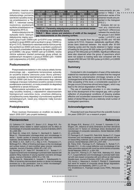
liczba cykli liczba po- Średnia minimum maksimum odch.std.<br />
żucia miarów n [µm] [µm] [µm] [µm]<br />
0 21 4,8 2,7 12,1 3,0<br />
30 000 45 5,6 0,4 28,5 7,5<br />
60 000 75 10,6 0,4 27,4 7,8<br />
100 000 50 11,9 1,5 30,0 6,2<br />
Praca naukowa finansowana ze środków na naukę w<br />
latach 2008-20011 jako projekt badawczy.<br />
piśmiennictwo<br />
taBela 1. parametry statystyczne pomiarów szerokości szczeliny<br />
brzeżnej na powierzchni żucia.<br />
taBle 1. mean values and statistics of width of the marginal<br />
gap measurements on the chewing surface.<br />
[1]. Calheiros C.F., Sadek F.T., Boaro L.C.C., Braga R.R., Polymerization<br />
stress related to radiant exposure and its effect on<br />
microleakage of composite restorations, Journal of dentistry (2007)<br />
35, p.946–952.<br />
[2]. Rosin M., Urban A.D., Gartner C., Bernhardt O., Spleith C.,<br />
Meyer G., Polymerization shrinkage-strain and microleakage in<br />
dentin – border cavites of chemical and light-cured restorative<br />
materials, Dental Materials 18 (2002) p.521–528.<br />
[3]. Wilder Jr. A.D., Swift Jr. E.J., May Jr. k.N., Thompsona J.y.,<br />
McDougal R.A., Effect of finishing technique on the microleakage<br />
and surface texture of resin-modified glass ionomer restorative<br />
materials, Journal of Dentistry (2000) 28, p.367–373.<br />
[4]. Fleminga G.J.P., Halla D.P., Shortalla A.C.C., Burkeb F.J.T,<br />
Cuspal movement and microleakage in premolar teeth restored with<br />
posterior filling materials of varying reported volumetric shrinkage<br />
values, Journal of Dentistry (2005) 33, p.139–146.<br />
[5]. Piemjaia M., Watanabeb A., Iwasakib y., Nakabayashib N.,<br />
Effect of remaining demineralised dentine on dental microleakage<br />
accessed by a dye penetration: how to inhibit microleakage?,<br />
Journal of Dentistry (2004) 32, p.495–501.<br />
measurements are presented<br />
in TABLE 1, the<br />
distribution series of the<br />
obtained results are presented<br />
in the histogram<br />
(FIG.2 ).<br />
Analysis did not reveal<br />
any statistically<br />
significant differences<br />
between the results from<br />
the groups 0 and 30000<br />
cycles ( p=0,9741) and<br />
between the results from the group 60 000 and 100 000<br />
cycles (p=0,7312). However, statistically significant differences<br />
were observed between the results after 30 000<br />
chewing cycles and the results obtained in higher ranges<br />
of loading for the group 60 000 cycles (p=0,00085) and the<br />
group of 100 000cycles (p=0,00069). Significant differences<br />
were also observed when the group of specimens which<br />
were not subject to loading tests was compared with the<br />
groups of 60 000 and 100 000 cycles (p=0,0043, p=0,00055<br />
respectively).<br />
summary<br />
Conducted in vitro investigation of wear of the restorative<br />
material bio-mechanical system revealed that the marginal<br />
gap formed by polymerization shrinkage remains on the<br />
unchanged level at the rate from 0 to 30 000 chewing cycles.<br />
After exceeding of this level, a considerable expansion of<br />
the gap occurs (it becomes almost twice bigger), which may<br />
lead to the clinical degradation of the filling.<br />
The use of mastication simulator in in vitro investigation<br />
of the development of the marginal gap, with a proper<br />
reflection of physiological conditions of chewing enables<br />
effective and accelerated assessment of functional degradation<br />
of dental fillings even at a relatively small number of<br />
investigated specimens.<br />
acknowledgements<br />
Presented work was financed from the scientific funds in<br />
the years 2008-2011 as a research project.<br />
references<br />
[6]. Bragaa R.R., Boaroa L.C.C., kuroeb T., Azevedoc C.L.N.,<br />
Singerc J.M., Influence of cavity dimensions and their derivatives<br />
(volume and ‘C’ factor) on shrinkage stress development and<br />
microleakage of composite restorations, Dental Materials (2006)<br />
22, p.818–823.<br />
[7]. Fleminga G.J.P., Carab R.R., Palin W.M., Burkec F.J.T., Cuspal<br />
movement and microleakage in premolar teeth restored with resin-based<br />
filling materials cured using a ‘soft-start’ polymerisation<br />
protocol, Dental Materials (2007) 23, p.637–643.<br />
[8]. Palina W.M., Fleminga G.J.P., Nathwania H., Burkeb F.J.T.,<br />
Randallc R.C., In vitro cuspal deflection and microleakage of maxillary<br />
premolars restored with novel low-shrink dental composites,<br />
Dental Materials (2005) 21, p.324–335.<br />
[9]. Cara R.R., Fleming G.J.P., Palin W.M., Walmsley A.D., Burke<br />
F.J.T., Cuspal deflection and microleakage in premolar teeth restored<br />
with resin-based composites with and without an intermediary<br />
flowable layer, Journal of Dentistry (2007) 35, p.482–4<strong>89</strong>.<br />
[10]. Hunicz J., Niewczas A., kordos P., Pieniak D., Experimental<br />
test stand for analisis of composite dental fillings degradation,<br />
Maintenance and Realiability, (2007) 2, p.37–43.<br />
[11]. Grosfeldowa O., Fizjologia narządu żucia, PZWL, Warszawa<br />
1981.<br />
249


