

89-91 - Polskie Stowarzyszenie Biomateriałów
89-91 - Polskie Stowarzyszenie Biomateriałów
89-91 - Polskie Stowarzyszenie Biomateriałów

You also want an ePaper? Increase the reach of your titles
YUMPU automatically turns print PDFs into web optimized ePapers that Google loves.
Oxidized cellulose has been widely used for many years<br />
as a wound-healing material due to its excellent properties,<br />
such as high absorbability, antibacterial and antiviral<br />
activity, non-toxicity and effects that prevent the formation<br />
of tissue adhesion after surgery [3-6]. Due to its ability to<br />
initiate or accelerate blood coagulation at the site where it is<br />
applied, oxidized cellulose has been used as a hemostatic<br />
material [7]. This material is also promising as a carrier for<br />
controlled drug delivery [2]. The possible use of various<br />
types and modifications of cellulose-based materials in<br />
tissue engineering is under extensive scientific research<br />
[8-10]. For example, cellulose fabrics [8] and injectable cellulose-based<br />
hydrogels [9] have been successfully tested<br />
as carriers for chondrocytes for cartilage regeneration in<br />
vitro and in vivo.<br />
In this study, we have investigated the adhesion, growth<br />
and morphology of vascular smooth muscle cells on various<br />
types of cellulose materials for potential use in soft tissue<br />
engineering. Four basic groups of materials were prepared:<br />
primal viscose (VIS), oxidized 6-carboxycellulose with<br />
2.1wt.% of –COOH groups (2.1), oxidized 6-carboxycellulose<br />
with 6.6% of –COOH groups (6.6) and dialdehyde cellulose<br />
from cotton knitting (DAC). The samples were further<br />
functionalized with arginine or chitosan. Positively-charged<br />
amine groups in these biomolecules were expected to support<br />
the adsorption of cell adhesion-mediating molecules<br />
and cell adhesion. At the same time, amino acids with the<br />
basic side chain (lysin, arginin), as well as chitosan, were<br />
expected to balance the relatively acid character of oxidized<br />
cellulose molecules [11].<br />
material and methods<br />
Preparation of biofunctionalized scaffolds<br />
Cellulose-based materials (listed above) in the form<br />
of woven fibrous scaffolds were exposed to solutions of<br />
arginine or chitosan (USA, Sigma-Aldrich) for 2 hours at<br />
laboratory temperature (20°C). The quantities of particular<br />
substances used for preparation of the solutions are<br />
summarized in TABLE 1. The samples were then washed<br />
twice for 2 hours in isopropyl alcohol, and were air-dried.<br />
Dialdehyde cellulose from cotton knit functionalized with<br />
arginine could not be created because of its high tendency<br />
to disintegrate.<br />
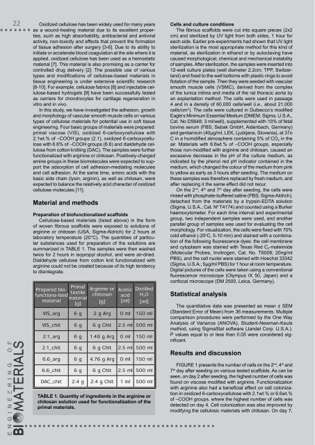
Prepared bio-<br />
functiona-lized<br />
material<br />
Primal<br />
textile<br />
material<br />
[g]<br />
Arginine or<br />
chitosan<br />
[g]<br />
Acetic<br />
acid<br />
[ml]<br />
Distilled<br />
H 2O<br />
[ml]<br />
VIS_arg 6 g 2 g Arg 0 ml 150 ml<br />
VIS_chit 6 g 6 g Chit 2.5 ml 500 ml<br />
2.1_arg 6 g 1.48 g Arg 0 ml 150 ml<br />
2.1_chit 6 g 6 g Chit 2.5 ml 500 ml<br />
6.6_arg 6 g 4.76 g Arg 0 ml 150 ml<br />
6.6_chit 6 g 6 g Chit 2.5 ml 500 ml<br />
DAC_chit 2.4 g 2.4 g Chit 1 ml 500 ml<br />
TaBlE 1. quantity of ingredients in the arginine or<br />
chitosan solution used for functionalization of the<br />
primal materials.<br />
Cells and culture conditions<br />
The fibrous scaffolds were cut into square pieces (2x2<br />
cm) and sterilized by UV light from both sides, 1 hour for<br />
each side. Earlier pre-experiments had shown that UV light<br />
sterilization is the most appropriate method for this kind of<br />
material, as sterilization in ethanol or by autoclaving have<br />
caused morphological, chemical and mechanical instability<br />
of samples. After sterilization, the samples were inserted into<br />
12-well culture plates (well diameter 2.2cm; TPP, Switzerland)<br />
and fixed to the well bottoms with plastic rings to avoid<br />
flotation of the sample. Then they were seeded with vascular<br />
smooth muscle cells (VSMC), derived from the complex<br />
of the tunica intima and media of the rat thoracic aorta by<br />
an explantation method. The cells were used in passage<br />
4 and in a density of 60,000 cells/well (i.e., about 21,000<br />
cells/cm 2 ). The cells were cultured in Dulbecco’s modified<br />
Eagle’s Minimum Essential Medium (DMEM; Sigma, U.S.A.,<br />
Cat. No D5648; 3 ml/well), supplemented with 10% of fetal<br />
bovine serum (FBS; Sebak GmbH, Aidenbach, Germany)<br />
and gentamicin (40µg/ml, LEK, Ljubljana, Slovenia), at 37o<br />
C in a humidified atmosphere containing 5% of CO 2 in the<br />
air. Materials with 6.6wt.% of –COOH groups, especially<br />
those non-modified with arginine and chitosan, caused an<br />
excessive decrease in the pH of the culture medium, as<br />
indicated by the phenol red pH indicator contained in the<br />
medium, which changed the colour of the medium from pink<br />
to yellow as early as 3 hours after seeding. The medium on<br />
these samples was therefore replaced by fresh medium, and<br />
after replacing it the same effect did not recur.<br />
On the 2 nd , 4 th and 7 th day after seeding, the cells were<br />
rinsed with phosphate-buffered saline (PBS; Sigma-Aldrich),<br />
detached from the materials by a trypsin-EDTA solution<br />
(Sigma, U.S.A., Cat. N 0 T4174) and counted using a Burker<br />
haemocytometer. For each time interval and experimental<br />
group, two independent samples were used, and another<br />
parallel group of samples was used for evaluating the cell<br />
morphology. For visualization, the cells were fixed with 70%<br />
cold ethanol (-20 o C, 5-10 min) and stained with a combination<br />
of the following fluorescence dyes: the cell membrane<br />
and cytoplasm was stained with Texas Red C 2-maleimide<br />
(Molecular Probes, Invitrogen, Cat. No. T6008; 20ng/ml<br />
PBS), and the cell nuclei were stained with Hoechst 33342<br />
(Sigma, U.S.A.; 5µg/ml PBS) for 1 hour at room temperature.<br />
Digital pictures of the cells were taken using a conventional<br />
fluorescence microscope (Olympus IX 50, Japan) and a<br />
confocal microscope (DM 2500, Leica, Germany).<br />
Statistical analysis<br />
The quantitative data was presented as mean ± SEM<br />
(Standard Error of Mean) from 36 measurements. Multiple<br />
comparison procedures were performed by the One Way<br />
Analysis of Variance (ANOVA), Student-Newman-Keuls<br />
method, using SigmaStat software (Jandel Corp. U.S.A.).<br />
P values equal to or less than 0.05 were considered significant.<br />
Results and discussion<br />
FIGURE 1 presents the number of cells on the 2 nd , 4 th and<br />
7 th day after seeding on various tested scaffolds. As can be<br />
seen, on day 2 after seeding, the highest number of cells was<br />
found on viscose modified with arginine. Functionalization<br />
with arginine also had a beneficial effect on cell colonization<br />
in oxidized 6-carboxycellulose with 2.1wt.% or 6.6wt.%<br />
of –COOH groups, where the highest number of cells was<br />
detected on day 4. Cell colonization was also improved by<br />
modifying the cellulosic materials with chitosan. On day 7,


