

89-91 - Polskie Stowarzyszenie Biomateriałów
89-91 - Polskie Stowarzyszenie Biomateriałów
89-91 - Polskie Stowarzyszenie Biomateriałów

You also want an ePaper? Increase the reach of your titles
YUMPU automatically turns print PDFs into web optimized ePapers that Google loves.
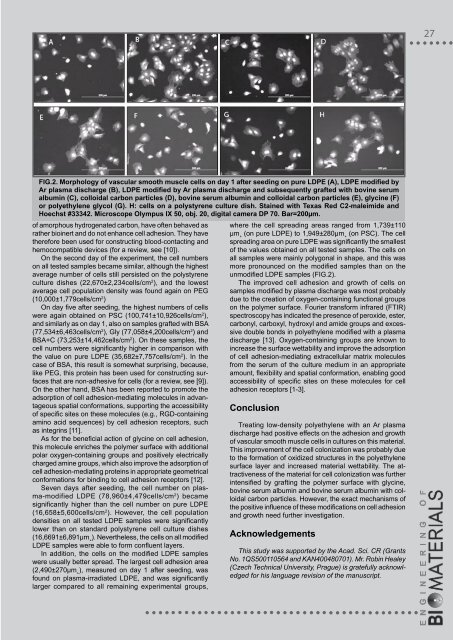
fIg.2. morphology of vascular smooth muscle cells on day 1 after seeding on pure ldPE (a), ldPE modified by<br />
ar plasma discharge (B), ldPE modified by ar plasma discharge and subsequently grafted with bovine serum<br />
albumin (C), colloidal carbon particles (d), bovine serum albumin and colloidal carbon particles (E), glycine (f)<br />
or polyethylene glycol (g). h: cells on a polystyrene culture dish. Stained with Texas Red C2-maleimide and<br />
hoechst #33342. microscope olympus Ix 50, obj. 20, digital camera dP 70. Bar=200µm.<br />
of amorphous hydrogenated carbon, have often behaved as<br />
rather bioinert and do not enhance cell adhesion. They have<br />
therefore been used for constructing blood-contacting and<br />
hemocompatible devices (for a review, see [10]).<br />
On the second day of the experiment, the cell numbers<br />
on all tested samples became similar, although the highest<br />
average number of cells still persisted on the polystyrene<br />
culture dishes (22,670±2,234cells/cm 2 ), and the lowest<br />
average cell population density was found again on PEG<br />
(10,000±1,779cells/cm 2 )<br />
On day five after seeding, the highest numbers of cells<br />
were again obtained on PSC (100,741±10,926cells/cm 2 ),<br />
and similarly as on day 1, also on samples grafted with BSA<br />
(77,534±6,463cells/cm 2 ), Gly (77,058±4,200cells/cm 2 ) and<br />
BSA+C (73,253±14,462cells/cm 2 ). On these samples, the<br />
cell numbers were significantly higher in comparison with<br />
the value on pure LDPE (35,682±7,757cells/cm 2 ). In the<br />
case of BSA, this result is somewhat surprising, because,<br />
like PEG, this protein has been used for constructing surfaces<br />
that are non-adhesive for cells (for a review, see [9]).<br />
On the other hand, BSA has been reported to promote the<br />
adsorption of cell adhesion-mediating molecules in advantageous<br />
spatial conformations, supporting the accessibility<br />
of specific sites on these molecules (e.g., RGD-containing<br />
amino acid sequences) by cell adhesion receptors, such<br />
as integrins [11].<br />
As for the beneficial action of glycine on cell adhesion,<br />
this molecule enriches the polymer surface with additional<br />
polar oxygen-containing groups and positively electrically<br />
charged amine groups, which also improve the adsorption of<br />
cell adhesion-mediating proteins in appropriate geometrical<br />
conformations for binding to cell adhesion receptors [12].<br />
Seven days after seeding, the cell number on plasma-modified<br />
LDPE (78,960±4,479cells/cm 2 ) became<br />
significantly higher than the cell number on pure LDPE<br />
(16,658±5,600cells/cm 2 ). However, the cell population<br />
densities on all tested LDPE samples were significantly<br />
lower than on standard polystyrene cell culture dishes<br />
(16,66<strong>91</strong>±6,<strong>89</strong>1µm˛). Nevertheless, the cells on all modified<br />
LDPE samples were able to form confluent layers.<br />
In addition, the cells on the modified LDPE samples<br />
were usually better spread. The largest cell adhesion area<br />
(2,490±270µm˛), measured on day 1 after seeding, was<br />
found on plasma-irradiated LDPE, and was significantly<br />
larger compared to all remaining experimental groups,<br />
where the cell spreading areas ranged from 1,739±110<br />
µm˛ (on pure LDPE) to 1,949±280µm˛ (on PSC). The cell<br />
spreading area on pure LDPE was significantly the smallest<br />
of the values obtained on all tested samples. The cells on<br />
all samples were mainly polygonal in shape, and this was<br />
more pronounced on the modified samples than on the<br />
unmodified LDPE samples (FIG.2).<br />
The improved cell adhesion and growth of cells on<br />
samples modified by plasma discharge was most probably<br />
due to the creation of oxygen-containing functional groups<br />
on the polymer surface. Fourier transform infrared (FTIR)<br />
spectroscopy has indicated the presence of peroxide, ester,<br />
carbonyl, carboxyl, hydroxyl and amide groups and excessive<br />
double bonds in polyethylene modified with a plasma<br />
discharge [13]. Oxygen-containing groups are known to<br />
increase the surface wettability and improve the adsorption<br />
of cell adhesion-mediating extracellular matrix molecules<br />
from the serum of the culture medium in an appropriate<br />
amount, flexibility and spatial conformation, enabling good<br />
accessibility of specific sites on these molecules for cell<br />
adhesion receptors [1-3].<br />
Conclusion<br />
Treating low-density polyethylene with an Ar plasma<br />
discharge had positive effects on the adhesion and growth<br />
of vascular smooth muscle cells in cultures on this material.<br />
This improvement of the cell colonization was probably due<br />
to the formation of oxidized structures in the polyethylene<br />
surface layer and increased material wettability. The attractiveness<br />
of the material for cell colonization was further<br />
intensified by grafting the polymer surface with glycine,<br />
bovine serum albumin and bovine serum albumin with colloidal<br />
carbon particles. However, the exact mechanisms of<br />
the positive influence of these modifications on cell adhesion<br />
and growth need further investigation.<br />
acknowledgements<br />
This study was supported by the Acad. Sci. CR (Grants<br />
No. 1QS500110564 and KAN400480701). Mr. Robin Healey<br />
(Czech Technical University, Prague) is gratefully acknowledged<br />
for his language revision of the manuscript.


