

Full Journal - Journal of Cell and Molecular Biology - Haliç Üniversitesi
Full Journal - Journal of Cell and Molecular Biology - Haliç Üniversitesi
Full Journal - Journal of Cell and Molecular Biology - Haliç Üniversitesi

You also want an ePaper? Increase the reach of your titles
YUMPU automatically turns print PDFs into web optimized ePapers that Google loves.
76 Seyhan Altun et al.<br />
increased a little <strong>and</strong> on the sixth day the cell number<br />
was found to be 110,750.<br />
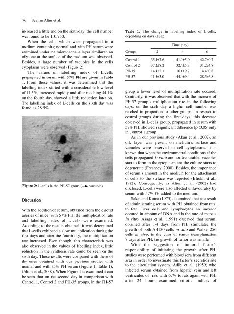
When the cells which were propagated in a<br />
medium containing normal <strong>and</strong> with PH serum were<br />
examined under the microscope, a layer similar to an<br />
oily one at the surface <strong>of</strong> the medium was observed.<br />
Besides, a large number <strong>of</strong> vacuoles in the cells<br />
cytoplasm were observed (Figure 2).<br />
The values <strong>of</strong> labelling index <strong>of</strong> L-cells<br />
propagated in serum with 57% PH are given in Table<br />
1. From these values, it was determined that the<br />
labelling index started with a considerable low level<br />
<strong>of</strong> 11.5%, increased rapidly <strong>and</strong> after reaching 44.1%<br />
on the fourth day, showed a little reduction later on.<br />
The labelling index <strong>of</strong> L-cells on the sixth day was<br />
found as 28.5%.<br />
Figure 2: L-cells in the PH-57 group ( vacuole).<br />
Discussion<br />
With the addition <strong>of</strong> serum, obtained from the carotid<br />
arteries <strong>of</strong> mice with 57% PH, the multiplication rate<br />
<strong>and</strong> labelling index <strong>of</strong> L-cells were examined.<br />
According to the results obtained, it was determined<br />
that L-cells exhibited a slow multiplication during the<br />
first days <strong>and</strong> after the fourth day, the multiplication<br />
rate increased. Even though, this characteristic was<br />
also observed in the values <strong>of</strong> labelling index, little<br />
reduction in the synthesis rate could be seen on the<br />
sixth day. These results were compared with those <strong>of</strong><br />
the ones obtained with our previous studies with<br />
normal <strong>and</strong> with 35% PH serum (Figure 1, Table 1),<br />
(Altun et al., 2002). When Figure 1 is examined it can<br />
be seen that on the second day in comparison with<br />
Control 1, Control 2 <strong>and</strong> PH-35 groups, in the PH-57<br />
Table 1: The change in labelling index <strong>of</strong> L-cells,<br />
depending on days (±SE).<br />
Groups 2<br />
Time (day)<br />
4 6<br />
Control 1 35.4±7.6 41.3±5.0 42.7±0.7<br />
Control 2 37.2±8.2 32.7±5.3 31.2±4.8<br />
PH-35 14.4±2.1 16.8±9.7 14.4±0.8<br />
PH-57 11.5±3.0 44.1±9.4 28.5±6.8<br />
group a lower level <strong>of</strong> multiplication rate occured.<br />
Contrarily, it was observed that with the increase <strong>of</strong><br />
PH-57 group’s multiplication rate in the following<br />
days, on the sixth day a higher cell number was<br />
reached in proportion to other groups. In respect to<br />
control groups during the first days, this decrease<br />
observed in L-cells group, propagated in serum with<br />
57% PH, showed a significant difference (p


