Report in English with a Dutch summary (KCE reports 63A)
Report in English with a Dutch summary (KCE reports 63A)
Report in English with a Dutch summary (KCE reports 63A)
You also want an ePaper? Increase the reach of your titles
YUMPU automatically turns print PDFs into web optimized ePapers that Google loves.
5<br />
10<br />
15<br />
20<br />
25<br />
30<br />
35<br />
40<br />
45<br />
146 Breast Cancer <strong>KCE</strong> <strong>reports</strong> 63<br />
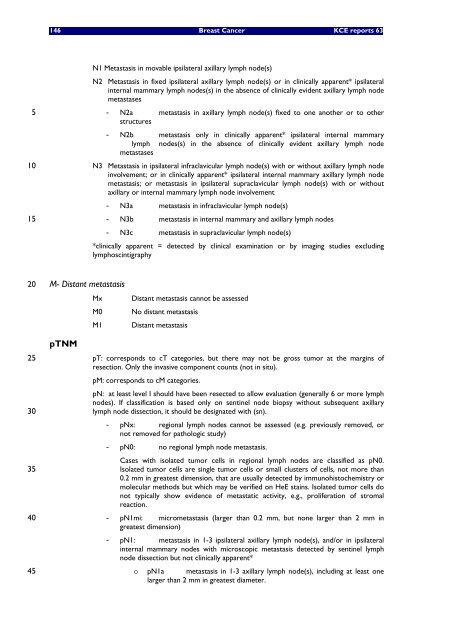
N1 Metastasis <strong>in</strong> movable ipsilateral axillary lymph node(s)<br />
N2 Metastasis <strong>in</strong> fixed ipsilateral axillary lymph node(s) or <strong>in</strong> cl<strong>in</strong>ically apparent* ipsilateral<br />
<strong>in</strong>ternal mammary lymph nodes(s) <strong>in</strong> the absence of cl<strong>in</strong>ically evident axillary lymph node<br />
metastases<br />
- N2a metastasis <strong>in</strong> axillary lymph node(s) fixed to one another or to other<br />
structures<br />
- N2b metastasis only <strong>in</strong> cl<strong>in</strong>ically apparent* ipsilateral <strong>in</strong>ternal mammary<br />
lymph nodes(s) <strong>in</strong> the absence of cl<strong>in</strong>ically evident axillary lymph node<br />
metastases<br />
N3 Metastasis <strong>in</strong> ipsilateral <strong>in</strong>fraclavicular lymph node(s) <strong>with</strong> or <strong>with</strong>out axillary lymph node<br />
<strong>in</strong>volvement; or <strong>in</strong> cl<strong>in</strong>ically apparent* ipsilateral <strong>in</strong>ternal mammary axillary lymph node<br />
metastasis; or metastasis <strong>in</strong> ipsilateral supraclavicular lymph node(s) <strong>with</strong> or <strong>with</strong>out<br />
axillary or <strong>in</strong>ternal mammary lymph node <strong>in</strong>volvement<br />
- N3a metastasis <strong>in</strong> <strong>in</strong>fraclavicular lymph node(s)<br />
- N3b metastasis <strong>in</strong> <strong>in</strong>ternal mammary and axillary lymph nodes<br />
- N3c metastasis <strong>in</strong> supraclavicular lymph node(s)<br />
*cl<strong>in</strong>ically apparent = detected by cl<strong>in</strong>ical exam<strong>in</strong>ation or by imag<strong>in</strong>g studies exclud<strong>in</strong>g<br />
lymphosc<strong>in</strong>tigraphy<br />
M- Distant metastasis<br />
Mx Distant metastasis cannot be assessed<br />
M0 No distant metastasis<br />
M1 Distant metastasis<br />
pTNM<br />
pT: corresponds to cT categories, but there may not be gross tumor at the marg<strong>in</strong>s of<br />
resection. Only the <strong>in</strong>vasive component counts (not <strong>in</strong> situ).<br />
pM: corresponds to cM categories.<br />
pN: at least level I should have been resected to allow evaluation (generally 6 or more lymph<br />
nodes). If classification is based only on sent<strong>in</strong>el node biopsy <strong>with</strong>out subsequent axillary<br />
lymph node dissection, it should be designated <strong>with</strong> (sn).<br />
- pNx: regional lymph nodes cannot be assessed (e.g. previously removed, or<br />
not removed for pathologic study)<br />
- pN0: no regional lymph node metastasis.<br />
Cases <strong>with</strong> isolated tumor cells <strong>in</strong> regional lymph nodes are classified as pN0.<br />
Isolated tumor cells are s<strong>in</strong>gle tumor cells or small clusters of cells, not more than<br />
0.2 mm <strong>in</strong> greatest dimension, that are usually detected by immunohistochemistry or<br />
molecular methods but which may be verified on HeE sta<strong>in</strong>s. Isolated tumor cells do<br />
not typically show evidence of metastatic activity, e.g., proliferation of stromal<br />
reaction.<br />
- pN1mi: micrometastasis (larger than 0.2 mm, but none larger than 2 mm <strong>in</strong><br />
greatest dimension)<br />
- pN1: metastasis <strong>in</strong> 1-3 ipsilateral axillary lymph node(s), and/or <strong>in</strong> ipsilateral<br />
<strong>in</strong>ternal mammary nodes <strong>with</strong> microscopic metastasis detected by sent<strong>in</strong>el lymph<br />
node dissection but not cl<strong>in</strong>ically apparent*<br />
o pN1a metastasis <strong>in</strong> 1-3 axillary lymph node(s), <strong>in</strong>clud<strong>in</strong>g at least one<br />
larger than 2 mm <strong>in</strong> greatest diameter.


