PDF (tesi dottorato ROTIROTI) - FedOA - Università degli Studi di ...
PDF (tesi dottorato ROTIROTI) - FedOA - Università degli Studi di ...
PDF (tesi dottorato ROTIROTI) - FedOA - Università degli Studi di ...

You also want an ePaper? Increase the reach of your titles
YUMPU automatically turns print PDFs into web optimized ePapers that Google loves.
The morphology of the porous silicon layers was investigated by variable angle<br />
spectroscopic ellipsometer (UVISEL, Horiba-Jobin-Yvon) and scanning electron<br />
microscope (SEM-FEG Gemini 300, Carl Zeiss).<br />
Both the analytical techniques have showed the presence of a top layer on the<br />
porous silicon structure of thickness between 20 nm and 100 nm and porosity of 20-<br />
40 % due to hydrogen contamination of the silicon wafer [31]. Such film prevents not<br />
only the pores from filling but also any biochemical interaction with the hydrogenated<br />
porous silicon surface after the etching process. For sensing purposes it is therefore<br />
mandatory to avoid the formation of the parasitic film by thermal treating the wafer at<br />
300 °C in nitrogen atmosphere before the electrochemical etch [32].<br />
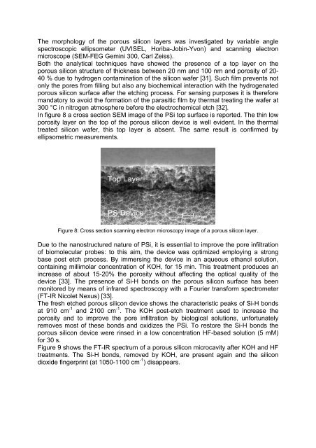
In figure 8 a cross section SEM image of the PSi top surface is reported. The thin low<br />
porosity layer on the top of the porous silicon device is well evident. In the thermal<br />
treated silicon wafer, this top layer is absent. The same result is confirmed by<br />
ellipsometric measurements.<br />
20 nm<br />
Figure 8: Cross section scanning electron microscopy image of a porous silicon layer.<br />
Due to the nanostructured nature of PSi, it is essential to improve the pore infiltration<br />
of biomolecular probes: to this aim, the device was optimized employing a strong<br />
base post etch process. By immersing the device in an aqueous ethanol solution,<br />
containing millimolar concentration of KOH, for 15 min. This treatment produces an<br />
increase of about 15-20% the porosity without affecting the optical quality of the<br />
device [33]. The presence of Si-H bonds on the porous silicon surface has been<br />
monitored by means of infrared spectroscopy with a Fourier transform spectrometer<br />
(FT-IR Nicolet Nexus) [33].<br />
The fresh etched porous silicon device shows the characteristic peaks of Si-H bonds<br />
at 910 cm -1 and 2100 cm -1 . The KOH post-etch treatment used to increase the<br />
porosity and to improve the pore infiltration by biological solutions, unfortunately<br />
removes most of these bonds and oxi<strong>di</strong>zes the PSi. To restore the Si-H bonds the<br />
porous silicon device were rinsed in a low concentration HF-based solution (5 mM)<br />
for 30 s.<br />
Figure 9 shows the FT-IR spectrum of a porous silicon microcavity after KOH and HF<br />
treatments. The Si-H bonds, removed by KOH, are present again and the silicon<br />
<strong>di</strong>oxide fingerprint (at 1050-1100 cm -1 ) <strong>di</strong>sappears.