Geological Survey of Denmark and Greenland Bulletin 26 ... - Geus
Geological Survey of Denmark and Greenland Bulletin 26 ... - Geus
Geological Survey of Denmark and Greenland Bulletin 26 ... - Geus

Create successful ePaper yourself
Turn your PDF publications into a flip-book with our unique Google optimized e-Paper software.
A<br />
200 nm<br />
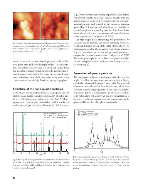
mode. Intact rock samples <strong>of</strong> small pieces <strong>of</strong> chalk or flint<br />
were glued onto gold-coated sample holders. In chalk samples,<br />
non-calcite minerals were identified <strong>and</strong> imaged from<br />
the insoluble residue. For such samples, the residue was dispersed<br />
ultrasonically in distilled water <strong>and</strong> the samples prepared<br />
by leaving a drop <strong>of</strong> the suspension to dry under room<br />
conditions on a block <strong>of</strong> highly oriented pyrolytic graphite.<br />
Structure <strong>of</strong> the nano-quartz particles<br />
AFM <strong>of</strong> non-calcite residues deposited on graphite showed<br />
that the nano-quartz consists predominantly <strong>of</strong> rather uniform,<br />
c. 600 Å large spherical particles (Fig. 2A). AFM images<br />
<strong>of</strong> intact flint surfaces showed that the flint consists <strong>of</strong><br />
similar spherical particles with a diameter <strong>of</strong> c. 500 Å or more<br />
B<br />
200 nm<br />
Fig. 2. Atomic force microscopy images <strong>of</strong> spherical grains <strong>of</strong> quartz. Noncontact<br />
mode, room conditions, force 0.175 nN, scanning speed 500 nm/s.<br />
A: Calcite-free residue deposited on graphite from well SA-1, 3344.15 m.<br />
B: Intact flint from Nana-1, 2135.7 m.<br />
(Fig. 2B) <strong>and</strong> some irregularly shaped particles. X-ray diffraction<br />
showed that the non-calcite residues <strong>and</strong> the flint <strong>and</strong><br />
quartz layers are composed <strong>of</strong> α-quartz having practically<br />
identical patterns <strong>and</strong> resembling the pattern <strong>of</strong> st<strong>and</strong>ard<br />
quartz (Fig. 3). It is remarkable that the quartz in all the examined<br />
samples <strong>of</strong> dispersed quartz <strong>and</strong> flint have almost<br />
identical unit cell a <strong>and</strong> c parameters <strong>and</strong> sizes <strong>of</strong> coherent<br />
scattering domains (Lindgreen et al. 2011).<br />
At high angles peak broadening was pronounced for<br />
the nano-quartz particles <strong>and</strong> careful recording revealed a<br />
broad <strong>and</strong> distorted pattern <strong>of</strong> the (212), (203) <strong>and</strong> (301) reflections<br />
compared to the reflection from st<strong>and</strong>ard quartz<br />
(Fig. 4). These distortions are due to larger a <strong>and</strong> c parameters<br />
compared to those <strong>of</strong> normal quartz (Lindgreen et al. 2011).<br />
The nano-quartz spheres had colloidal properties <strong>and</strong> flocculated<br />
in suspensions with sufficient ionic strength, such as<br />
sea water (Fig. 5).<br />
Formation <strong>of</strong> quartz particles<br />
The nano-quartz spheres are anticipated to be <strong>of</strong> a type that<br />
might crystallise in a marine environment which is slightly<br />
enriched in silicon (Williams & Crerar 1985). The source <strong>of</strong><br />
silicon was probably opal-A from radiolarians, which were<br />
the main silica-bearing organism in the chalk sea (Maliva<br />
& Dickson 1992). It is important that the non-crystalline<br />
Si in radiolarians will dissolve at the low concentration <strong>of</strong><br />
Si, which is sufficient to precipitate fine quartz, <strong>and</strong> that the<br />
quartz will be the first silica phase to crystallise.<br />
Si<br />
Si<br />
St<strong>and</strong>ard Merck quartz<br />
SA 3344.15 m quartz in matrix<br />
Nana 2135.7 m quartz in flint<br />
Si<br />
St<strong>and</strong>ard Merck quartz<br />
SA 3344.15 m quartz in matrix<br />
Nana 2135.7 m quartz in flint<br />
212α 1<br />
203α 1<br />
203α 2<br />
301α 1<br />
C<br />
A<br />
212α 2<br />
301α 2<br />
B<br />
B<br />
C<br />
A<br />
20 30 40 50 60 70 80 90<br />
°2Θ<br />
Fig. 3. A: X-ray diffraction pattern <strong>of</strong> st<strong>and</strong>ard Merck quartz. B: <strong>of</strong> calcitefree<br />
residue from chalk, South Arne Field, well SA-1, 3344.15 m, C: <strong>and</strong> <strong>of</strong><br />
flint layer in Halfdan Field, well Nana-1, 2135.7 m. Co-K α radiation, 5%<br />
Si added as internal st<strong>and</strong>ard.<br />
80 81<br />
°2Θ<br />
Fig. 4. X-ray diffraction patterns. A: Region <strong>of</strong> (212), (203) <strong>and</strong> (301) reflections<br />
<strong>of</strong> st<strong>and</strong>ard Merck quartz. B: The same region for calcite-free<br />
residue from chalk, South Arne Field, well SA-1, 3344.15 m. C: The same<br />
region for flint layer in Halfdan Field, well Nana-1, 2135.7 m. Co-K α radiation.<br />
82<br />
10