Congenital malformations - Edocr
Congenital malformations - Edocr
Congenital malformations - Edocr

Create successful ePaper yourself
Turn your PDF publications into a flip-book with our unique Google optimized e-Paper software.
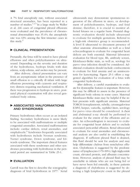
160 PART IV RESPIRATORY MALFORMATIONS<br />
A 7% fetal aneuploidy rate, without associated<br />
structural anomalies, has been reported in a<br />
number of studies. 1,4,5 In a large study by Waller<br />
et al, 246 cases of congenital pleural effusions<br />
were evaluated and the prevalence of chromosomal<br />
abnormalities was 35.4%; the aneuploidy<br />
rate was 63% among the first trimester cases in<br />
this study. 6<br />
CLINICAL PRESENTATION<br />
Prenatally, the fetus will be noted to have pleural<br />
effusions and often polyhydramnios on ultrasound.<br />
Depending on the severity and duration<br />
of the hydrothorax, hydrops fetalis with skin<br />
edema, scalp edema, and ascites may be present.<br />
After delivery, clinical presentation can vary<br />
from an asymptomatic infant in the presence of<br />
small effusion to a critically ill infant with large<br />
effusions presenting with cyanosis and respiratory<br />
distress requiring mechanical ventilation. If<br />
there was progression to hydrops in utero, postnatal<br />
physical examination will also reveal generalized<br />
body edema.<br />
ASSOCIATED MALFORMATIONS<br />
AND SYNDROMES<br />
Primary hydrothorax often occurs as an isolated<br />
finding. Secondary hydrothorax is more likely<br />
to be associated with <strong>malformations</strong> or multiple<br />
congenital anomalies. Associated <strong>malformations</strong><br />
include: cardiac defects, renal anomalies, and<br />
omphalocele. 6 Syndromes frequently associated<br />
with hydrothorax include Noonan syndrome,<br />
Turner syndrome, Down syndrome, and Edwards<br />
syndrome. The important clinical features<br />
associated with these syndromes and other syndromes<br />
presenting with hydrothorax in the perinatal<br />
period are summarized in Table 25-1.<br />
EVALUATION<br />
Carroll was the first to describe the sonographic<br />
diagnosis of fetal hydrothorax in 1977. 7 Serial<br />
ultrasounds may demonstrate spontaneous regression<br />
of the effusion in utero, or development<br />
of polyhydramnios, hydrops, and fetal<br />
demise; therefore, it is crucial to monitor affected<br />
fetuses on a regular basis. Prenatal diagnostic<br />
evaluation should include ultrasound<br />
to evaluate for multiple gestations. Referral to<br />
a high-risk obstetrics group is recommended.<br />
A level II ultrasound to document presence of<br />
other anatomic abnormalities as well as a fetal<br />
echocardiogram to evaluate for congenital heart<br />
defects are both essential. Maternal laboratory<br />
evaluation for blood type, Rh, antibody screen,<br />
Kleinhauer-Betke stain, as well as, serology for<br />
parvo virus infection should be considered. Additional<br />
evaluation should include cordocentesis<br />
to evaluate for fetal anemia and an amniocentesis<br />
for karyotyping. Figure 25-1 offers a suggested<br />
algorithm for evaluation of a fetus with<br />
congenital hydrothorax.<br />
After delivery, a careful examination to evaluate<br />
for dysmorphic features is important. However,<br />
this may be difficult to assess in the presence of<br />
significant body edema in some cases. Maternal<br />
Kleinhauer-Betke stain may be helpful if the infant<br />
presents with significant anemia. Maternal<br />
TORCH (toxoplasmosis, rubella, cytomegalovirus<br />
[CMV], herpes, varicella, syphilis) titers, serology<br />
for parvo virus should be considered. A chest<br />
and abdominal x-ray should be performed to<br />
evaluate for the extent of the effusions and ascites.<br />
An echocardiogram is necessary to evaluate<br />
for congenital heart defects and to exclude<br />
associated pericardial effusion. Renal ultrasound<br />
to evaluate for renal anomalies and chromosomal<br />
analysis are also useful in establishing the<br />
diagnosis. If sufficient pleural fluid is drained,<br />
this fluid should be sent for analysis and may<br />
help differentiate chylous from nonchylous effusion.<br />
Chylothorax is suggested by the predominance<br />
of lymphocytes (>70–90%), high triglyceride<br />
count, elevated protein, and albumin concentrations.<br />
However, analysis of pleural fluid may be<br />
unreliable in infants who are not being fed or<br />
have never been fed enterally, in these patients<br />
a diagnosis of chylothorax is suggested by detecting<br />
a high lymphocyte count in the pleural