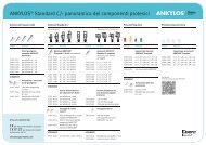
success is... - DENTSPLY Friadent
success is... - DENTSPLY Friadent
success is... - DENTSPLY Friadent

You also want an ePaper? Increase the reach of your titles
YUMPU automatically turns print PDFs into web optimized ePapers that Google loves.
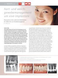
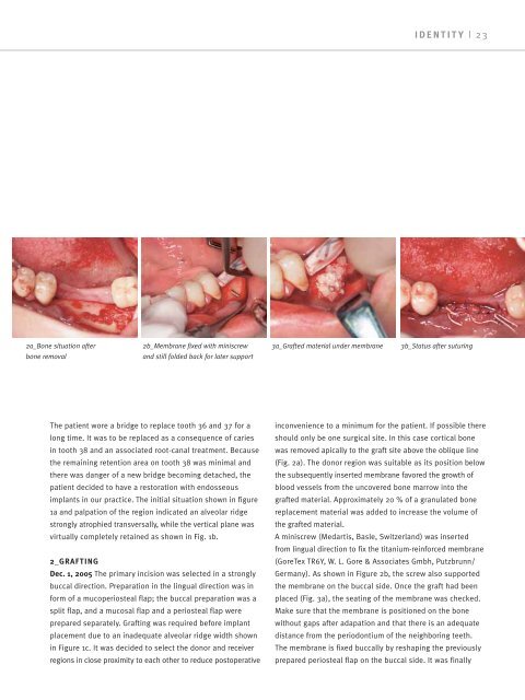
2a_Bone situation after<br />
bone removal<br />
2b_Membrane fixed with min<strong>is</strong>crew<br />
and still folded back for later support<br />
The patient wore a bridge to replace tooth 36 and 37 for a<br />
long time. It was to be replaced as a consequence of caries<br />
in tooth 38 and an associated root-canal treatment. Because<br />
the remaining retention area on tooth 38 was minimal and<br />
there was danger of a new bridge becoming detached, the<br />
patient decided to have a restoration with endosseous<br />
implants in our practice. The initial situation shown in figure<br />
1a and palpation of the region indicated an alveolar ridge<br />
strongly atrophied transversally, while the vertical plane was<br />
virtually completely retained as shown in Fig. 1b.<br />
2_GRAFTING<br />
Dec. 1, 2005 The primary inc<strong>is</strong>ion was selected in a strongly<br />
buccal direction. Preparation in the lingual direction was in<br />
form of a mucoperiosteal flap; the buccal preparation was a<br />
split flap, and a mucosal flap and a periosteal flap were<br />
prepared separately. Grafting was required before implant<br />
placement due to an inadequate alveolar ridge width shown<br />
in Figure 1c. It was decided to select the donor and receiver<br />
regions in close proximity to each other to reduce postoperative<br />
3a_Grafted material under membrane<br />
IDENTITY | 23<br />
3b_Status after suturing<br />
inconvenience to a minimum for the patient. If possible there<br />
should only be one surgical site. In th<strong>is</strong> case cortical bone<br />
was removed apically to the graft site above the oblique line<br />
(Fig. 2a). The donor region was suitable as its position below<br />
the subsequently inserted membrane favored the growth of<br />
blood vessels from the uncovered bone marrow into the<br />
grafted material. Approximately 20 % of a granulated bone<br />
replacement material was added to increase the volume of<br />
the grafted material.<br />
A min<strong>is</strong>crew (Medart<strong>is</strong>, Basle, Switzerland) was inserted<br />
from lingual direction to fix the titanium-reinforced membrane<br />
(GoreTex TR6Y, W. L. Gore & Associates Gmbh, Putzbrunn/<br />
Germany). As shown in Figure 2b, the screw also supported<br />
the membrane on the buccal side. Once the graft had been<br />
placed (Fig. 3a), the seating of the membrane was checked.<br />
Make sure that the membrane <strong>is</strong> positioned on the bone<br />
without gaps after adapation and that there <strong>is</strong> an adequate<br />
d<strong>is</strong>tance from the periodontium of the neighboring teeth.<br />
The membrane <strong>is</strong> fixed buccally by reshaping the previously<br />
prepared periosteal flap on the buccal side. It was finally