- Page 2 and 3:
Modern Polymer Spectroscopy Edited
- Page 4 and 5:
Modern Polymer Spectroscopy Edited
- Page 6 and 7:
Preface For unfortunate reasons the
- Page 8 and 9:
However, theory and calculations yi
- Page 10 and 11:
Contents 1 Two-Dimensional Infrared
- Page 12 and 13:
Con ten t~ xi Index 5.2 Force Field
- Page 14 and 15:
Contributors M. Del Zoppo Dipartime
- Page 16 and 17:
1 Two-Dimensional Infrared (2D IR)
- Page 18 and 19:
1.2 Brick~qrorrnrl 3 . Figure 1-3.
- Page 20 and 21:
1.0 , Figure 1-6. The in-phtrse and
- Page 22 and 23:
1.2 Bcrckgroinizd 7 where AA (i~) i
- Page 24 and 25:
1.2 Background 9 1.2.5 Two-Dimensio
- Page 26 and 27:
1.3 Bnsic Properties of 20 IR Corre
- Page 28 and 29:
2D IR spectrometer coupled with a d
- Page 30 and 31:
1.5 Applirtrtions 15 Unlike a dispe
- Page 32 and 33:
\ '\ Melliyleiie 1'7- -3000 Posiliv
- Page 34 and 35:
11 Amorphous Crystalline ,...." I F
- Page 36 and 37:
1.5 Applications 21 / I 1430 Figure
- Page 38 and 39:
1.5 Appliccitioiis 23 Methylene Fig
- Page 40 and 41:
1.5 Ajqdicrrtions 25 3024 Figure 1-
- Page 42 and 43:
1.5 Applicutions 27 Furthermore. th
- Page 44 and 45:
1.7 Coizclirsions 29 derived in a s
- Page 46 and 47:
1.9 References 31 [9] Colthup, N. B
- Page 48 and 49:
2 Segmental Mobility of Liquid Crys
- Page 50 and 51:
SRMPLE/DETECTOR e3 ‘retardation
- Page 52 and 53:
2.4 Srvuc me-Dependenr Alignment 37
- Page 54 and 55:
2.5 Electric Field-Innductid Orirnt
- Page 56 and 57:
2.5 Electric Field-nclticetl Orient
- Page 58 and 59:
a -@ COWER SRHPLE ._ 2.5 Elec tric
- Page 60 and 61:
2.5 Electric Field-Itzduced Orienta
- Page 62 and 63:
2.5 Electric Field-IwrElrced Orient
- Page 64 and 65:
2.5 Electric Field-Im/irced Orientu
- Page 66 and 67:
2.5 Electric Field-Induced Orientli
- Page 68 and 69:
2.5 Electric Field-Induced Orientat
- Page 70 and 71:
2.5 Electric Field-Induced Orienfat
- Page 72 and 73:
2.5 Elrc tric Field-Iiidircwl Orim
- Page 74 and 75:
2500 2008 I s00 WRVtNdMRtR CM-I Fig
- Page 76 and 77:
-3.6 Aligmient OJ' Side- Clinin Liy
- Page 78 and 79:
~ polyester 2.6 Alignnient qf Side-
- Page 80 and 81:
I." 0.9 0.8 Figure 2-34. FTlR 0.7 p
- Page 82 and 83:
induced alignment of the investigat
- Page 84 and 85:
2.7 Orientation oj Liquid Ci:i,stal
- Page 86 and 87:
2.7 Orieritation of Liquid Ciysttrl
- Page 88 and 89:
0 8 18 16 1 a W u z a m a 0 Ln m Q
- Page 90 and 91:
2.7 Orientation oj Liquid Crystals
- Page 92 and 93:
2.7 Orientation of Liquid Crjvtals
- Page 94 and 95:
2.7 Orientatioiz of Liquid Crystals
- Page 96 and 97:
2.8 Conclusions 81 strain. As A0 is
- Page 98 and 99:
3. I0 Refcreiice.r 83 [I 71 Hoffina
- Page 100 and 101:
2.10 References 85 [92] Wiesner, U.
- Page 102 and 103:
t Order/Disorder in Chain Molecules
- Page 104 and 105:
onds which hold atoms together thro
- Page 106 and 107:
3.2 The Dyriamical Case qf Simll ar
- Page 108 and 109:
3.2 The Dyrinniical Case of Sinnll
- Page 110 and 111:
3.4 S11or.f- and Loizg-Rcrizye Vibr
- Page 112 and 113:
3.4 Slzort- and Loiig-Range Vibrati
- Page 114 and 115:
eyularity), e.g., 2. During the pol
- Page 116 and 117:
3.5 Towards Lnrger Molecules: From
- Page 118 and 119:
3.5 TOIIYW~ Laiyer Molertiles: Fvoi
- Page 120 and 121:
3.5 Towmu" Larger Molecules: F~om O
- Page 122 and 123:
1700 - CIO --- ca c 22 _- c 12 l l
- Page 124 and 125:
3.6 From Dynamics to Vibrational Sp
- Page 126 and 127:
3.6 Froin Dynaiiiics to T’ihratio
- Page 128 and 129:
3.7 The Case of Isotactic Polypropy
- Page 130 and 131:
3.7 The Cuse of Isotactic Polypvop.
- Page 132 and 133:
3.8 Density of Vibrational States a
- Page 134 and 135:
3.8 Dtvz.sit,v of L’ihrntioizal S
- Page 136 and 137:
3.8 Driisity of Vibratioiid StLitrs
- Page 138 and 139:
3 9 Mouiiiq ToIvmds Rerrlitv: From
- Page 140 and 141:
3.9 Moving Towards Reality: From Or
- Page 142 and 143:
3.9 Moving Towards Reality: Froin O
- Page 144 and 145:
3.10 Wlmt Do We Learn from Cnlcailc
- Page 146 and 147:
+ 3.11 A Very Siriiple Ccrse: Latti
- Page 148 and 149:
3 I1 A Yey) Siiiiple Case: LLittice
- Page 150 and 151:
3.11 A Vq> Siiiiplc Crrw: Luttice D
- Page 152 and 153:
Figure 3-22. Sample eigenvectors in
- Page 154 and 155:
3.12 CIS-trans Opening @the Double
- Page 156 and 157:
3.13 Defect Modes cis Structtrr.al
- Page 158 and 159:
3.13 Defect Mo&s cis Structurcil Pr
- Page 160 and 161:
0 3.14 Case Studies N 0 d - Y N o m
- Page 162 and 163:
3.14 Case Studies 147 OC 60 58 57.
- Page 164 and 165:
3.14 Case Studies 149 GI A V E N U
- Page 166 and 167:
3.14 Case Studies 15 1 correspond t
- Page 168 and 169:
3.14 Case Studies 153 (I10 + 200) +
- Page 170 and 171:
3.14 Case Studies 155 1500 1480 146
- Page 172 and 173:
3.14 Case Studies 157 the molecular
- Page 174 and 175:
17E (ir 1, R): (iii) z = 4, t = 1,
- Page 176 and 177:
3.16 Stnictural Znlioi~zogeneity an
- Page 178 and 179:
3.1 7 Fermi Resonancrs 163 stants.
- Page 180 and 181:
3.1 7 Femi Resorinnces 165 2 L 1 29
- Page 182 and 183:
lowing. The CHl-bending mode 6 [cer
- Page 184 and 185:
3.17 Ferini Re.sonrriire.s 169 a Fi
- Page 186 and 187:
3. I7 Fernii Resoiiances 17 1 terin
- Page 188 and 189:
3.18 Band Broadening and Confonnati
- Page 190 and 191:
3.18 Bmd Brourleiziriy md Cmforrnat
- Page 192 and 193:
3.18 Band Bvoaderiing arid Conjonna
- Page 194 and 195:
3.18 Band Broadening and Confornmti
- Page 196 and 197:
3.19 A Worked E.utinple 181 pre-mel
- Page 198 and 199:
3.19 A Wovh-ecl E.uanzple 183
- Page 200 and 201:
3.19 A Worked Example 185 19 5°C 2
- Page 202 and 203:
3.19 A Workrd Example 187 Figure 3-
- Page 204 and 205:
3.19 A Worked E.rcnniplr 189 LAM I
- Page 206 and 207:
3.19 A Worked E.xuniple 191 Figure
- Page 208 and 209:
3.19 A Worked E.vnn~yle 193 Figure
- Page 210 and 211:
3.19 A Worked E.vnrqde 195 009.1 00
- Page 212 and 213:
3.19 A Worked Example 197 existence
- Page 214 and 215:
3.1 9 A Worked Example 199 The soli
- Page 216 and 217:
3.20 References 201 very important,
- Page 218 and 219:
3.20 References 203 [41] M. Gussoni
- Page 220 and 221:
3.20 Refeeveiicrs 205 [I 171 P. Jon
- Page 222 and 223:
4 Vibrational Spectroscopy of Intac
- Page 224 and 225:
4.3 Georrietvy oj Intuct Po1vniev.r
- Page 226 and 227:
1'45 4.4 Geometric Chunges I?zditce
- Page 228 and 229:
4.4 Geometric Chmges Iiid~lrcecl bj
- Page 230 and 231:
4 5 Mer1iodolog.v of Raiii~11 Studi
- Page 232 and 233:
4.7 Poly(p-pheiq.lene) 217 with an
- Page 234 and 235:
4.7 Pol)~(p-pheiz~~lene) 219 ENERGY
- Page 236 and 237:
is worthwhile pointing out that the
- Page 238 and 239:
~ ~ ~ ~ ~ ~ ~ ~ ~ ~ ~ 801 Table 4-3
- Page 240 and 241:
4.7 Poly(y-phenylene) 225 Figure 4-
- Page 242 and 243: Table 4-4. Observed Raman frequenci
- Page 244 and 245: 4.8 Other Polwieys 229 Table 4-5. T
- Page 246 and 247: under various models. According to
- Page 248 and 249: 4. I0 Mechnriism oj Charge. Transpo
- Page 250 and 251: 4.12 Reftrences 235 [ 131 Kuzmany,
- Page 252 and 253: 4.12 References 237 [98j Matsunuma,
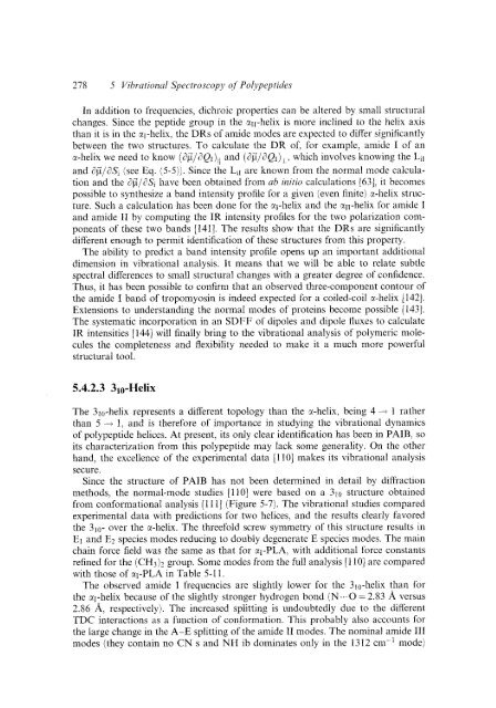
- Page 254 and 255: 5 Vibrational Spectroscopy of Polyp
- Page 256 and 257: 5.2 FOYCC Fields 241 such calculati
- Page 258 and 259: 5.2 Force Fields 243 accounts for o
- Page 260 and 261: 5.2 Force Fields 245 centers of the
- Page 262 and 263: 5.2 Force Fields 247 ture with conf
- Page 264 and 265: 5.3 Amide Modes 249 to the nh initi
- Page 266 and 267: 5.3 Ainide Modes 251 Table 5-1. Ami
- Page 268 and 269: Table 5-2. Some amide modes of four
- Page 270 and 271: 5.3 Amidc Modes 255 Figure 5-1. Opt
- Page 272 and 273: 5.4 Polypeptides 257 nonhydrogen-bo
- Page 274 and 275: 5.4 Polvpeptida 259 Figure 5-2. Ant
- Page 276 and 277: I 5.4 Polypeptides 261 )I .- + $ 80
- Page 278 and 279: 1226M 1222s (1 1165W 1167s (1 1120V
- Page 280 and 281: 135s 9 1 Ms1i 122Wbr 147 NH ob(37)
- Page 282 and 283: 5.4 Polypeptides 267 glycine residu
- Page 284 and 285: 5.4 Polypeptides 269 Raman bands in
- Page 286 and 287: 5.4 Polypeptides 271 Figure 5-7. St
- Page 288 and 289: Frequency (cm-'1 100% b 700 500 300
- Page 290 and 291: 5.4 Polypptidt~s 215 Table 5-9. Obs
- Page 294 and 295: 5.4 Poliyeptides 219 Table 5-11. Co
- Page 296 and 297: Table 5-12. Aniide mode frequencies
- Page 298 and 299: 1351 Lifson, S., Stern, P. S., J. C
- Page 300 and 301: 5.6 Refeverices 285 [130] Tiffany,
- Page 302 and 303: Index Amide modes 249 Amorphous pol
- Page 304 and 305: Step-scan FTIR spectroscopy 14,34 -