

Vol 44 # 4 December 2012 - Kma.org.kw
Vol 44 # 4 December 2012 - Kma.org.kw
Vol 44 # 4 December 2012 - Kma.org.kw

You also want an ePaper? Increase the reach of your titles
YUMPU automatically turns print PDFs into web optimized ePapers that Google loves.
<strong>December</strong> <strong>2012</strong><br />
KUWAIT MEDICAL JOURNAL 304<br />
technique for diagnosis and abscess drainage. Lunsford<br />
et al [9] reported good results with overall bacteriologic<br />
identification of 97% and cure rates of 72% in patients<br />
with brain abscesses. Stereotactic aspirations of<br />
intracerebral hypertensive hemorrhages (acute or<br />
subacute) have shown encouriging results [10] . Catheter<br />
reservoirs may be implanted into the hematoma,<br />
followed with streptokinase or tissue plasminogen<br />
activator injections to lyse the clot [11] .<br />
Although it is a minimally invasive technique, yet<br />
complications have been reported. Sometimes, sample<br />
of the brain tissue taken for biopsy may be nondiagnostic<br />
and may warrant a repeat biopsy. Other<br />
risks include intracranial hemorrhage, infection or<br />
seizures.<br />
Appuzo et al [12] reported 1% morbidity (intracranial<br />
hemorrhage, infection, increased neurological deficit<br />
and seizures) and 0.2 % mortality. Lunsford et al [13]<br />
reported postoperative complications in 2.9% (77)<br />
patients in a series of 2651 patients who underwent<br />
different stereotactic procedures. Out of those,<br />
intracranial hemorrhage occurred in 55 (2.07%)<br />
patients; 11 (0.41%) had local infections at pin sites;<br />
11 (0.41%) patients had post-procedural seizures.<br />
Two (0.075%) patients died from the complications of<br />
the procedure. Although some centers are currently<br />
migrating to frameless stereotactic procedures, their<br />
complication rates are yet to be reported [13] .<br />
PATIENTS AND METHODS<br />
Between 2005 and 2011, we have managed 40<br />
patients with stereotactic surgery. The patients were<br />
selected on the basis of following diagnostic and<br />
therapeutic indications.<br />
Diagnostic<br />
1. Tumors not correlating with their natural history<br />
2. Multiple brain lesions<br />
3. Small and deeply located tumors<br />
4. Patients in good clinical condition (Karnofsky score<br />
> 70)<br />
Therapeutic<br />
5. Cystic lesions that need aspiration<br />
6. Intracranial abscesses<br />
The mean age of the patients was 47 years (range 9<br />
- 70 years). There were 26 male and 14 female patients.<br />
Most of the patients presented with occasional<br />
headaches followed by nausea. Five patients<br />
presented with focal seizures and 12 with neurological<br />
deficits. Twenty patients had lesions, located in the<br />
supratentorial region, 12 in the thalamic and basal<br />
ganglia area, two in the suprasellar region and seven<br />
had multiple intracranial lesions. Stereotactic surgery<br />
was performed with computed tomography (CT)/<br />
magnetic resonance imaging (MRI) - guided Leksell<br />
stereotactic system® and Leksell SurgiPlan® software<br />
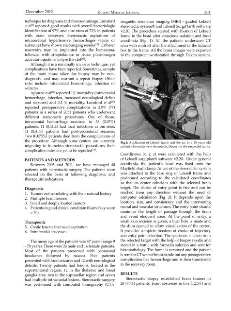
v2.20. The procedure started with fixation of Leksell<br />
frame to the head after conscious sedation and local<br />
anesthesia (Fig. 1). All the patients underwent CT<br />
scan with contrast after the attachment of the fiducial<br />
box to the frame. All the brain images were exported<br />
to the computer workstation through Dicom system.<br />
Fig.1: Application of Leksell frame and the arc in a 45 years old<br />
patient who underwent stereotactic biopsy for the suspected tumor<br />
Coordinates (x, y, z) were calculated with the help<br />
of Leksell surgiplan® software v2.20. Under general<br />
anesthesia, the patient’s head was fixed onto the<br />
Mayfield skull clamp. An arc of the stereotactic system<br />
was attached to the base ring of Leksell frame and<br />
positioned according to the calculated coordinates<br />
so that its center coincides with the selected brain<br />
target. The choice of entry point is free and can be<br />
reached from any direction without the need of<br />
computer calculation (Fig. 2). It depends upon the<br />
location, size, and consistency and the intervening<br />
neural and vascular structures. The entry point should<br />
minimize the length of passage through the brain<br />
and avoid eloquent areas. At the point of entry, a<br />
small skin incision is given, a burr hole is made and<br />
the dura opened to allow visualization of the cortex.<br />
It provides complete freedom of choice of trajectory<br />
and entry point selection. The specimen is taken from<br />
the selected target with the help of biopsy needle and<br />
stored in a bottle with formalin solution and sent for<br />
histopathology. The frame is removed and the patient<br />
is sent for CT scan of brain to rule out any postoperative<br />
complication like hemorrhage and is then transferred<br />
to the recovery room.<br />
RESULTS<br />
Stereotactic biopsy established brain tumors in<br />
28 (70%) patients, brain abscesses in five (12.5%) and


