

Vol 44 # 4 December 2012 - Kma.org.kw
Vol 44 # 4 December 2012 - Kma.org.kw
Vol 44 # 4 December 2012 - Kma.org.kw

Create successful ePaper yourself
Turn your PDF publications into a flip-book with our unique Google optimized e-Paper software.
<strong>December</strong> <strong>2012</strong><br />
KUWAIT MEDICAL JOURNAL 332<br />
Case Report<br />
Oncocytic and Clear Cell Areas in a Solid Pseudopapillary<br />
Tumor of the Pancreas: A Case Report<br />
Anuradha C K Rao, Manna Valiathan, Padmapriya Jaiprakash<br />
Department of Pathology, Kasturba Medical College, Manipal, Karnataka, India<br />
Kuwait Medical Journal <strong>2012</strong>; <strong>44</strong> (4): 332 - 334<br />
ABSTRACT<br />
Solid pseudopapillary tumor (SPT) of pancreas is a rare,<br />
distinct, low-grade malignant neoplasm. Variable areas<br />
like clear cell foci can exist in this tumor, which can easily<br />
mislead the pathologist. Diagnosis of the same is important<br />
to distinguish from other clear cell lesions in the pancreas.<br />
Oncocytoid differentiation is rarer still and can raise doubts<br />
about the diagnosis, especially on trucut biopsies. Herein,<br />
we discuss a case of SPT of the pancreas exhibiting variable<br />
cellular morphology, of which very few cases have been<br />
reported in world literature.<br />
KEY WORDS: oncocytic, pancreas, solid pseudopapillary tumor<br />
INTRODUCTION<br />
Solid pseudopapillary tumor (SPT) is an uncommon,<br />
low-grade malignant neoplasm accounting for 1% of<br />
all exocrine pancreatic tumors. Exact histogenesis of<br />
this tumor remains uncertain [1] . As the various names<br />
for the tumor suggest, it is a solid and cystic pancreatic<br />
neoplasm with characteristic pseudopapillae and sheets<br />
of intermediate and small sized polygonal cells with<br />
minimal morphological deviation. Herein, we report a<br />
case of oncocytic variant of this tumor on account of its<br />
rarity and the few cases reported till date.<br />
CASE HISTORY<br />
A 29-year-old lady presented to the surgical outpatient<br />
department, with a history of swelling in the<br />
upper abdomen of one month duration, which was not<br />
associated with pain, vomiting or abdominal distention.<br />
Patient had a past history of exploratory laparotomy<br />
10 years ago, for palliative gastrojejunostomy and<br />
enterostomy in view of biopsy reported as suspected<br />
neuroendocrine tumor of pancreas, which, however,<br />
was not removed. On examination, a firm to hard mass<br />
of 10 x 7 cm, moving with respiration was palpated<br />
in the left hypochondrial region. All routine laboratory<br />
investigations were within normal range except an ESR<br />
of 40. Ultrasound abdomen was normal and upper GI<br />
endoscopy revealed extraluminal compression on<br />
stomach. A CT-scan revealed a cystic neoplasm arising<br />
from the head of pancreas and a clinical diagnosis<br />
of cystic neoplasm of mucinous origin of pancreas<br />
was made, in view of which the patient underwent<br />
Whipple’s pancreatectomy. Post-operative period<br />
was uneventful and the patient was discharged on<br />
the tenth post-operative day. The patient had no fresh<br />
complaints on follow-up after two months. However,<br />
she presented with amenorrhea after four months,<br />
post-operatively. On ultrasonography, polycystic<br />
ovaries were detected and she was advised for weight<br />
reduction. The patient was lost to follow-up after that.<br />
Pathological findings<br />
Gross: The tumor was well-demarcated, nodular and<br />
unencapsulated, weighing 911 grams and measuring<br />
16 x 11 x 10 cm. Cut section showed a multiloculated<br />
cystic tumor with variegated appearance comprising<br />
grey-white, friable irregular solid areas, yellow areas<br />
and hemorrhagic foci and peripheral rim of pancreatic<br />
tissue.<br />
Microscopy: Sections showed a well demarcated<br />
but unencapsulated tumor composed of sheets and<br />
trabeculae of loosely cohesive polygonal cells with<br />
central ovoid nuclei, few with eccentrically placed<br />
nuclei and finely stippled or granular chromatin.<br />
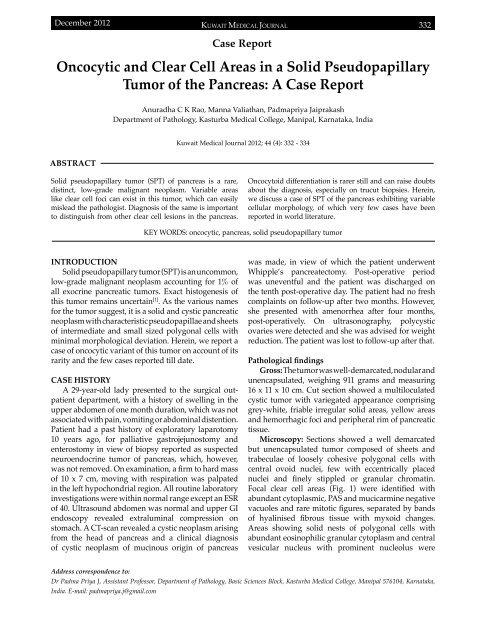
Focal clear cell areas (Fig. 1) were identified with<br />
abundant cytoplasmic, PAS and mucicarmine negative<br />
vacuoles and rare mitotic figures, separated by bands<br />
of hyalinised fibrous tissue with myxoid changes.<br />
Areas showing solid nests of polygonal cells with<br />
abundant eosinophilic granular cytoplasm and central<br />
vesicular nucleus with prominent nucleolus were<br />
Address correspondence to:<br />
Dr Padma Priya J, Assistant Professor, Department of Pathology, Basic Sciences Block, Kasturba Medical College, Manipal 576104, Karnataka,<br />
India. E-mail: padmapriya.j@gmail.com


