Tubes - BD
Tubes - BD
Tubes - BD

You also want an ePaper? Increase the reach of your titles
YUMPU automatically turns print PDFs into web optimized ePapers that Google loves.
7<br />
insert systems<br />
drug discovery and development Bd Biosciences - Discovery Labware<br />
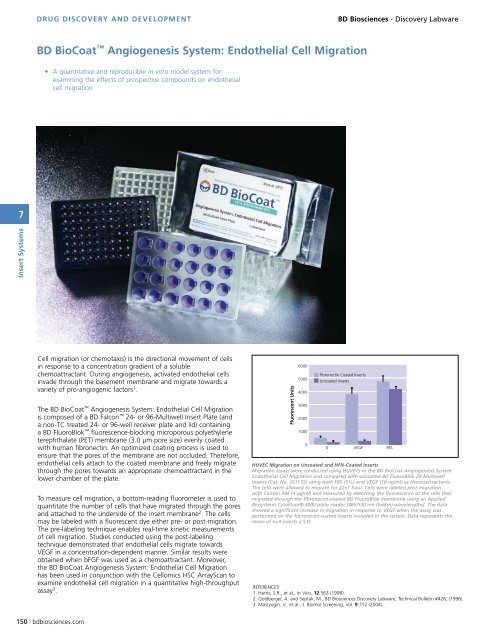
Bd Biocoat angiogenesis system: endothelial cell migration<br />
• A quantitative and reproducible in vitro model system for<br />
examining the effects of prospective compounds on endothelial<br />
cell migration<br />
Cell migration (or chemotaxis) is the directional movement of cells<br />
in response to a concentration gradient of a soluble<br />
chemoattractant. During angiogenesis, activated endothelial cells<br />
invade through the basement membrane and migrate towards a<br />
variety of pro-angiogenic factors 1 .<br />
The <strong>BD</strong> BioCoat Angiogenesis System: Endothelial Cell Migration<br />
is composed of a <strong>BD</strong> Falcon 24- or 96-Multiwell Insert Plate (and<br />
a non-TC treated 24- or 96-well receiver plate and lid) containing<br />
a <strong>BD</strong> FluoroBlok fluorescence-blocking microporous polyethylene<br />
terephthalate (PET) membrane (3.0 µm pore size) evenly coated<br />
with human fibronectin. An optimized coating process is used to<br />
ensure that the pores of the membrane are not occluded. Therefore,<br />
endothelial cells attach to the coated membrane and freely migrate<br />
through the pores towards an appropriate chemoattractant in the<br />
lower chamber of the plate.<br />
To measure cell migration, a bottom-reading fluorometer is used to<br />
quantitate the number of cells that have migrated through the pores<br />
and attached to the underside of the insert membrane 2 . The cells<br />
may be labeled with a fluorescent dye either pre- or post-migration.<br />
The pre-labeling technique enables real-time kinetic measurements<br />
of cell migration. Studies conducted using the post-labeling<br />
technique demonstrated that endothelial cells migrate towards<br />
VEGF in a concentration-dependent manner. Similar results were<br />
obtained when bFGF was used as a chemoattractant. Moreover,<br />
the <strong>BD</strong> BioCoat Angiogenesis System: Endothelial Cell Migration<br />
has been used in conjunction with the Cellomics HSC ArrayScan to<br />
examine endothelial cell migration in a quantitative high-throughput<br />
assay 3 .<br />
150 | bdbiosciences.com<br />
Fluorescent Units<br />
6000<br />
5000<br />
4000<br />
3000<br />
2000<br />
1000<br />
0<br />
Fibronectin Coated Inserts<br />
Uncoated Inserts<br />
0<br />
HUVEC Migration on Uncoated and HFN-Coated Inserts<br />
Migration assays were conducted using HUVECs in the <strong>BD</strong> BioCoat Angiogenesis System:<br />
Endothelial Cell Migration and compared with uncoated <strong>BD</strong> FluoroBlok 24-Multiwell<br />
Inserts (Cat . No . 351155) using both FBS (5%) and VEGF (10 ng/ml) as chemoattractants .<br />
The cells were allowed to migrate for 22±1 hour . Cells were labeled post-migration<br />
with Calcein AM (4 µg/ml) and measured by detecting the fluorescence of the cells that<br />
migrated through the fibronectin-coated <strong>BD</strong> FluoroBlok membrane using an Applied<br />
Biosystems CytoFluor® 4000 plate reader [485/530 nm (Ex/Em) wavelengths] . The data<br />
showed a significant increase in migration in response to VEGF when the assay was<br />
performed on the fibronectin-coated inserts included in the system . Data represents the<br />
mean of n=3 inserts ± S .D .<br />
VEGF<br />
REFERENCES:<br />
1. Harris, S.R., et al., In Vivo, 12:563 (1998).<br />
2. Goldberger, A. and Septak, M., <strong>BD</strong> Biosciences Discovery Labware, Technical Bulletin #428, (1998).<br />
3. Mastyugin, V., et al., J. Biomol Screening, Vol. 9:712 (2004).<br />
FBS