Diffusion Processes with Hidden States from ... - FU Berlin, FB MI
Diffusion Processes with Hidden States from ... - FU Berlin, FB MI
Diffusion Processes with Hidden States from ... - FU Berlin, FB MI

Create successful ePaper yourself
Turn your PDF publications into a flip-book with our unique Google optimized e-Paper software.
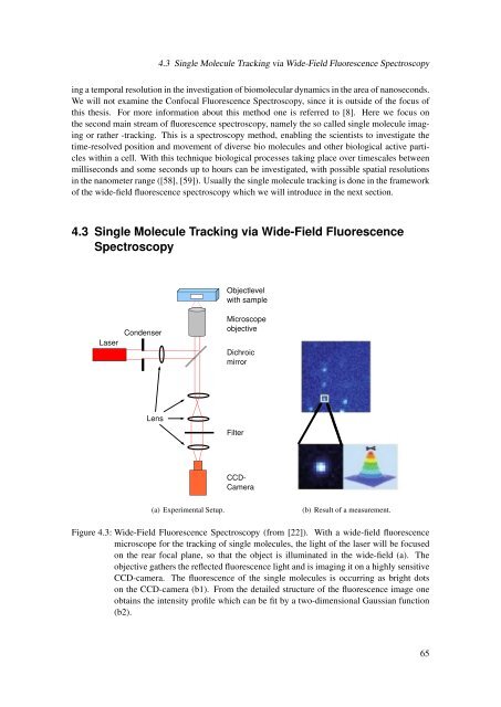
4.3 Single Molecule Tracking via Wide-Field Fluorescence Spectroscopying a temporal resolution in the investigation of biomolecular dynamics in the area of nanoseconds.We will not examine the Confocal Fluorescence Spectroscopy, since it is outside of the focus ofthis thesis. For more information about this method one is referred to [8]. Here we focus onthe second main stream of fluorescence spectroscopy, namely the so called single molecule imagingor rather -tracking. This is a spectroscopy method, enabling the scientists to investigate thetime-resolved position and movement of diverse bio molecules and other biological active particles<strong>with</strong>in a cell. With this technique biological processes taking place over timescales betweenmilliseconds and some seconds up to hours can be investigated, <strong>with</strong> possible spatial resolutionsin the nanometer range ([58], [59]). Usually the single molecule tracking is done in the frameworkof the wide-field fluorescence spectroscopy which we will introduce in the next section.4.3 Single Molecule Tracking via Wide-Field FluorescenceSpectroscopyObjectlevel<strong>with</strong> sampleLaserCondenserMicroscopeobjectiveDichroicmirrorLensFilterCCDCamera(a) Experimental Setup.(b) Result of a measurement.Figure 4.3: Wide-Field Fluorescence Spectroscopy (<strong>from</strong> [22]). With a wide-field fluorescencemicroscope for the tracking of single molecules, the light of the laser will be focusedon the rear focal plane, so that the object is illuminated in the wide-field (a). Theobjective gathers the reflected fluorescence light and is imaging it on a highly sensitiveCCD-camera. The fluorescence of the single molecules is occurring as bright dotson the CCD-camera (b1). From the detailed structure of the fluorescence image oneobtains the intensity profile which can be fit by a two-dimensional Gaussian function(b2).65