170 COMPARATIVE PARASITOLOGY, <strong>67</strong>(2), JULY <strong>2000</strong> (deposited at the Institut Agronomique et Veterinaire Hassan II, Rabat, Morocco), was made by one of us (S.B.). The second collection, from a single specimen of T. hennanni from Catalonia, Spain, was made by C. Feliu, Barcelona, Spain, and deposited at the Barcelona Zoo, Spain. Nematodes were preserved in 70% ethanol before being cleared with lactophenol for study. Figures were made with the aid of a drawing tube. Nematodes were dehydrated by passage through progressive ethanol concentrations to absolute ethanol and critical-point-dried (M scope 500, Hitachi, Japan). The scanning electron microscope used was a Hitachi S 520, Hitachi, Japan at 20 kV. Measurements given are for the holotype male and the allotype female. Measurements in parentheses are the ranges of paratype males and females. All measurements are in micrometers. Results Thaparia thapari thapari (Dubinina, 1949) (Figs. 1-10) Redescription GENERAL: The material examined consisted of 6 males and 15 females. Body medium-sized, stout. Mouth triangular, with 3 transparent lips. Buccal cavity with denticles. Cephalic sense organs consisting of inner circle of 6 nerve endings, papillae not pedunculate (Figs. 2, 3, 6, 7), the outer circle not observed, and amphids present. Esophagus divided into 2 portions: anterior muscular part, and comparatively longer posterior glandular part terminating in valvular bulb; 17 chitinoid pieces surrounding anterior end of esophagus (Figs. 1, 3). Excretory pore postesophageal. MALE: Length 2,754-3,169; maximum thickness 191-229. In worm measuring 2,773, esophagus 388: corpus 180 and isthmus plus bulb 208. Nerve ring and excretory pore 161 and 889, respectively, from anterior end. Posterior extremity truncated. Tail <strong>67</strong> long. Spicule needle-shaped, 100 long. Gubernaculum Vshaped. Three pairs of caudal papillae: 2 circumcloacal (1 pair preanal and 1 pair postanal) and 1 pair at tail end. Preanal membrane, present with 6 lobes (Figs. 4, 5, 8, 9, 10), and caudal alae absent. FEMALE: Length 4,282-4,716; maximum thickness 356-378. In a worm measuring 4,600, esophagus 615: corpus 240 and isthmus plus bulb 375. Nerve ring, excretory pore, and vulva at 180, 1,282, and 2,264, respectively, from anterior end. Tail 270 long. Taxonomic summary HOST: Spur-thighed tortoise, Testudo graeca Linnaeus, 1758. SITE IN HOST: Cecum. TYPE LOCALITY/COLLECTION DATE: Settat, Morocco, 32°30'45"N, 7°45'30"W, 22 July 1999, by S.B. SPECIMENS DEPOSITED: Museum National d'Histoire Naturelle, Paris, France. Number 825 HE Remarks Thapar (1925) described the female of this species from Testudo graeca, as Oxyuris sp. Dubinina (1949) studied males, the structures of which made it possible to include the species in Tachygonetria Wedl, 1862. Fetter (1966) redescribed and transferred this species to the genus Thaparia and divided it in 2 subspecies: T. thapari thapari and T. thapari australis. The emended diagnosis characterizes T. thapari thapari with 17 chitinoid pieces, whereas Petter (1961, 1966) cited 6 chitinoid pieces. Cephalic sense organs consist of an inner circle of 6 nerve endings (papillae not pedunculate), 4 submedian and 2 lateral close to amphids; outer papillae were not observed. Description Copyright © 2011, The Helminthological Society of Washington Thaparia carlosfeliui sp. n. (Figs. 11-23) GENERAL: The material examined consisted of 20 males and 40 females. Nematoda, Oxyuroidea, Pharyngodonidae, Thaparia. Robust worms of small size. Mouth surrounded by 3 lips. Esophagus in 2 parts, with elongated isthmus. Amphids prominent. Differs from the diagnosis of the genus in the number of caudal papillae. MALE: (holotype and 3 paratypes): Mouth surrounded by 3 V-shaped cut lips (Figs. 13, 21). Six oral papillae, arranged in 3 pairs (Fig. 13). Buccal cavity without denticles. Esophagus lobes visible. No chitinoid pieces visible. Tail without alae. Structure of caudal region complex (Figs. 15, 16, 22, 23). One pair of preanal papillae, 1 pair of large postanal elongated papillae. Posterior lip of anus with central nipple. Surrounding ventral membrane present lateral and anterior to anus, and second preanal membrane situated posterior to first membrane. Anterior lip of anus with 2 lobes, extremity of spic-
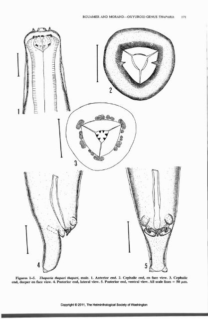
BOUAMER AND MORAND—OXYUROID GENUS THAPARIA 171 Figures 1-5. Thaparia thapari thapari, male. 1. Anterior end. 2. Cephalic end, en face view. 3. Cephalic end, deeper en face view. 4. Posterior end, lateral view. 5. Posterior end, ventral view. All scale lines = 50 |un. Copyright © 2011, The Helminthological Society of Washington
- Page 1 and 2: July 2000 Number 2 Comparative Para
- Page 3 and 4: Comp. Parasitol. 67(2), 2000 pp. 14
- Page 5 and 6: Table 1. Unidentified diplectanids
- Page 7 and 8: preferences, separation of Lateroca
- Page 9 and 10: ward, 1996). Vincent's sillago, Sil
- Page 11 and 12: (see Johnston and Tiegs, 1922; Murr
- Page 13 and 14: Sphyraena chiysotaenia Klunzinger,
- Page 15 and 16: figures of the internal anatomy (wh
- Page 17 and 18: KRITSKY ET AL.—DIPLECTANIDS FROM
- Page 19 and 20: 2)] long, slender, fusiform; greate
- Page 21 and 22: creation of paraphyletic taxa when
- Page 23 and 24: Comp. Parasitol. 67(2), 20(K) pp. 1
- Page 25 and 26: DAILEY AND GOLDBERG—LANGERONIA BU
- Page 27: Comp. Parasitol. 67(2), 2000 pp. 16
- Page 31 and 32: Figure 10. Thaparia thaparia thapar
- Page 33 and 34: BOUAMER AND MORAND—OXYUROID GENUS
- Page 35 and 36: BOUAMER AND MORAND—OXYUROID GENUS
- Page 37 and 38: of Professor Robert Bourgat (Univer
- Page 39 and 40: Comp. Parasitol. 67(2), 2000 pp. 18
- Page 41 and 42: Discussion Twelve parasite species
- Page 43 and 44: Table 2. Continued. Parasite Tricho
- Page 45 and 46: ter and fiberglass or concrete pond
- Page 47 and 48: Sutherland, D. R. 1999. Assessing t
- Page 49 and 50: Table 1. Wild hosts for Neobenedeni
- Page 51 and 52: CB < •£ a o '= 2 I BULLARD ET AL
- Page 53 and 54: Laboratory (GCRL), Ocean Springs, M
- Page 55 and 56: Comp. Parasitol. 67(2), 2000 pp. 19
- Page 57 and 58: Table 2. Presence of Hymenolepis na
- Page 59 and 60: tones ungiiiculatus). Canadian Vete
- Page 61 and 62: BOLEK AND COGGINS—HELMINTH COMMUN
- Page 63 and 64: BOLEK AND COGGINS—HELMINTH COMMUN
- Page 65 and 66: ence of Rhabdias bufonis Schrank, 1
- Page 67 and 68: Iowa I. Trematodes of amphibians. A
- Page 69 and 70: MACHADO ET AL.—ECOLOGY OF ENDOHEL
- Page 71 and 72: 40- 35- 30- 25- 20- 15- 10- 5- n .
- Page 73 and 74: MACHADO ET AL.—ECOLOGY OF ENDOHEL
- Page 75 and 76: MACHADO ET AL.—ECOLOGY OF ENDOHEL
- Page 77 and 78: Table 1. Summary of results of gros
- Page 79 and 80:
LYONS ET AL.—HOOKWORMS IN NORTHER
- Page 81 and 82:
lands in the Bering Sea. Journal of
- Page 83 and 84:
River, Kobe, Hyogo Prefecture, Japa
- Page 85 and 86:
HASEGAWA ET AL.—LIFE HISTORY OF S
- Page 87 and 88:
larvae of 5. contortus or S. japoni
- Page 89 and 90:
is. Using histochemical methods our
- Page 91 and 92:
1.2 n 1.0- |T. spiralis |T. pseudos
- Page 93 and 94:
G. A. Schaub, and F. Brombacher. 19
- Page 95 and 96:
Table 1. Infectivity and distributi
- Page 97 and 98:
FUJINO ET AL.—EXPULSION OF ECHINO
- Page 99 and 100:
Comp. Parasitol. 67(2), 2000 pp. 24
- Page 101 and 102:
0) 0> O 20- O 2 3 4 Weeks post infe
- Page 103 and 104:
vae from Temazcal and 3 from Culiac
- Page 105 and 106:
KOGA ET AL.—SURFACE ULTRASTRUCTUR
- Page 107 and 108:
ales del Institute de Biologfa de l
- Page 109 and 110:
CANARIS AND KINSELLA—RESEARCH NOT
- Page 111 and 112:
Comp. Parasitol. 67(2), 2000 pp. 25
- Page 113 and 114:
Comp. Parasitol. 67(2), 2()(X) pp.
- Page 115 and 116:
1866; and Hyla wrightorum (Taylor,
- Page 117 and 118:
Several horses, all with unknown an
- Page 119 and 120:
Comp. Parasitol. 67(2), 2000 pp. 26
- Page 121 and 122:
ANNIVERSARY AWARD 263 laboratory on
- Page 123 and 124:
Comp. Parasitol. 67(2), 2000 p. 265
- Page 125 and 126:
Diplectanum sillagonum, 145 Diplect
- Page 127 and 128:
Philichthyidae, 253 Phodopus sungor
- Page 129 and 130:
Name: MEMBERSHIP APPLICATION 271 AP
- Page 131 and 132:
*Edna M. Buhrer * Mildred A. Doss *