

Comparative Parasitology 67(2) 2000 - Peru State College
Comparative Parasitology 67(2) 2000 - Peru State College
Comparative Parasitology 67(2) 2000 - Peru State College

Create successful ePaper yourself
Turn your PDF publications into a flip-book with our unique Google optimized e-Paper software.
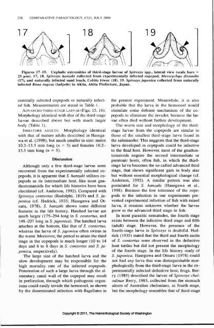
228 COMPARATIVE PARASITOLOGY, <strong>67</strong>(2), JULY <strong>2000</strong><br />
Figures 17-19. Cephalic extremities of third-stage larvae of Spiroxys spp., lateral view (scale bars =<br />
25 (Jim). 17, 18. Spiroxys hanzaki collected from experimentally infected copepod, Mesocyclops dissimilis<br />
(17), and naturally infected sand loach, Cobitis biwae (18). 19. Spiroxys japonica collected from naturally<br />
infected Rana rugosa (tadpole) in Akita, Akita Prefecture, Japan.<br />
imentally infected copepods or naturally infected<br />
fish. Measurements are stated in Table 1.<br />
ADVANCED THIRD-STAGE LARVAE (Figs. 15, 16):<br />
Morphology identical with that of the third-stage<br />
larvae described above but with much larger<br />
body (Table 1).<br />
IMMATURE ADULTS: Morphology identical<br />
with that of mature adults described in Hasegawa<br />
et al. (1998), but much smaller in size: males<br />
10.2-13.3 mm long (« = 5) and females 10.2-<br />
15.5 mm long (n = 5).<br />
Discussion<br />
Although only a few third-stage larvae were<br />
recovered from the experimentally infected copepods,<br />
it is apparent that 5. hanzaki utilizes copepods<br />
as its intermediate host, like most gnathostomatoids<br />
for which life histories have been<br />
elucidated (cf. Anderson, 1992). Compared with<br />
Spiroxys contortus (Rudolphi, 1819) and S. japonica<br />
(cf. Hedrick, 1935; Hasegawa and Otsuru,<br />
1978), S. hanzaki shows some different<br />
features in the life history. Hatched larvae are<br />
much larger (175-294 long in S. contortus, and<br />
148-207 long in S. japonica). The hatched larva<br />
attaches at the bottom, like that of 5. contortus,<br />
whereas the larva of S. japonica often swims in<br />
the water. Moreover, the period to attain the third<br />
stage in the copepods is much longer (10 to 14<br />
days and 6 to 8 days in S. contortus and S. japonica,<br />
respectively).<br />
The large size of the hatched larva and the<br />
slow development may be responsible for the<br />
high mortality rate of the infected copepods.<br />
Penetration of such a large larva through the alimentary<br />
canal wall of the copepod may result<br />
in perforation, through which pathogenic organisms<br />
could easily invade the hemocoel, as shown<br />
by the disseminated infection with flagellates in<br />
Copyright © 2011, The Helminthological Society of Washington<br />
the present experiment. Meanwhile, it is also<br />
probable that the larva in the hemocoel would<br />
stimulate some defense mechanism of the copepods<br />
to eliminate the invader, because the larvae<br />
often died without further development.<br />
The worm size and morphology of the thirdstage<br />
larvae from the copepods are similar to<br />
those of the smallest third-stage larva found in<br />
the salamander. This suggests that the third-stage<br />
larva developed in copepods could be infective<br />
to the final host. However, most of the gnathostomatoids<br />
require the second intermediate or<br />
paratenic hosts, often fish, in which the thirdstage<br />
larva becomes the so-called advanced third<br />
stage, that shows significant gain in body size<br />
but without essential morphological change (cf.<br />
Anderson, 1992). A similar pattern was also<br />
postulated for S. hanzaki (Hasegawa et al.,<br />
1998). Because the low tolerance of the copepods<br />
to the infection in our experiments prevented<br />
experimental infection of fish with raised<br />
larva, it remains unknown whether the larvae<br />
grow to the advanced third stage in fish.<br />
In most parasitic nematodes, the fourth stage<br />
exists between the infective third stage and fifth<br />
(adult) stage. However, the presence of the<br />
fourth-stage larva in Spiroxys is doubtful. Hedrick<br />
(1935) stated that the third and fourth molts<br />
of S. contortus were observed in the definitive<br />
host turtles but did not present the morphology<br />
of the fourth stage. In the life history study of<br />
S. japonica, Hasegawa and Otsuru (1978) could<br />
not find any larva that was distinguishable morphologically<br />
from the third-stage larva in the experimentally<br />
infected definitive host, frogs. Berry<br />
(1985) described the larvae of Spiroxys chelodinae<br />
Berry, 1985, collected from the stomach<br />
ulcers of Australian chelonians, as fourth stage,<br />
but the morphology resembles that of third-stage


