160 PEARSON, J. M. H., ROSS, F. W. Nerve involvement in leprosy - pathology, differencial diagnosis and principles of management. Lepr. Rev., v.46, p.199 - 212. 1975. PFATZGRAFF, R. E., RAMU, G. In: HASTINGS, R. C. Leprosy. 2.ed. Lon<strong>do</strong>n: Churchill Livingstone, 1994. p.237 - 87. PITRES, A., TESTUT, L. Les nerfs en schémas. Paris: Octave Doin, 1925, v.5. p. 344. POIRIER, R. , CHARPY, A., CUNÉO, B. Abrégé d'anatomie. Paris: Masson, 1908. v.2, p.710 -1, 1004. ROHEN, J., YOKOCHI, C. Anatomia humana. 2.ed. São Paulo: Manole, 1987. v.2. p.48. ROMANES, G. J. CUNNINGHAM'S textbook of anatomy. 10.ed. Lon<strong>do</strong>n: Oxford, 1964. p.735, 928 - 929. ROSS, W. F., PEARSON, J. M. H. The recognition and management of nerve damage under field conditions. Lepr. Rev., v.46, p.231 - 4, 1975. ROUVIÈRE, A. Anatomia humana descriptiva y topográfica. 5.ed. Madrid: Bailly, 1959. v.3, p.160 - 1, 191, 233. SABIN, T. D., SWIFT, T. R., JACOBSON, R. R. Leprosy. In: DYCK, P. J., THOMAS, P. K, GRIFFIN, J. W., LOW, A. P., PODUSLO, J. F. Peripheral neuropathy. 3.ed. Mexico: W.B. Saunders, 1993. v. 2, p.1354 - 79. SAPPEY, P. H. C. Traité d'anatomie descriptive. 4.ed. Paris: Lecrosnier et Babé, 1889. v.3, p.425, 6. SCHAEFFER, J. P. MORRIS' human anatomy. 1953. p.772 – 773, 1141 - 1142. New York: Mc Graw Hill, SILVA, F. B. El sindrome neural leproso. Ensayo e sistematizacion. Rev. Peru. Salud Publica., v.5, p.250 - 62, 1957. SKJELDAL, O. H., STOKKE, O., REFSUM, S. Phytanic acid storage disease. In: DYCK, P. J., THOMAS, P. K, GRIFFIN, J. W., LOW, A. P., PODUSLO, J.F. Peripheral neuropathy. 3.ed. Mexico: W.B. Saunders, 1993. v.2, p.1149 - 60. SOBOTTA, J., FERNER, H., STAUBESAND, J. Atlas de anatomia humana. 18 ed. Rio de Janeiro: Guanabara Koogan, 1982, v.1, p.302, 339.
161 SOBOTTA, J., PUTZ, R., PABST, R. Atlas de anatomia humana. 20 ed. Rio de Janeiro: Guanabara Koogan, 1993, v.1, p.243 - 44. SPALTEHOLZ, W. Atlas de anatomia humana. Barcelona: Labor, 1950. v.2, p.555. SPALTEHOLZ, W., SPANNER, R. Atlas de anatomia humana. 16.ed. São Paulo: Roca, 1988. v.2, p.393. SRINIVAS, H. V., LAKHANI, R., MEHTA, L. M, ANTIA, N. H. Study of tickened nerves in a leprosy endemic region. Part I. Lepr. India, v.52, p.52 - 64, 1980. STEWART, J. D. Compression and entrapment neuropathies. In: DYCK, J. P., THOMAS, P. K. GRIFFIN, J. W. LOW, P. A. PODUSLO, J. F. Peripheral neuropathy. 3.ed. Mexico: W.B. Saunders, 1993. v.2,. p.961 - 79. TAGA, R., STIPP, A. C. M. Manual prático de morfometria ao microscópio óptico. Bauru: USP - Departamento de Morfologia, 1994. p.22. TALHARI, S., NEVES, R. G., OLIVEIRA, S. G. Manifestações <strong>nervo</strong>sas e diagnóstico diferencial. In: TALHARI, S., NEVES, R. G. Hansenologia. Manaus: Funcomiz, 1984. p.35 - 53. TALWAR, S., JHA., P.K, TIWARI, V. D. Neuritic leprosy: epidemiology and therapeutic responsiveness. Lepr. Rev., v.63, p.263 - 8, 1992. TESTUT, L. Traité d'anatomie humaine. 7.ed. Paris: Octave Doin, 1922a. v.2, p.291 - 2. TESTUT, L. Traité d'anatomie humaine. 7.ed. Paris: Octave Doin, 1922b. v.3, p.186-7. TESTUT, L., JACOB, O. Traité d'anatomie topographique. 4.ed. Paris: Octave Doin, 1922. v.2, p.740, 781, 791, 799 - 808, 855. TESTUT, L., LATARJET, A. Trata<strong>do</strong> de anatomia humana. 9.ed. Barcelona: Salvat, 1958. v.2, p.438 - 42. TESTUT, L., LATARJET, A. Trata<strong>do</strong> de anatomia humana. 9.ed. Barcelona: Salvat, 1959. v.3, p.294. TILLAUX, P. Traité d'anatomie topographique. Paris: Asselin et Houzeau, 1908. p.565 - 70.
- Page 1 and 2:
ROSEMARI BACCARELLI ESTUDO DO RAMO
- Page 3:
Quanto mais fundamente penso, mais
- Page 7 and 8:
1. INTRODUÇÃO A hanseníase é um
- Page 9 and 10:
uma ou poucas lesões cutâneas bem
- Page 11 and 12:
dos nervos, com tendência à simet
- Page 13 and 14:
deposição de imunocomplexos, nos
- Page 15 and 16:
agravamento das lesões neurológic
- Page 17 and 18:
18 Pfaltzgraaf & Ramu (1994) relata
- Page 19 and 20:
de hanseníase, e os resultados dos
- Page 21 and 22:
fazem o diagnóstico e o tratamento
- Page 23 and 24:
trabalho com a finalidade de observ
- Page 25 and 26:
27 Logo em seguida, Laehr (apud Mur
- Page 27 and 28:
(88,9%) apresentaram um ou mais ner
- Page 29 and 30:
no interior de macrófagos, célula
- Page 31 and 32:
unilaterais e 84 bilaterais). Quant
- Page 33 and 34:
35 Browne (1975) refere que vários
- Page 35 and 36:
anestésica, fraqueza ou paralisia
- Page 37 and 38:
espectivamente para os duplas de ex
- Page 39 and 40:
veia metacarpal dorsal, veia cefál
- Page 41 and 42:
Testut, 1925, Testut & Latarjet, 19
- Page 43 and 44:
Schaeffer, 1953, Rouvière, 1959, T
- Page 45 and 46:
acrescentam a região dorsal do qua
- Page 47 and 48:
49 Os limites da região posterior
- Page 49 and 50:
51 De acordo com Tillaux (1908), Te
- Page 51 and 52:
aquiorradial e extensor radial long
- Page 53 and 54:
4. MATERIAL E MÉTODO distribuiçã
- Page 55 and 56:
4.1.2. Método A área anatômica a
- Page 57 and 58:
61 As respostas às questões de um
- Page 59 and 60:
Foi efetuada a dissecção do ramo
- Page 61 and 62:
observar as relações anatômicas
- Page 63 and 64:
5. RESULTADOS 5.1 Estudo Clínico A
- Page 65 and 66:
superfície foram excluídos da amo
- Page 67 and 68:
72 Os valores referidos pelo examin
- Page 69 and 70:
haverem sido considerados não palp
- Page 71 and 72:
Os valores do teste do qui-quadrado
- Page 73 and 74:
RSNR manteve trajetória ao longo d
- Page 75 and 76:
do rádio em ramos lateral e medial
- Page 77 and 78:
AQUI82 • em 1 (5,00%) peça, foi
- Page 79 and 80:
egião posterolateral da extremidad
- Page 81 and 82:
5.2.3.2.3. Nervo sobre um conjunto
- Page 83 and 84:
5.2.4.2. Relação anatômica da ve
- Page 85 and 86:
FIGURA 2 - Vista lateral do antebra
- Page 87 and 88:
FIGURA 4 - Vista lateral do antebra
- Page 89 and 90:
FIGURA 6 - Vista lateral do antebra
- Page 91 and 92:
FIGURA 8 - Vista lateral do antebra
- Page 93 and 94:
FIGURA 10 - Vista lateral do antebr
- Page 95 and 96:
FIGURA 12.1 - Vista lateral do ante
- Page 97 and 98:
FIGURA 13 - Vista lateral do antebr
- Page 99 and 100:
FIGURA 15 - Vista lateral do antebr
- Page 101 and 102: FIGURA 17 - Vista lateral doantebra
- Page 103 and 104: 108 FIGURA 19 - Vista lateral do an
- Page 105 and 106: 5.3. Anatomia Microscópica Conside
- Page 107 and 108: músculos abdutor longo e extensor
- Page 109 and 110: Quanto ao posicionamento, as veias
- Page 111 and 112: 116 FIGURA 21 - Corte histológico
- Page 113 and 114: FIGURA 25 - Corte histológico tran
- Page 115 and 116: 120
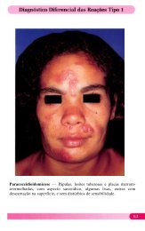
- Page 117 and 118: 123 A classificação da hansenías
- Page 119 and 120: mesmo quando as alterações hipert
- Page 121 and 122: 127 aos resultados da palpação do
- Page 123 and 124: médias da espessura dos RSNR exami
- Page 125 and 126: ao acaso. Os resultados entre 0 e 0
- Page 127 and 128: espessamento dos nervos, o qual se
- Page 129 and 130: 135 (Adamson et al., 1966, Gama Net
- Page 131 and 132: Nesta pesquisa, verificou-se ramos
- Page 133 and 134: lateral, segundo Töndury (1958), C
- Page 135 and 136: do ramo lateral do RSNR refletiu-se
- Page 137 and 138: elações anatômicas do RSNR com a
- Page 139 and 140: Profundamente a ele, de lateral par
- Page 141 and 142: elações anatômicas de proximidad
- Page 143 and 144: conjunto com o nervo. Nesse caso, o
- Page 145 and 146: 7. CONCLUSÕES Os resultados obtido
- Page 147 and 148: 8. REFERÊNCIAS BIBLIOGRÁFICAS* AB
- Page 149 and 150: GARDNER, E., GRAY, D. J., O'RAHILLY
- Page 151: 159 MEHTA, L. M., ANTIA, N. H., LAK
- Page 157 and 158: 165
- Page 159 and 160: 167
- Page 161 and 162: 169
- Page 163 and 164: 171
- Page 165 and 166: RESUMO Este trabalho objetiva contr
- Page 167 and 168: ABSTRACT The purpose of this paper