Nutritional Secondary Hyperparathyroidism in the Horse
Nutritional Secondary Hyperparathyroidism in the Horse
Nutritional Secondary Hyperparathyroidism in the Horse

You also want an ePaper? Increase the reach of your titles
YUMPU automatically turns print PDFs into web optimized ePapers that Google loves.
10 The Anatomy of <strong>the</strong> Parathyroid Glands <strong>in</strong> <strong>the</strong> <strong>Horse</strong><br />
ESTES’ (1907) description clearly established that three ma<strong>in</strong> types of epi<strong>the</strong>lial<br />
cells occur <strong>in</strong> <strong>the</strong> equ<strong>in</strong>e parathyroid, viz., chief cells, water-clear cells, and<br />
oxyphil cells. This should be emphasized <strong>in</strong> view of <strong>the</strong> fact that even modern<br />
textbooks and papers deal<strong>in</strong>g with general parathyroid histology stress <strong>the</strong><br />
existence of water-clear cells and oxyphil cells as a characteristic of <strong>the</strong> human<br />
parathyroid gland only.<br />
LITTY (1907) dist<strong>in</strong>guished two types of epi<strong>the</strong>lial cells <strong>in</strong> <strong>the</strong> equ<strong>in</strong>e parathyroid.<br />
In addition to <strong>the</strong> predom<strong>in</strong>at<strong>in</strong>g light chief cells (accord<strong>in</strong>g to modern<br />
nomenclature), he described large cells with well-def<strong>in</strong>ed borders, a clear cytoplasm,<br />
and a nucleus hav<strong>in</strong>g variable sta<strong>in</strong><strong>in</strong>g properties. These cells usually<br />
occurred <strong>in</strong> restricted areas <strong>in</strong> small groups, but <strong>in</strong> rare cases <strong>the</strong>y predom<strong>in</strong>ated<br />
<strong>in</strong> <strong>the</strong> microscopic field.<br />
BOBEAU (1911) contributed a detailed description of <strong>the</strong> histology of <strong>the</strong><br />
equ<strong>in</strong>e parathyroids. He recognized four epi<strong>the</strong>lial cell types, viz., light chief cells<br />
(celldes normales) with a longitud<strong>in</strong>al diameter of 12 to 16 p and a nucleus of 5 to<br />
7 p; water-clear cells (celhles claires) with no sta<strong>in</strong>able cytoplasm (“one has <strong>the</strong><br />
impression of nuclei float<strong>in</strong>g <strong>in</strong> an empty space”); dark oxyphil cells (cellules<br />
sombres) with a longitud<strong>in</strong>al diameter of 10 p and deeply eos<strong>in</strong>ophilic cytoplasm;<br />
and pale oxyphil cells (grosses celldes ci prod& gramdegx ozt colloid).<br />
BOBEAU also exam<strong>in</strong>ed <strong>the</strong> parathyroids for fat. He stated that fat was present<br />
<strong>in</strong> <strong>the</strong> cytoplasm of <strong>the</strong> light chief cells and, more rarely, <strong>in</strong> <strong>the</strong> dark oxyphil cells<br />
but never <strong>in</strong> <strong>the</strong> water-clear and pale oxyphil cells. Unfortunately, BOBEAU did<br />
not give <strong>the</strong> age of any of his horses, and <strong>the</strong> correlation between age and <strong>the</strong><br />
degree of fat present <strong>in</strong> <strong>the</strong> cells is <strong>the</strong>refore lack<strong>in</strong>g.<br />
VERMEULEN (1917) described two ma<strong>in</strong> epi<strong>the</strong>lial cell types, viz., chief cells<br />
and oxyphil cells. He also proposed a classification of <strong>the</strong> parathyroid glands<br />
accord<strong>in</strong>g to <strong>the</strong> amount and arrangement of connective tissue with<strong>in</strong> <strong>the</strong> glands.<br />
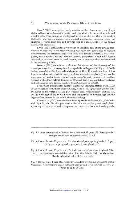
Fig. 3. Lower parathyroids of horses, both male and 12 years old. Parathyroid at<br />
straight arrow, cyst at curved arrows, x 4.5.<br />
Fig. 4. <strong>Horse</strong>, female, 22 years old. Relative size of parathyroid glands. Left part<br />
of figure: upper gland; right part: lower gland, x 5.<br />
Fig. 5. <strong>Horse</strong>, female, 17 years old. Typical structure of parathyroid gland. Th<strong>in</strong><br />
connective tissue septa subdivid<strong>in</strong>g gland <strong>in</strong>to f<strong>in</strong>e lobuli. Rich vascularization.<br />
Ma<strong>in</strong>ly light chief cells. H & E, x 275.<br />
Fig. 6. <strong>Horse</strong>, male, 1 year old. Relatively abundant stroma <strong>in</strong> parathyroid gland.<br />
Numerous KURSTEINER’S canals (straight arrow) and cysts (curved arrows) at<br />
hilus. H & E, x 22.5.<br />
Downloaded from<br />
vet.sagepub.com by guest on April 14, 2010