Nutritional Secondary Hyperparathyroidism in the Horse
Nutritional Secondary Hyperparathyroidism in the Horse
Nutritional Secondary Hyperparathyroidism in the Horse

Create successful ePaper yourself
Turn your PDF publications into a flip-book with our unique Google optimized e-Paper software.
18 'I'hc Anatomy of <strong>the</strong> Parathyroid Glands <strong>in</strong> <strong>the</strong> <strong>Horse</strong><br />
def<strong>in</strong>ed than that of any o<strong>the</strong>r cell (Figs. 8, 9, and 10). In <strong>in</strong>termediate<br />
stages <strong>the</strong> basophilic cytoplasmic structures coalesced to form small<br />
spherical bodies, crescentshaped flakes, or more irregular formations.<br />
The spherical bodies could occur at a juxtanuclear position, but when<br />
<strong>the</strong>y were multiple, as was often <strong>the</strong> case, <strong>the</strong>y could also occur <strong>in</strong> <strong>the</strong><br />
extreme peripheiy of <strong>the</strong> cell. The flakes occurred irregularly <strong>in</strong> <strong>the</strong><br />
cytoplasm. The more complete <strong>the</strong> development of <strong>the</strong> waterclear<br />
cells was, <strong>the</strong> more peripheral was <strong>the</strong>ir location. The well-def<strong>in</strong>ed<br />
membrane of <strong>the</strong> water-clear cells was due to <strong>the</strong> presence of such<br />
coalesced flakes glued like tapestry to <strong>the</strong> cell membrane. The cyto-<br />
plasm conta<strong>in</strong>ed no sta<strong>in</strong>able fat. The nucleus was smaller than it was<br />
<strong>in</strong> <strong>the</strong> light chief cell and <strong>the</strong> cytoplasm to nucleus ratio, <strong>the</strong>refore,<br />
<strong>in</strong>creased (Table 11).<br />
Pale oxyphil cells were rare. They were present <strong>in</strong> horses 10 years<br />
of age and older. The <strong>in</strong>cidence did not <strong>in</strong>crease with <strong>in</strong>creas<strong>in</strong>g age;<br />
<strong>the</strong>y were always rare. Usually <strong>the</strong>y occurred s<strong>in</strong>gly, but sometimes<br />
<strong>the</strong>y were <strong>in</strong> pairs (Fig. 9) or, very rarely, <strong>in</strong> islands of three or more<br />
cells. The cytoplasm of <strong>the</strong>se huge cells (Table 11) was slightly granular<br />
and <strong>in</strong>tensely p<strong>in</strong>k. The nucleus was circular and deeply basophilic,<br />
and <strong>the</strong> nucleoli were sharply demarcated. The pale oxyphil cells did<br />
not conta<strong>in</strong> sta<strong>in</strong>able fat.<br />
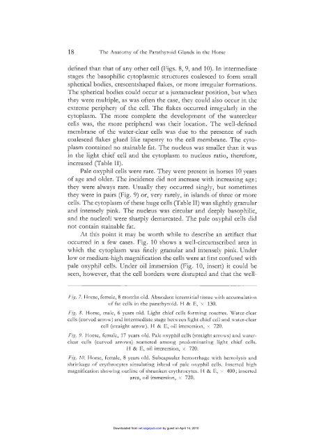
At this po<strong>in</strong>t it may be worth while to describe an artifact that<br />
occurred <strong>in</strong> a few cases. Fig. 10 shows a well-circumscribed area <strong>in</strong><br />
which <strong>the</strong> cytoplasm was f<strong>in</strong>ely granular and <strong>in</strong>tensely p<strong>in</strong>k. Under<br />
low or medium-high magnification <strong>the</strong> cells were at first confused with<br />
pale oxyphil cells. Under oil immersion (Fig. 10, <strong>in</strong>sert) it could be<br />
seen, however, that <strong>the</strong> cell borders were disrupted and that <strong>the</strong> well-<br />
/;(