Nutritional Secondary Hyperparathyroidism in the Horse
Nutritional Secondary Hyperparathyroidism in the Horse
Nutritional Secondary Hyperparathyroidism in the Horse

Create successful ePaper yourself
Turn your PDF publications into a flip-book with our unique Google optimized e-Paper software.
lioentgcnologic Ohscrvntions 55<br />
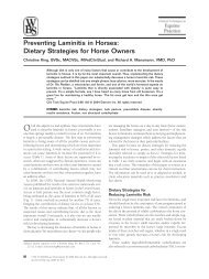
I,'(?. 20. Post-mortcm lateral vicu of rostra1 end of left mandiblc. Control colt<br />
upper, NSI-I 2 lo\vcr. Note radioluccnt iiiiliary mottliiia <strong>in</strong> NSll 2 comparcd to<br />
dcnsc cortical Ipattcrn <strong>in</strong> control, also cxtcnsivc resorption of Imnc surround<strong>in</strong>g<br />
<strong>in</strong>cisor tccth <strong>in</strong> NSk 1 2 comparcd to control.<br />
generalized throughout <strong>the</strong> head, ribs, and limbs (<strong>the</strong> vertebral column<br />
and pelvis were not radiographed). These changes were similar to<br />
those seen dur<strong>in</strong>g life but much more clearly def<strong>in</strong>ed. The contrast<br />
between <strong>the</strong> teeth and surround<strong>in</strong>g bone was strik<strong>in</strong>g (Fig. 19). The<br />
cortical bone along <strong>the</strong> ramus of <strong>the</strong> mandible <strong>in</strong> NSH 2 was mottled<br />
and th<strong>in</strong>ned to <strong>the</strong> po<strong>in</strong>t of nonesistence. The mandibular canals <strong>in</strong><br />
NSf I2 and 3 blended with <strong>the</strong> surround<strong>in</strong>g bone <strong>in</strong> sharp contrast to<br />
<strong>the</strong> control's mandible, <strong>in</strong> which <strong>the</strong> canal was clearly def<strong>in</strong>ed. There<br />
was also a loss of contrast between diploe and lam<strong>in</strong>a <strong>in</strong> <strong>the</strong> roof<br />
of <strong>the</strong> cranium. The bone had a radiolucent moth-eaten appearance<br />
(Figs. 19 and 20).<br />
The cortices of <strong>the</strong> 1-cm.-thick transverse sections from <strong>the</strong> mid-<br />
diaphysis of <strong>the</strong> long bones were radiolucently stippled throughout<br />
(Fig. 21). There was also a radiolucent mottled appearance <strong>in</strong> <strong>the</strong><br />
endosteal area, becom<strong>in</strong>g less evident toward <strong>the</strong> periosteum. In some<br />
cases <strong>the</strong> periphery of <strong>the</strong> medullary cavity had a dist<strong>in</strong>ct scalloped<br />
appearance (Fig. 21).<br />
Downloaded from<br />
vet.sagepub.com by guest on April 14, 2010