- Page 1 and 2:
-i^-;*«SE5L ^Ci«K3C;< t'x: KSKSC^
- Page 3:
-» >jj>>i> : 5> zssc^. ^j>> ,>aa>
- Page 8 and 9:
niVTED BT TATI.OB AND CO., tlTTM! Q
- Page 10:
IV LIST OF CONTRIBUTORS. W. Mitten,
- Page 13 and 14:
THE JOURNAL OF BOTANY, BEITISH AND
- Page 15 and 16:
NOTICE OF A FOSSIL LYCOPODIACEOUS F
- Page 17 and 18:
NOTICE OF A FOSSIL LYCOFODIACEOUS F
- Page 19 and 20:
NOTICE OF A FOSSIL LYCOPOUIACEOUS F
- Page 21 and 22:
9 NOTES ON LEMXACEiE AND ON THE DIS
- Page 23 and 24:
ON LEMXACEiE AND THE RAPHTDIAN CHAR
- Page 25 and 26:
ON lemnacea: and the RAPHIDIAN CHAB
- Page 27 and 28:
ON THE PHOENIX OF THE HONGKONG FLOR
- Page 29 and 30:
CARL FRIEURICH PHILLIPP VON MARTIUS
- Page 31 and 32:
CARL FRIEDRICH PHILLIPP VON MARTIUS
- Page 33 and 34:
CARL FRIEDRICH PHILLIPP VON MARTIUS
- Page 35 and 36:
THE LEAF-FIBRE OF NEW ZEALAND FLAX.
- Page 37 and 38:
THE LEAF-FIBRE OF NEW ZEALAND FLAX.
- Page 39 and 40:
THE LEAF-PIBKE OF NEW ZEALAND FLAX.
- Page 41 and 42: THE LEAF-FIBRE OF NE^V ZEALAND FLAX
- Page 43 and 44: BOTANICAL NEWS. 31 till it can be s
- Page 46 and 47: K ^ \k\h^ vX \:w ,-^v^/ '? \ ' /^ar
- Page 48 and 49: 34 STATIONS OF SOME PLYMOUTH KUBI.
- Page 50 and 51: 36 STATIONS OF SOME PLYMODTH RUBI.
- Page 52 and 53: 38 STATIONS OF SOME PLYMOUTH UUBI.
- Page 54 and 55: 40 STATIONS or SOME PL'S MOUTH KUBI
- Page 56 and 57: 42 NOTE ON THESIUM DECUKRENS AND T.
- Page 58 and 59: 44 THE LEAF-FIBRE OF NEW ZEALAND FL
- Page 60 and 61: 46 THE LEAF-FIBUE OF NEW ZEALAND FL
- Page 62 and 63: 48 NEW BRITISH LICHENS. By the Rev.
- Page 64 and 65: 50 NEW BRITISH LICHENS. Oa serpenti
- Page 66 and 67: 52 JAMES BACKHOUSE. religious body
- Page 68 and 69: 54 JAMES BACKHOUSE. was to Westbury
- Page 70 and 71: 56 JAMES BACKHOUSE. where we liave
- Page 72 and 73: 58 ' >iEW PUBLICATIOKS. spheres of
- Page 74: . 60 BOTANICAL NEWS. — — Red Pi
- Page 77 and 78: 61 NEW AND KAEE BRITISH HYMEXOMYCET
- Page 79 and 80: Pers. ; 4, spores, X 700diam. — N
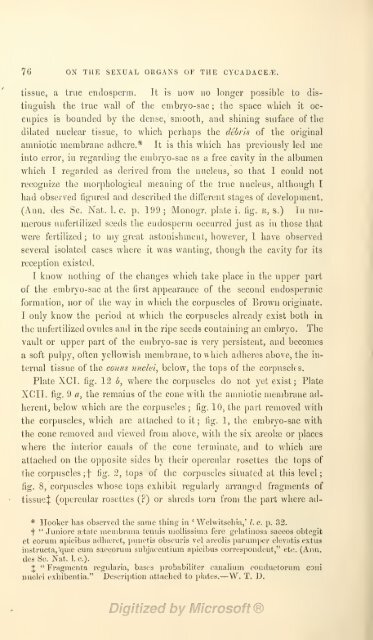
- Page 81 and 82: ON THE SEXUAL ORGANS OF THE CYCADAC
- Page 83 and 84: ON THE SEXUAL OKGANS OF THE CYCADAC
- Page 85 and 86: ON THE SEXUAL ORGANS OF THE CYCADAC
- Page 87 and 88: ON THE SEXUAL ORGANS OF THE CYCADAC
- Page 89 and 90: ON THE SEXUAL ORGANS OF THE CYCADAC
- Page 91: ON THE SEXUAL ORGANS OF THE CYCADAC
- Page 95 and 96: VAKIATIONS IN EPIG.EA REPENS. 79 no
- Page 97 and 98: VARIATIONS IN EPIGyFA REPENS. 81 Oq
- Page 99 and 100: PLANT REMAINS IN NORTH AMERICA. S3
- Page 101 and 102: CORRESl'ONDENCE. 85 vegetation of t
- Page 103 and 104: BOTANICAL NEWS. 87 Genus Thuja, Lin
- Page 105 and 106: BOTAN.'CAL XEWS. 89 coccineutn, Gei
- Page 107 and 108: BOTANICAL NEWS. 91 flowers was bloo
- Page 110: Tcub. 92. W.G. SiTuth.liLh. Vincent
- Page 114 and 115: G-, Smitii, del et lith . TcLb9G. V
- Page 116 and 117: 94 ON THE SEXUAL OKGANS OF THE CYCA
- Page 118 and 119: 96 ON THE SEXUAL ORGANS OF THE CYCA
- Page 120 and 121: 98 ON THE SEXUAL ORGANS OF THE CYCA
- Page 122 and 123: 100 ON THE SEXUAL ORGANS OF THE CYC
- Page 124 and 125: 102 ON THE SEXUAL OEGANS OF THE CYC
- Page 126 and 127: 104 ON THE SEXUAL OUGANS OF THE CYC
- Page 128 and 129: 106 NEW BRITISH LICHENS. tliouli it
- Page 130 and 131: 108 ON THE FLOKA OF SKYE. Lecnnora
- Page 132 and 133: 110 ON THE FLORA OF SKYE. C. alpinu
- Page 134 and 135: 112 ON THE FLOBA OF SKYE. Euphrasia
- Page 136 and 137: 114 DE NOVA RHAMNI SPECIE. Equisetu
- Page 138 and 139: 116 NOTE ON THE CHINESE NAME OF ELE
- Page 140 and 141: 118 A BOTANICAL TOUR AMONG THE SOUT
- Page 142:
120 A BOTANICAL TOUR AMONG THE SOUT
- Page 145 and 146:
121 A BOTANICAL TOUR AMONG THE SOUT
- Page 147 and 148:
A BOTANICAL TOUR AMONG THE SOUTH SE
- Page 149 and 150:
A BOTANICAL TOUR AMONG THE SOUTH SE
- Page 151 and 152:
A BOTANICAL TOUR AMONG THE SOUTH SE
- Page 153 and 154:
A BOTANICAL TOUR AMONG THE SOUTH SE
- Page 155 and 156:
A BOTANICAL TOUE AMONG THE SOUTH SE
- Page 157 and 158:
A BOTANICAL TOUU AMONG THE SOUTH SE
- Page 159 and 160:
— A BOTANICAL TOUR AMONG THE SOUT
- Page 161 and 162:
REPORT OF THE LONDON BOTANICAL EXCH
- Page 163 and 164:
REPORT OF THE LONDON BOTANICAL EXCH
- Page 165 and 166:
REPORT OF THE LONDON BOTANICAL EXCH
- Page 167 and 168:
REPORT OF THE LONDON BOTANICAL EXCH
- Page 169 and 170:
REPORT OF THE LONDON BOTANICAL EXCH
- Page 171 and 172:
REPORT OF THE LONDON BOTANICAL EXCH
- Page 173 and 174:
NOTES ON RANGE IN DEPTH OF MARINE A
- Page 175 and 176:
NOTES ON RANGE IX DEPTH OF MABINE A
- Page 177 and 178:
153 ON THE GENUS KNORRIA, Stemb. By
- Page 179 and 180:
REPORT ON THE CULTIVATION OF CHINCH
- Page 181 and 182:
REPORT ON THE CULTIVATION OF CHINCH
- Page 183 and 184:
REPORT ON THE CULTIVATION OF CHINCH
- Page 185 and 186:
161 ON HABENARIA MIERSIANA, Champ.
- Page 187 and 188:
SERTULUM CHINENSE QUARTUM. 163 Liiu
- Page 189 and 190:
SEKTULUM CHINENSE QUARTUM. 165 fulv
- Page 191 and 192:
SERTULUM CHINENSE QUARTUM. 167 AVil
- Page 193 and 194:
HORACE MANN. 169 year so full of ha
- Page 195 and 196:
STATISTICS, ETC., OF HAWAIIAN PLANT
- Page 197 and 198:
-Saxifragaceae. -Haloragese. -Begon
- Page 199 and 200:
STATISTICS, ETC., OF HAWAIIAN PLANT
- Page 201 and 202:
Erythrsea sabseoides, Gray. Laborde
- Page 203 and 204:
STATISTICS, ETC., OF HAWAIIAN PLANT
- Page 205 and 206:
Scaevola sericea. Plumbago Zeylanic
- Page 207 and 208:
REPORT OF THE VICTORIAN GOVERNMENT
- Page 209 and 210:
REPOET OF THE VICTORIAN GOVERNMENT
- Page 211 and 212:
REPORT OF THE VICTORIAN GOVERNMENT
- Page 213 and 214:
KEPORT OF THE YICTOKIAN GOVERNMENT
- Page 215 and 216:
REPORT or THE VICTORIAN GOVERNMENT
- Page 217 and 218:
REPORT OF THE VICTORIAN GOVERNMENT
- Page 219 and 220:
REPORT OF THE VICTORIAN GOVERNMENT
- Page 221 and 222:
• REPORT OF THE VICTORIAN GOVERNM
- Page 223 and 224:
BErOKT OF THE VICTOKIAN GOVERNMENT
- Page 225 and 226:
REPORT OF THE VICTORIAN GOVERNMENT
- Page 227 and 228:
REVISION OF THE GENUS SANGDISORBA.
- Page 229 and 230:
REVISION OF THE GENUS SANGUISOEBA.
- Page 231 and 232:
NEW PUBLICATIONS. 207 tubus fructif
- Page 233 and 234:
NEW PUBLICATIONS. 209 Aqarlcm bulbo
- Page 235 and 236:
NEW PUBLICATIONS. 211 Tab. 40. A. c
- Page 237 and 238:
CORRESPONDENCE. 213 than two montli
- Page 239 and 240:
BOTANICAL NEWS. 215 He asserts that
- Page 242 and 243:
Tai- 94.
- Page 245 and 246:
217 ON THE GENUS SYMBOLANTHUS. By J
- Page 247 and 248:
•219 INDEX CRITICUS BUTOMACKARUM,
- Page 249 and 250:
ALISMACEARUM JUXCAGINACEARL'MQUE. 2
- Page 251 and 252:
ALISMACEARUM JUNCAGINACEAllUMaUE. 2
- Page 253 and 254:
ALISMACEARUM JUNCAGINACEARUMQUE. 22
- Page 255 and 256:
ALISMACEARUM JUNCAGINACEARIJMQUE. 2
- Page 257 and 258:
ALISMACEARUM JUNCAGINACEAEUMQUE. 22
- Page 259 and 260:
ALISMACEARUM JUNCAGIKACEARUMCIUE. 2
- Page 261 and 262:
NEW BRITISH LICHEXS. 233 Oil the de
- Page 263 and 264:
NOTES ON THBv FERN-FLORA OF CHINA.
- Page 265 and 266:
NOTES ON THE FERN-FLORA OF CHINA. 2
- Page 267 and 268:
NOTES ON THE FEKN-FLORA OF CHINA. 2
- Page 269 and 270:
NEW PUBLICATIONS. 241 ^ inch long.
- Page 271 and 272:
MEW PIJBLICATIONS. 243 covery in th
- Page 273 and 274:
NEW PUBLICATIONS. 245 Die Lemnacoen
- Page 275 and 276:
BOTANICAL NEWS. 247 to class it wit
- Page 278 and 279:
Tah. 9'^
- Page 280 and 281:
250 NKW AND KARK BRITISH HYMENOMYCK
- Page 282 and 283:
252 NOTES ON SOME COMPOSITE OF OTAG
- Page 284 and 285:
254 NOTES ON SOME COMPOSURE OF OTAG
- Page 286 and 287:
25G NOTES ON SOME COMPOSITiE OF OTA
- Page 288 and 289:
358 NOTES ON SOME COMPOSITiE OF OTA
- Page 290 and 291:
260 NOTES ON SOME COMPOSIT.E OF OTA
- Page 292 and 293:
262 NOTES ON SOME COMPOSIT.E OF OTA
- Page 294 and 295:
264 NOTES ON SOME COMPOSIT.E OF OTA
- Page 296 and 297:
266 OFFICIAL llEPORT ON THE BOTANIC
- Page 298 and 299:
268 OBITUARY OF FREDEUICK SCHEER. 3
- Page 300 and 301:
270 OBITUARY OF FREDERICK SCHEER. A
- Page 302 and 303:
273 NEW PUBLICATION. other scented
- Page 304 and 305:
274: NEW PUBLICATION. splendens (wi
- Page 306 and 307:
276 NEW PUBLICATION. that of which
- Page 308 and 309:
278 NEW PUBLICATION. (which ill man
- Page 310 and 311:
280 BOTANICAL NEWS. Wendland, who f
- Page 312 and 313:
282 BRITISH ASSOCIATION, MEETING AT
- Page 314 and 315:
284 BRITISH ASSOCIATION, MEETING AT
- Page 316 and 317:
286 BRITISH ASSOCIATION, MEETING AT
- Page 318 and 319:
388 BKITISH ASSOCIATION, MEETING AT
- Page 320 and 321:
290 BRITISH ASSOCIATION, MEETING AT
- Page 322 and 323:
292 BRITISH ASSOCIATION, MEETING AT
- Page 324 and 325:
294 BKITISH ASSOCIATION, MEETING AT
- Page 326 and 327:
296 NOTE ON MELJSTOMA REPENS, Desro
- Page 328 and 329:
298 THE NOKTHERN LIMIT OF EDIBLE BE
- Page 330 and 331:
300 LOUD Howe's island. It is somew
- Page 332 and 333:
302 LOUD Howe's island. where the s
- Page 334 and 335:
304 NEVT PUB),ICATIONS. common to a
- Page 336 and 337:
•306 NEW PUBLICATIONS. from one a
- Page 338 and 339:
308 NEW PUBLICATIONS. individualize
- Page 340 and 341:
310 NEW PUBLICATIONS. nience of ref
- Page 342 and 343:
312 BOTANICAL NEWS. dress in silenc
- Page 344 and 345:
314 ON THE GIGANTIC NEW AROIDEA FRO
- Page 346 and 347:
316 NOTES ON ISLE OF WIGHT PLANTS.
- Page 348 and 349:
ai8 NOTES RESPECTING SOME PLYMOUTH
- Page 350 and 351:
320 NOTES ON SOME PLANTS OF QTAGO,
- Page 352 and 353:
322 NOTES ON SOME PLANTS OF OTAGO,
- Page 354 and 355:
324 NOTES ON SOME PLANTS OF OTAGO,
- Page 356 and 357:
326 NOTES ON SOME PLANTS OF OTAGO,
- Page 358 and 359:
328 NOTES ON SOME PLANTS OF OTAGO,
- Page 360 and 361:
330 NOTES ON SOME PLANTS OF OTAGO,
- Page 362 and 363:
332 DESCRIPTION OF TWO NEW SPECIES
- Page 364 and 365:
334 ON VERNACULAR NAME3. nations, w
- Page 366 and 367:
336 NOTE ON ABfiUS CANTONIENSIS. as
- Page 368 and 369:
338 NOTE SUE LA FAMILLE DES EQXJISE
- Page 370 and 371:
340 EPILOBIUM OBSCURUM IN ORKNEY OE
- Page 372 and 373:
342 MEMORANDA. venient; and several
- Page 374:
344 high, and the air frequently su
- Page 378 and 379:
Tcub. 99. .1 T T Tj.1^ ViTir.f»nt
- Page 380 and 381:
346 WHAT IS THE TIIAMES-SIDE BRASSI
- Page 382 and 383:
348 WHAT IS THE THAMES-SIDE BRASSIC
- Page 384 and 385:
350 ON A NEW SPECIES OF OREOPANAX.
- Page 386 and 387:
352 NOTE ON AIRA SETACEA, Hudson (A
- Page 388 and 389:
354 SOME ACCOUNT OF CHESHIKE KTJBI.
- Page 390 and 391:
356 SOME ACCOUNT OF CHESHIRE RUBI.
- Page 392 and 393:
358 SOME ACCOUNT OF CHESHIRE IIUBI.
- Page 394 and 395:
360 CORRESPONDENCE. 30. R. ulth(jei
- Page 396 and 397:
362 CORREbPO:JfDENCE. names worth r
- Page 398 and 399:
364 NEW PUBLICATIONS. It is perhaps
- Page 400 and 401:
366 NEW PUBLICATIONS. cially in mit
- Page 402 and 403:
368 NEW PUBLICATIONS. The deficienc
- Page 404 and 405:
370 BOTANICAL NEWS. We have to reco
- Page 406 and 407:
372 INDEX. Britten, J., On Epilobiu
- Page 408 and 409:
374 INDEX. Holland, Mr. R., CoUecti
- Page 410 and 411:
376 INDEX. Polygonum aAdculare, 317
- Page 412:
PRINTED BY XAYLOB AND CO,, LITTLE Q
- Page 417 and 418:
Da. SEEiiA>->-'s ' JOUENAL- OF BOTA
- Page 419:
The ' Journal op Botany ' will he p
- Page 422:
5>W • ?^ S^: t 5i '^^^a^^.