Download (5Mb) - Covenant University Repository
Download (5Mb) - Covenant University Repository
Download (5Mb) - Covenant University Repository

Create successful ePaper yourself
Turn your PDF publications into a flip-book with our unique Google optimized e-Paper software.
1144<br />
used for the culture of fungi. A required amount of PDA<br />
was taken in conical flasks separately and was sterilized<br />
by autoclave (121 ° C, 15 psi) for 15 minutes. Purified<br />
lectin (in PBS solution) was mixed with sterilized melted<br />
PDA medium to have 100 µg/ml in PDA and this was<br />
poured (about 20 ml/plate) in sterilized petridishes. At the<br />
center of each plate, 5 days old fungal mycelial block (4<br />
mm in diameter) was inoculated and incubated at 27 ° C. A<br />
control set was also maintained in each experiment.<br />
Linear mycelial growth of fungus was measured after 3-5<br />
days of incubation in triplicate. The average of two<br />
measurements was taken as mycelial colony diameter of<br />
the fungus in milimeter. All the antifungal results were<br />
compared with the standard antifungal antibiotic nystatin<br />
(100 µg/ml) in PDA. Galactose was used as negative<br />
control. The percentage inhibition of radial mycelial<br />
growth of the test fungus was calculated as follows:<br />
% Inhibition = (C-T/C) × 100<br />
Where, C = diameter of the fungal colony in the control<br />
Canadian Journal of Pure and Applied Sciences<br />
petridish and T = diameter of the fungal colony in the<br />
treated petridish.<br />
RESULTS AND DISCUSSION<br />
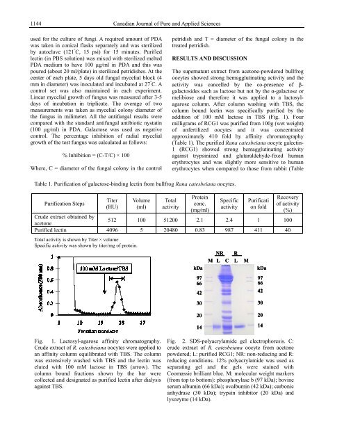
Table 1. Purification of galactose-binding lectin from bullfrog Rana catesbeiana oocytes.<br />
Purification Steps<br />
Titer<br />
(HU)<br />
Volume<br />
(ml)<br />
Total<br />
activity<br />
The supernatant extract from acetone-powdered bullfrog<br />
oocytes showed strong hemagglutinating activity and the<br />
activity was cancelled by the co-presence of βgalactosides<br />
such as lactose but not by the α-galactose or<br />
melibiose and therefore it was applied to a lactosylagarose<br />
column. After column washing with TBS, the<br />
column bound lectin was specifically purified by the<br />
addition of 100 mM lactose in TBS (Fig. 1). Four<br />
milligrams of RCG1 was purified from 100g (wet weight)<br />
of unfertilized oocytes and it was concentrated<br />
approximately 410 fold by affinity chromatography<br />
(Table 1). The purified Rana catesbeiana oocyte galectin-<br />
1 (RCG1) showed strong hemagglutinating activity<br />
against trypsinized and glutaraldehyde-fixed human<br />
erythrocytes and was slightly more sensitive to human<br />
erythrocytes when compared to those from rabbit (Table<br />
Protein<br />
conc.<br />
(mg/ml)<br />
Specific<br />
activity<br />
Purificati<br />
on fold<br />
Recovery<br />
of activity<br />
(%)<br />
Crude extract obtained by<br />
acetone<br />
512 100 51200 2.1 2.4 1 100<br />
Purified lectin 4096 5 20480 0.83 987 411 40<br />
Total activity is shown by Titer × volume<br />
Specific activity was shown by titer/mg of protein.<br />
Fig. 1. Lactosyl-agarose affinity chromatography.<br />
Crude extract of R. catesbeiana oocytes were applied to<br />
an affinity column equilibrated with TBS. The column<br />
was extensively washed with TBS and the lectin was<br />
eluted with 100 mM lactose in TBS (arrow). The<br />
column bound fractions shown by the bar were<br />
collected and designated as purified lectin after dialysis<br />
against TBS.<br />
kDa<br />
97<br />
66<br />
42<br />
30<br />
20<br />
14<br />
NR R<br />
M L C L M<br />
kDa<br />
97<br />
66<br />
42<br />
30<br />
20<br />
14<br />
Fig. 2. SDS-polyacrylamide gel electrophoresis. C:<br />
crude extract of R. catesbeiana oocyte from acetone<br />
powdered; L: purified RCG1; NR: non-reducing and R:<br />
reducing conditions. 12% polyacrylamide was used as<br />
separating gel and the gels were stained with<br />
Coomassie brilliant blue. M: molecular weight markers<br />
(from top to bottom): phosphorylase b (97 kDa); bovine<br />
serum albumin (66 kDa); ovalbumin (42 kDa); carbonic<br />
anhydrase (30 kDa); trypsin inhibitor (20 kDa) and<br />
lysozyme (14 kDa).