

SPHENOPHRYNE - American Museum of Natural History
SPHENOPHRYNE - American Museum of Natural History
SPHENOPHRYNE - American Museum of Natural History

Create successful ePaper yourself
Turn your PDF publications into a flip-book with our unique Google optimized e-Paper software.
108 BULLETIN AMERICAN MUSEUM OF NATURAL HISTORY NO. 253<br />
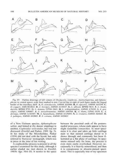
Fig. 65. Outline drawings <strong>of</strong> left vomers <strong>of</strong> Oxydactyla, Liophryne, Austrochaperina, and Sphenophryne<br />
in ventral aspect; scale lines marked in mm. Curved line at right <strong>of</strong> each figure marks the lingual<br />
border <strong>of</strong> the maxillary shelf. A. O. stenodactyla, AMNH A92800. B. O. alpestris, AMNH A65299. C.<br />
O. coggeri, AMS R22822. D. A. brevipes, AMNH A130527. E. L. allisoni, BPBM 9631. F. L. rhododactyla,<br />
BPBM 9793. G. L. dentata, UPNG 2641. H. L. schlaginhaufeni, AMNH A78183. I. A. gracilipes,<br />
AMNH A90407. J. A. novaebritanniae, AMNH A88569. K. A. blumi, UPNG 9559. L. A.<br />
derongo, AMNH A145507. M. A. basipalmata, AMNH A129495. N. A. rivularis, AMNH A84445. O.<br />
A. palmipes, AMNH A92805. P. S. cornuta, AMNH A92803.<br />
<strong>of</strong> a New Guinean species, Aphantophryne<br />
sabini, and hinted at (by denser stippling) in<br />
another, Cophixalus verecundus, but was not<br />
discussed (Zweifel and Parker, 1989: fig. 5).<br />
In his study <strong>of</strong> the Microhylidae, Parker<br />
(1934) did not deal with the hyoid, but only<br />
referred to Trewavas’ monograph, which was<br />
in press at the time <strong>of</strong> his writing.<br />
A coathooklike process is present in all the<br />
species I examined for this study, although it<br />
earlier eluded me (not shown in Zweifel,<br />
1985b: figs. 50A–D). It nestles in the space<br />
between the proximal ends <strong>of</strong> the posteromedial<br />
processes, whose ends appear to have<br />
slight socketlike concavities. In most specimens<br />
it is clear and takes up little cartilage<br />
stain so that stained cartilage dorsal to it<br />
shows through and commonly has been illustrated<br />
as a flat plane <strong>of</strong> cartilage. In specimens<br />
stained only for bone, the process is<br />
even more easily overlooked. However, occasionally<br />
it is heavily mineralized, and then<br />
it is conspicuous in alizarin-stained specimens.<br />
This is especially true <strong>of</strong> my specimen


