

Vol 43 # 3 September 2011 - Kma.org.kw
Vol 43 # 3 September 2011 - Kma.org.kw
Vol 43 # 3 September 2011 - Kma.org.kw

Create successful ePaper yourself
Turn your PDF publications into a flip-book with our unique Google optimized e-Paper software.
178<br />
Osteoarthritis of the Knee: Review of Risk Factors and Treatment Programs ...<br />
<strong>September</strong> <strong>2011</strong><br />
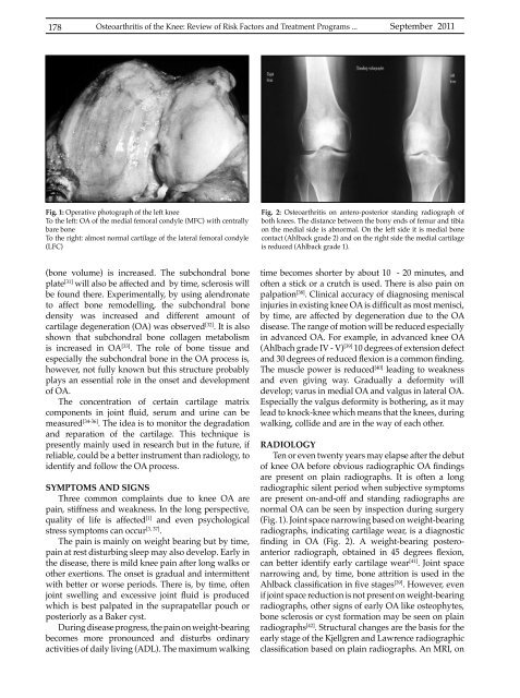
Fig. 1: Operative photograph of the left knee<br />
To the left: OA of the medial femoral condyle (MFC) with centrally<br />
bare bone<br />
To the right: almost normal cartilage of the lateral femoral condyle<br />
(LFC)<br />
Fig. 2: Osteoarthritis on antero-posterior standing radiograph of<br />
both knees. The distance between the bony ends of femur and tibia<br />
on the medial side is abnormal. On the left side it is medial bone<br />
contact (Ahlback grade 2) and on the right side the medial cartilage<br />
is reduced (Ahlback grade 1).<br />
(bone volume) is increased. The subchondral bone<br />
plate [31] will also be affected and by time, sclerosis will<br />
be found there. Experimentally, by using alendronate<br />
to affect bone remodelling, the subchondral bone<br />
density was increased and different amount of<br />
cartilage degeneration (OA) was observed [32] . It is also<br />
shown that subchondral bone collagen metabolism<br />
is increased in OA [33] . The role of bone tissue and<br />
especially the subchondral bone in the OA process is,<br />
however, not fully known but this structure probably<br />
plays an essential role in the onset and development<br />
of OA.<br />
The concentration of certain cartilage matrix<br />
components in joint fluid, serum and urine can be<br />
measured [34-36] . The idea is to monitor the degradation<br />
and reparation of the cartilage. This technique is<br />
presently mainly used in research but in the future, if<br />
reliable, could be a better instrument than radiology, to<br />
identify and follow the OA process.<br />
SYMPTOMS AND SIGNS<br />
Three common complaints due to knee OA are<br />
pain, stiffness and weakness. In the long perspective,<br />
quality of life is affected [1] and even psychological<br />
stress symptoms can occur [3, 37] .<br />
The pain is mainly on weight bearing but by time,<br />
pain at rest disturbing sleep may also develop. Early in<br />
the disease, there is mild knee pain after long walks or<br />
other exertions. The onset is gradual and intermittent<br />
with better or worse periods. There is, by time, often<br />
joint swelling and excessive joint fluid is produced<br />
which is best palpated in the suprapatellar pouch or<br />
posteriorly as a Baker cyst.<br />
During disease progress, the pain on weight-bearing<br />
becomes more pronounced and disturbs ordinary<br />
activities of daily living (ADL). The maximum walking<br />
time becomes shorter by about 10 - 20 minutes, and<br />
often a stick or a crutch is used. There is also pain on<br />
palpation [38] . Clinical accuracy of diagnosing meniscal<br />
injuries in existing knee OA is difficult as most menisci,<br />
by time, are affected by degeneration due to the OA<br />
disease. The range of motion will be reduced especially<br />
in advanced OA. For example, in advanced knee OA<br />
(Ahlbach grade IV - V) [39] 10 degrees of extension defect<br />
and 30 degrees of reduced flexion is a common finding.<br />
The muscle power is reduced [40] leading to weakness<br />
and even giving way. Gradually a deformity will<br />
develop; varus in medial OA and valgus in lateral OA.<br />
Especially the valgus deformity is bothering, as it may<br />
lead to knock-knee which means that the knees, during<br />
walking, collide and are in the way of each other.<br />
RADIOLOGY<br />
Ten or even twenty years may elapse after the debut<br />
of knee OA before obvious radiographic OA findings<br />
are present on plain radiographs. It is often a long<br />
radiographic silent period when subjective symptoms<br />
are present on-and-off and standing radiographs are<br />
normal OA can be seen by inspection during surgery<br />
(Fig. 1). Joint space narrowing based on weight-bearing<br />
radiographs, indicating cartilage wear, is a diagnostic<br />
finding in OA (Fig. 2). A weight-bearing posteroanterior<br />
radiograph, obtained in 45 degrees flexion,<br />
can better identify early cartilage wear [41] . Joint space<br />
narrowing and, by time, bone attrition is used in the<br />
Ahlback classification in five stages [39] . However, even<br />
if joint space reduction is not present on weight-bearing<br />
radiographs, other signs of early OA like osteophytes,<br />
bone sclerosis or cyst formation may be seen on plain<br />
radiographs [42] . Structural changes are the basis for the<br />
early stage of the Kjellgren and Lawrence radiographic<br />
classification based on plain radiographs. An MRI, on


