

Vol 43 # 3 September 2011 - Kma.org.kw
Vol 43 # 3 September 2011 - Kma.org.kw
Vol 43 # 3 September 2011 - Kma.org.kw

You also want an ePaper? Increase the reach of your titles
YUMPU automatically turns print PDFs into web optimized ePapers that Google loves.
202<br />
Characterization of Acrylamide Mediated Testicular Toxicity in Rat: Light and Electron ...<br />
<strong>September</strong> <strong>2011</strong><br />
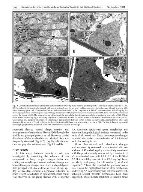
Fig. 8: (A) Part of seminiferous tubule from control rat testis showing many normal spermatogenetic (arrows) and Sertoli cells (S) x 1100,<br />
(B) Control rat testis showing Sertoli cell with prominent nucleolus (long arrow) and two characteristic bodies near it (small arrow) x 4600,<br />
Insert showing apical cleft of the nucleus (arrow) and mitochondria (M) in the cytoplasm x 10,500, (C) rat testes treated with 45 mg/kg AA<br />
showing degenerated spermatogonia (sg) with large lipid droplet (LD),degenerated peritubular myoid cell (MC) and vacuole (V) at the basal<br />
part of the tubule x 3400. The insert showing widening of the intercellular junctions (arrow) of the two adjacent germ cells x 5800, (D) rat<br />
testes treated with 45 mg/kg AA showing degenerated Sertoli cell nucleus (N) with condensed chromatin and shrunken nucleolus (arrow).<br />
Cytoplasmic lipid droplets (small arrow), widening of tight junctions between Sertoli cell and the surrounding spermatocytes (star) were<br />
observed. Detachment of Sertoli cell from the basal lamina (double head arrow) was also observed x 7900. The insert showing spermatid<br />
(arrow) phagocytozed by the Sertoli cell x 7900. Transmission electron microscope<br />
spermatid showed normal shape, number and<br />
arrangements of outer dense fibers (ODF) through the<br />
middle and principal piece of its tail. However, partial<br />
dissolution of fibrous sheath in the principal piece was<br />
frequently observed (Fig. 9 D). Leydig cells showed<br />
clear atrophy after AA treatment (Fig. 9 A and B).<br />
DISCUSSION<br />
In this study, testicular toxicity of AA was<br />
investigated by examining the influence of this<br />
compound on body weight changes, testis and<br />
epididymal weight, sperm count and morphology and<br />
histopathological changes in rat testis and epididymis.<br />
Rats gavaged with AA at doses of 45 or 60 mg/kg/<br />
day for five days showed a significant reduction in<br />
body weight. A reduction in epididymal sperm count<br />
was observed in the group treated with 60 mg/kg<br />
AA. Abnormal epididymal sperm morphology and<br />
abnormal histopathological findings were noted in the<br />
testis of all treated rats. These dose response changes<br />
provided the initial characterization of AA induced<br />
testicular toxicity in rats.<br />
Gross observational and behavioural changes<br />
and neurotoxicity observed on rats treated with AA<br />
at doses of 45 and 60 mg/kg were entirely consistent<br />
with the previous study conducted by Hashimoto et<br />
al [8] who treated mice with 0.2 to 0.5 of the LD50 of<br />
AA (1.5 mmol/kg equivalent to 106.6 mg/kg) twice<br />
weekly by oral gavage for 8-10 weeks. Tyl et al and<br />
Lopachin [17,18] have also reported this phenomenon in<br />
rats. It must be highlighted that no clear mechanism<br />
underlying AA neurotoxicity has yet been uncovered,<br />
although several possible mechanisms have been<br />
suggested. These include inhibition of kinesin-based


