

Vol 43 # 3 September 2011 - Kma.org.kw
Vol 43 # 3 September 2011 - Kma.org.kw
Vol 43 # 3 September 2011 - Kma.org.kw

Create successful ePaper yourself
Turn your PDF publications into a flip-book with our unique Google optimized e-Paper software.
200<br />
Characterization of Acrylamide Mediated Testicular Toxicity in Rat: Light and Electron ...<br />
<strong>September</strong> <strong>2011</strong><br />
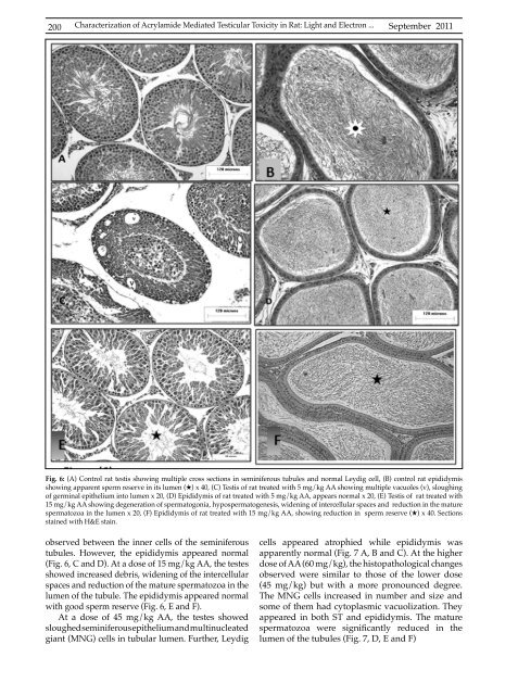
Fig. 6: (A) Control rat testis showing multiple cross sections in seminiferous tubules and normal Leydig cell, (B) control rat epididymis<br />
showing apparent sperm reserve in its lumen (H) x 40, (C) Testis of rat treated with 5 mg/kg AA showing multiple vacuoles (v), sloughing<br />
of germinal epithelium into lumen x 20, (D) Epididymis of rat treated with 5 mg/kg AA, appears normal x 20, (E) Testis of rat treated with<br />
15 mg/kg AA showing degeneration of spermatogonia, hypospermatogenesis, widening of intercellular spaces and reduction in the mature<br />
spermatozoa in the lumen x 20, (F) Epididymis of rat treated with 15 mg/kg AA, showing reduction in sperm reserve (H) x 40. Sections<br />
stained with H&E stain.<br />
observed between the inner cells of the seminiferous<br />
tubules. However, the epididymis appeared normal<br />
(Fig. 6, C and D). At a dose of 15 mg/kg AA, the testes<br />
showed increased debris, widening of the intercellular<br />
spaces and reduction of the mature spermatozoa in the<br />
lumen of the tubule. The epididymis appeared normal<br />
with good sperm reserve (Fig. 6, E and F).<br />
At a dose of 45 mg/kg AA, the testes showed<br />
sloughed seminiferous epithelium and multinucleated<br />
giant (MNG) cells in tubular lumen. Further, Leydig<br />
cells appeared atrophied while epididymis was<br />
apparently normal (Fig. 7 A, B and C). At the higher<br />
dose of AA (60 mg/kg), the histopathological changes<br />
observed were similar to those of the lower dose<br />
(45 mg/kg) but with a more pronounced degree.<br />
The MNG cells increased in number and size and<br />
some of them had cytoplasmic vacuolization. They<br />
appeared in both ST and epididymis. The mature<br />
spermatozoa were significantly reduced in the<br />
lumen of the tubules (Fig. 7, D, E and F)


