SCIENTIFIC REPORT 2004 - Sylvester Comprehensive Cancer Center
SCIENTIFIC REPORT 2004 - Sylvester Comprehensive Cancer Center
SCIENTIFIC REPORT 2004 - Sylvester Comprehensive Cancer Center

You also want an ePaper? Increase the reach of your titles
YUMPU automatically turns print PDFs into web optimized ePapers that Google loves.
S H A R E D R E S O U R C E S<br />
S H A R E D R E S O U R C E S<br />
A N A L Y T I C A L I M A G I N G C O R E<br />
MANAGER<br />
Alberto Pugliese, M.D.<br />
Associate Professor of Medicine<br />
CO-MANAGER<br />
Beata R. Frydel, Ph.D.<br />
Associate Scientist of Neurosurgery<br />
PURPOSE<br />
Many research endeavors rely on state-of-theart<br />
imaging and molecular histology techniques<br />
requiring the use of complex and costly<br />
equipment not practical for the individual investigator<br />
to acquire and maintain. The Analytical<br />
Imaging Core is a campus-wide resource spearheaded<br />
by the Diabetes Research Institute and<br />
UM/<strong>Sylvester</strong>, with seed support from the dean<br />
of the University of Miami School of Medicine.<br />
The Juvenile Diabetes Research Foundation<br />
awarded additional support for a five-year period<br />
in December 2003. The core’s main goals are to:<br />
• Provide access to sophisticated, modern instrumentation<br />
for imaging and molecular analysis<br />
of tissue and cellular specimens to investigators.<br />
• Provide expertise, guidance, and training to<br />
core users and help them optimize protocols for<br />
their applications that can be shared with other<br />
investigators.<br />
SERVICES<br />
This core provides the following services:<br />
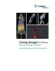
1) Confocal Microscopy: Confocal microscopy offers<br />
many advantages over standard fluorescent<br />
microscopy including increased sensitivity,<br />
resolution, and the ability to image relatively<br />
thick, fluorescently labeled biological specimens<br />
in two or three dimensions. Confocal<br />
microscopy creates an “optical section” of the<br />
cells or tissues being imaged and an increase in<br />
effective resolution due to a large increase in<br />
signal-to-noise ratio. As a result, outstanding<br />
images can be collected from cells and tissue<br />
sections that would otherwise yield little or no<br />
information.<br />
The workhorse instrument for confocal microscopy<br />
is the Zeiss LSM-510, which can detect<br />
up to five channels and four fluorescent<br />
channels simultaneously—from UV to far red,<br />
plus a separate detector for transmitted light.<br />
The outstanding beam control afforded by the<br />
Zeiss LSM-510 makes it an ideal instrument<br />
for other advanced fluorescent applications<br />
such as fluorescence resonance energy transfer<br />
(FRET), fluorescence recovery after photo<br />
bleaching (FRAP), or ratio-imaging for<br />
fluorescence quantitation. The core also is<br />
equipped with an Atto Instruments spinning<br />
disk confocal microscope (CARV), a confocal<br />
instrument particularly suited for live cell<br />
analysis, and video-rate (30 frames per second)<br />
imaging.<br />
2) Standard Epifluorescence Microscopy: The core<br />
also is equipped with a Leica DMIRB inverted<br />
microscope capable of performing triple fluorescence,<br />
phase contrast, and light microscopy, etc.<br />
UM/<strong>Sylvester</strong> <strong>Comprehensive</strong> <strong>Cancer</strong> <strong>Center</strong> Scientific Report <strong>2004</strong> 143