6th International Workshop on Breast Densitometry and Breast ...
6th International Workshop on Breast Densitometry and Breast ...
6th International Workshop on Breast Densitometry and Breast ...
- No tags were found...

You also want an ePaper? Increase the reach of your titles
YUMPU automatically turns print PDFs into web optimized ePapers that Google loves.
6 th <str<strong>on</strong>g>Internati<strong>on</strong>al</str<strong>on</strong>g> <str<strong>on</strong>g>Workshop</str<strong>on</strong>g> <strong>on</strong> <strong>Breast</strong> <strong>Densitometry</strong><br />
<strong>and</strong> <strong>Breast</strong> Cancer Risk Assessment<br />
P2 COMPARISON OF BREAST DENSITY MEASUREMENTS WITH A<br />
MAMMOGRAPHIC VOLUMETRIC AND AREA ALGORITHM AND MAGNETIC<br />
RESONANCE IMAGING<br />
O Al<strong>on</strong>zo-Proulx 1 , J Mainprize 1 , E. Warner 1 , J. Harvey 2 , M. Yaffe 1<br />
1 Sunnybrook Health Sciences Centre, 2075 Bayview Ave., Tor<strong>on</strong>to, Ontario Canada, M4N 3M5 ;<br />
2 University of Virginia Health System, Charlottesville, Virginia<br />
Introducti<strong>on</strong>: Area <strong>and</strong> volumetric breast density (ABD <strong>and</strong> VBD) is typically determined from twodimensi<strong>on</strong>al<br />
x-ray projecti<strong>on</strong> mammograms. In this study we compare the values of ABD, VBD, dense<br />
volume <strong>and</strong> total breast volume estimated in this way, with measurements obtained directly from a threedimensi<strong>on</strong>al<br />
breast MRI image dataset.<br />
ABSTRACTS<br />
Materials <strong>and</strong> Methods: Our VBD algorithm, CUMULUS V, has been described earlier [1,2]. A<br />
calibrati<strong>on</strong> surface relating the imaging signal to the compositi<strong>on</strong> <strong>and</strong> thickness of breast phantoms is<br />
obtained experimentally, <strong>and</strong> corrected using simulati<strong>on</strong> to compensate for the attenuati<strong>on</strong> difference<br />
between the phantom materials <strong>and</strong> breast tissue. The resp<strong>on</strong>se of the thickness readout of the<br />
mammography machine is characterized to allow for an accurate estimate of the breast thickness under<br />
compressi<strong>on</strong>. The ABD measurements were d<strong>on</strong>e using the CUMULUS area algorithm. The MR images<br />
were obtained from a study of high-risk women [3]. The images were segmented in four steps. 1) the<br />
inhomogeneities arising from the coil sensitivity were corrected semi-automatically in each slice; 2) a<br />
user delineated the chest wall <strong>and</strong> air boundaries; 3) a user manually selected the signal peaks due to fatty<br />
<strong>and</strong> fibrogl<strong>and</strong>ular tissue; 4) a soft-threshold functi<strong>on</strong> was applied between the peaks to transform the<br />
image into fracti<strong>on</strong>al density c<strong>on</strong>tent. Images from 80 patients (left <strong>and</strong> right breast) were analyzed in this<br />
study.<br />
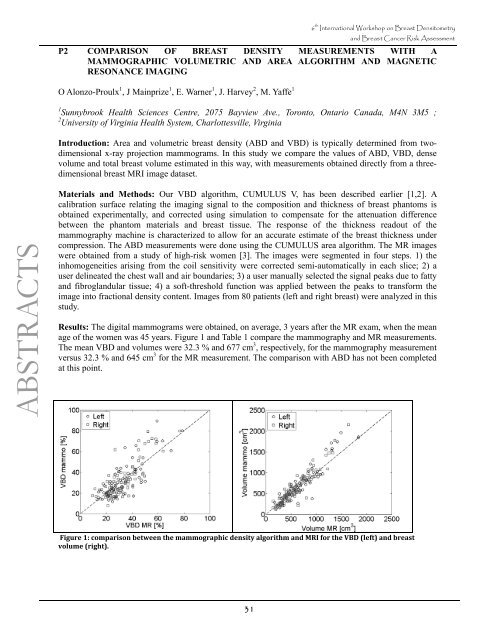
Results: The digital mammograms were obtained, <strong>on</strong> average, 3 years after the MR exam, when the mean<br />
age of the women was 45 years. Figure 1 <strong>and</strong> Table 1 compare the mammography <strong>and</strong> MR measurements.<br />
The mean VBD <strong>and</strong> volumes were 32.3 % <strong>and</strong> 677 cm 3 , respectively, for the mammography measurement<br />
versus 32.3 % <strong>and</strong> 645 cm 3 for the MR measurement. The comparis<strong>on</strong> with ABD has not been completed<br />
at this point.<br />
Figure 1: comparis<strong>on</strong> between the mammographic density algorithm <strong>and</strong> MRI for the VBD (left) <strong>and</strong> breast<br />
volume (right).<br />
31