6th International Workshop on Breast Densitometry and Breast ...
6th International Workshop on Breast Densitometry and Breast ...
6th International Workshop on Breast Densitometry and Breast ...
- No tags were found...
Create successful ePaper yourself
Turn your PDF publications into a flip-book with our unique Google optimized e-Paper software.
6 th <str<strong>on</strong>g>Internati<strong>on</strong>al</str<strong>on</strong>g> <str<strong>on</strong>g>Workshop</str<strong>on</strong>g> <strong>on</strong> <strong>Breast</strong> <strong>Densitometry</strong><br />
<strong>and</strong> <strong>Breast</strong> Cancer Risk Assessment<br />
P48<br />
A FULLY-AUTOMATED METHOD FOR QUANTIFYING FIBROGLANDULAR<br />
TISSUE AND BACKGROUND PARENCHYMAL ENHANCEMENT IN BREAST MRI<br />
Sh<strong>and</strong><strong>on</strong>g Wu, Susan P. Weinstein, Emily F. C<strong>on</strong>ant, Despina K<strong>on</strong>tos<br />
Department of Radiology, University of Pennsylvania, Philadelphia PA USA<br />
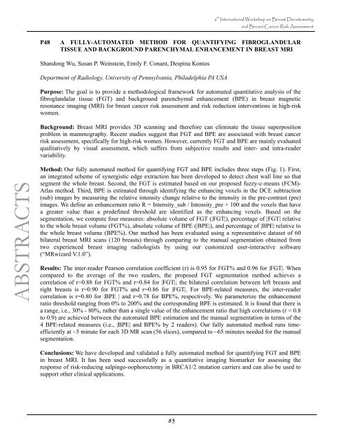
Purpose: The goal is to provide a methodological framework for automated quantitative analysis of the<br />
fibrogl<strong>and</strong>ular tissue (FGT) <strong>and</strong> background parenchymal enhancement (BPE) in breast magnetic<br />
res<strong>on</strong>ance imaging (MRI) for breast cancer risk assessment <strong>and</strong> risk reducti<strong>on</strong> interventi<strong>on</strong>s in high-risk<br />
women.<br />
Background: <strong>Breast</strong> MRI provides 3D scanning <strong>and</strong> therefore can eliminate the tissue superpositi<strong>on</strong><br />
problem in mammography. Recent studies suggest that FGT <strong>and</strong> BPE are associated with breast cancer<br />
risk assessment, specifically for high-risk women. However, currently FGT <strong>and</strong> BPE are mainly evaluated<br />
qualitatively by visual assessment, which suffers from subjective results <strong>and</strong> inter- <strong>and</strong> intra-reader<br />
variability.<br />
ABSTRACTS<br />
Method: Our fully automated method for quantifying FGT <strong>and</strong> BPE includes three steps (Fig. 1). First,<br />
an integrated scheme of synergistic edge extracti<strong>on</strong> has been developed to detect chest wall line so that<br />
segment the whole breast. Sec<strong>on</strong>d, the FGT is estimated based <strong>on</strong> our proposed fuzzy-c-means (FCM)-<br />
Atlas method. Third, BPE is estimated through identifying the enhancing voxels in the DCE subtracti<strong>on</strong><br />
(sub) images by measuring the relative intensity change relative to the intensity in the pre-c<strong>on</strong>trast (pre)<br />
images. We define an enhancement ratio R = Intensity_sub / Intensity_pre × 100 <strong>and</strong> the voxels that have<br />
a greater value than a predefined threshold are identified as the enhancing voxels. Based <strong>on</strong> the<br />
segmentati<strong>on</strong>, we compute four measures: absolute volume of FGT (|FGT|), percentage of |FGT| relative<br />
to the whole breast volume (FGT%), absolute volume of BPE (|BPE|), <strong>and</strong> percentage of |BPE| relative to<br />
the whole breast volume (BPE%). Our method has been evaluated using a representative dataset of 60<br />
bilateral breast MRI scans (120 breasts) through comparing to the manual segmentati<strong>on</strong> obtained from<br />
two experienced breast imaging radiologists by using our customized user-interactive software<br />
(“MRwizard V.1.0”).<br />
Results: The inter-reader Pears<strong>on</strong> correlati<strong>on</strong> coefficient (r) is 0.95 for FGT% <strong>and</strong> 0.96 for |FGT|. When<br />
compared to the average of the two readers, the proposed FGT segmentati<strong>on</strong> method achieves a<br />
correlati<strong>on</strong> of r=0.88 for FGT% <strong>and</strong> r=0.84 for |FGT|; the bilateral correlati<strong>on</strong> between left breasts <strong>and</strong><br />
right breasts is r=0.90 for FGT% <strong>and</strong> r=0.86 for |FGT|. For BPE-related measures, the inter-reader<br />
correlati<strong>on</strong> is r=0.80 for |BPE | <strong>and</strong> r=0.78 for BPE%, respectively. We parameterize the enhancement<br />
ratio threshold ranging from 0% to 200% <strong>and</strong> the corresp<strong>on</strong>ding BPE is estimated. It is found that there is<br />
a range, i.e., 30% - 80%, rather than a single value of the enhancement ratio that high correlati<strong>on</strong>s (r = 0.8<br />
to 0.9) are achieved between the automated BPE estimati<strong>on</strong> <strong>and</strong> the manual segmentati<strong>on</strong> in terms of the<br />
4 BPE-related measures (i.e., |BPE| <strong>and</strong> BPE% by 2 readers). Our fully automated method runs timeefficiently<br />
at ~5 minute for each 3D MR scan (56 slices), compared to ~65 minutes needed for the manual<br />
segmentati<strong>on</strong>.<br />
C<strong>on</strong>clusi<strong>on</strong>s: We have developed <strong>and</strong> validated a fully automated method for quantifying FGT <strong>and</strong> BPE<br />
in breast MRI. It has been used successfully as a quantitative imaging biomarker for assessing the<br />
resp<strong>on</strong>se of risk-reducing salpingo-oophorectomy in BRCA1/2 mutati<strong>on</strong> carriers <strong>and</strong> can also be used to<br />
support other clinical applicati<strong>on</strong>s.<br />
83


