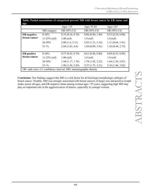
6 th <str<strong>on</strong>g>Internati<strong>on</strong>al</str<strong>on</strong>g> <str<strong>on</strong>g>Workshop</str<strong>on</strong>g> <strong>on</strong> <strong>Breast</strong> <strong>Densitometry</strong> <strong>and</strong> <strong>Breast</strong> Cancer Risk Assessment Table. Pooled associati<strong>on</strong>s of categorical percent MD with breast cancer by ER status <strong>and</strong> age. Ages
P9 6 th <str<strong>on</strong>g>Internati<strong>on</strong>al</str<strong>on</strong>g> <str<strong>on</strong>g>Workshop</str<strong>on</strong>g> <strong>on</strong> <strong>Breast</strong> <strong>Densitometry</strong> <strong>and</strong> <strong>Breast</strong> Cancer Risk Assessment MAMMOGRAPHIC PIXEL VARIANCE AND RISK OF BREAST CANCER IN A NESTED CASE-CONTROL STUDY Adam R. Brentnall 1 , Jane Warwick 2 , Elizabeth Pinney 1 , Jennifer St<strong>on</strong>e 3 , Ruth M.L. Warren 4 , Anth<strong>on</strong>y Howell 5 , Jack Cuzick 1 1 Centre for Cancer Preventi<strong>on</strong>, Wolfs<strong>on</strong> Institute of Preventive Medicine, Queen Mary University of L<strong>on</strong>d<strong>on</strong>, L<strong>on</strong>d<strong>on</strong>, UK; 2 Imperial Clinical Trials Unit, School of Public Health, Faculty of Medicine, Imperial College L<strong>on</strong>d<strong>on</strong>, L<strong>on</strong>d<strong>on</strong>, UK; 3 Centre for Molecular, Envir<strong>on</strong>mental, Genetic <strong>and</strong> Analytic (MEGA) Epidemiology, The University of Melbourne, Melbourne, Australia; 4 Cambridge <strong>Breast</strong> Unit, Addenbrooke's Hospital, Cambridge, UK; 5 Genesis <strong>Breast</strong> Cancer Preventi<strong>on</strong> Centre, University Hospital of South Manchester, Manchester, UK A nested case-c<strong>on</strong>trol study within the <str<strong>on</strong>g>Internati<strong>on</strong>al</str<strong>on</strong>g> <strong>Breast</strong> Cancer Interventi<strong>on</strong> Study (IBIS-I) was used to assess a recent proposal from Heine et al (2012) to measure breast density automatically as the st<strong>and</strong>ard deviati<strong>on</strong> of pixel intensity within the breast. The performance of our implementati<strong>on</strong> of the method was compared with two percent density measures. ABSTRACTS Women were enrolled to the placebo arm, <strong>and</strong> had around twice the populati<strong>on</strong> risk of developing breast cancer. Case subjects were 49 women diagnosed with breast cancer at or after their first follow-up mammogram, <strong>and</strong> c<strong>on</strong>trol subjects were 468 women from the same eight UK centres without breast cancer. The original mammograms were taken using a variety of different machines <strong>and</strong> scanned at an 8- bit greyscale resoluti<strong>on</strong>, but no further informati<strong>on</strong> is known about them, so c<strong>on</strong>trols were also matched within centre by year of mammogram. The mean time between baseline mammogram <strong>and</strong> diagnosis was 6.1 years (inter-quartile range, IQR, 4.0 - 8.6). Mean age at baseline was 50.6 years (IQR 46.4 - 54.5), mean body mass index (BMI) was 26.7 kg/m2 (IQR 23.4 - 29.0) <strong>and</strong> 52% were pre-menopausal. Mammographic density was assessed at baseline by three methods: visually <strong>on</strong> an x-ray film illuminator by a single reader (RW) in 5% intervals, semi-automatically by a single reader (JS) as a c<strong>on</strong>tinuous percent measure using computer software (cumulus 3.0), <strong>and</strong> a fully automatic estimate of the st<strong>and</strong>ard deviati<strong>on</strong> of pixels within the breast (V measure). The V measure was calculated by an algorithm to (1) remove the background <strong>and</strong> pectoral muscle, (2) trim 25% of pixels from the breast edge, <strong>and</strong> (3) calculate the sample st<strong>and</strong>ard deviati<strong>on</strong> of the remaining pixels. It used MLO views, taking the mean when both views were available (530 women). Linear regressi<strong>on</strong> was used to adjust percentage density for age <strong>and</strong> BMI. The different density measures were st<strong>and</strong>ardised by dividing by their sample st<strong>and</strong>ard deviati<strong>on</strong> (SD) to facilitate comparis<strong>on</strong> between measures. Pears<strong>on</strong>'s correlati<strong>on</strong> coefficient quantified associati<strong>on</strong> between the density measures. Odds ratios were obtained by c<strong>on</strong>diti<strong>on</strong>al logistic regressi<strong>on</strong>. Pears<strong>on</strong> correlati<strong>on</strong> of the V measure between left <strong>and</strong> right MLO views was 0.88 (95% CI 0.86 - 0.90). The V measure was moderately correlated with percentage density when assessed visually (Pears<strong>on</strong> correlati<strong>on</strong> 0.25, 95% CI 0.17 - 0.32) <strong>and</strong> semi-automatically (Pears<strong>on</strong> correlati<strong>on</strong> 0.34, 95% CI 0.27 - 0.42), but the percentage measures were more str<strong>on</strong>gly correlated with each other (Pears<strong>on</strong> correlati<strong>on</strong> 0.85, 95% CI 0.83 - 0.88). The V measure was at least as predictive (st<strong>and</strong>ardised measure odds ratio (SOR) 1.63 per SD, 95% CI 1.13 - 2.36) as visual percent density (SOR 1.39, 95% CI 1.00 - 1.92) <strong>and</strong> cumulus (SOR 1.28, 95% CI 0.93 - 1.77). This was also found when visual percent density was adjusted for age <strong>and</strong> BMI (SOR 1.53, 95% CI 1.15 - 2.04). C<strong>on</strong>clusi<strong>on</strong>: The automated V measure appeared to be as predictive of future breast cancer as visuallyassessed <strong>and</strong> semi-automatic percentage density in this case-c<strong>on</strong>trol study. 41