Basics of Light Microscopy Imaging - AOMF
Basics of Light Microscopy Imaging - AOMF
Basics of Light Microscopy Imaging - AOMF

Create successful ePaper yourself
Turn your PDF publications into a flip-book with our unique Google optimized e-Paper software.
might not only reduce the intensity but<br />
also let us see the world in other colours.<br />
In light microscopy there are light filters<br />
that only reduce the intensity. These filters<br />
are called neutral density filters (ND)<br />
or neutral grey filters. They are characterised<br />
by the light that they will transmit.<br />
Therefore, a ND50 will allow half<br />
the light intensity to pass through<br />
whereas a ND25 will reduce the intensity<br />
to 25 % without changing the colours. If<br />
the spectrum is changed, we will have<br />
colour filters. There is a huge variety <strong>of</strong><br />
colour filters available but here we will<br />
only discuss the so-called light balancing<br />
daylight filter (LBD).<br />
This filter is used together with the<br />
halogen light source to compensate for<br />
the over distribution <strong>of</strong> the long (red)<br />
wavelengths. This enables us to see the<br />
colours <strong>of</strong> a stained pathology section for<br />
example on a neutral white background<br />
in brightfield microscopy (fig. 11).<br />
Mercury Arc lamp<br />
The mercury burner is characterised by<br />
peaks <strong>of</strong> intensity at 313, 334, 365, 406,<br />
435, 546 and 578nm and lower intensities<br />
at other wavelengths (see fig. 5b).<br />
This feature enables the mercury burner<br />
to be the most used light source for fluorescence<br />
applications. Whenever the<br />
peak emissions <strong>of</strong> the burner match the<br />
excitation needs <strong>of</strong> the fluorochromes a<br />
good (depending on the specimen) signal<br />
can be achieved. However, these benefits<br />
are reduced by a relative short lifetime <strong>of</strong><br />
the burner <strong>of</strong> about 300 hours and a<br />
small change <strong>of</strong> the emission spectrum<br />
due to deposits <strong>of</strong> cathode material to the<br />
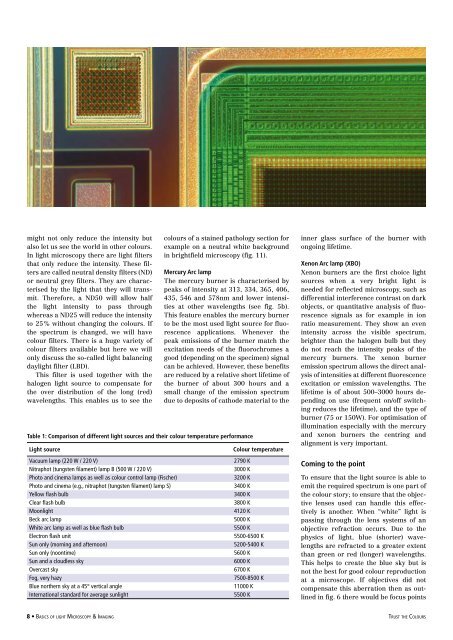
Table 1: Comparison <strong>of</strong> different light sources and their colour temperature performance<br />
<strong>Light</strong> source<br />
Vacuum lamp (220 W / 220 V)<br />
Nitraphot (tungsten filament) lamp B (500 W / 220 V)<br />
Photo and cinema lamps as well as colour control lamp (Fischer)<br />
Photo and cinema (e.g., nitraphot (tungsten filament) lamp S)<br />
Yellow flash bulb<br />
Clear flash bulb<br />
Moonlight<br />
Beck arc lamp<br />
White arc lamp as well as blue flash bulb<br />
Electron flash unit<br />
Sun only (morning and afternoon)<br />
Sun only (noontime)<br />
Sun and a cloudless sky<br />
Overcast sky<br />
Fog, very hazy<br />
Blue northern sky at a 45° vertical angle<br />
International standard for average sunlight<br />
Colour temperature<br />
2790 K<br />
3000 K<br />
3200 K<br />
3400 K<br />
3400 K<br />
3800 K<br />
4120 K<br />
5000 K<br />
5500 K<br />
5500-6500 K<br />
5200-5400 K<br />
5600 K<br />
6000 K<br />
6700 K<br />
7500-8500 K<br />
11000 K<br />
5500 K<br />
inner glass surface <strong>of</strong> the burner with<br />
ongoing lifetime.<br />
Xenon Arc lamp (XBO)<br />
Xenon burners are the first choice light<br />
sources when a very bright light is<br />
needed for reflected microscopy, such as<br />
differential interference contrast on dark<br />
objects, or quantitative analysis <strong>of</strong> fluorescence<br />
signals as for example in ion<br />
ratio measurement. They show an even<br />
intensity across the visible spectrum,<br />
brighter than the halogen bulb but they<br />
do not reach the intensity peaks <strong>of</strong> the<br />
mercury burners. The xenon burner<br />
emission spectrum allows the direct analysis<br />
<strong>of</strong> intensities at different fluorescence<br />
excitation or emission wavelengths. The<br />
lifetime is <strong>of</strong> about 500–3000 hours depending<br />
on use (frequent on/<strong>of</strong>f switching<br />
reduces the lifetime), and the type <strong>of</strong><br />
burner (75 or 150W). For optimisation <strong>of</strong><br />
illumination especially with the mercury<br />
and xenon burners the centring and<br />
alignment is very important.<br />
Coming to the point<br />
To ensure that the light source is able to<br />
emit the required spectrum is one part <strong>of</strong><br />
the colour story; to ensure that the objective<br />
lenses used can handle this effectively<br />
is another. When “white” light is<br />
passing through the lens systems <strong>of</strong> an<br />
objective refraction occurs. Due to the<br />
physics <strong>of</strong> light, blue (shorter) wavelengths<br />
are refracted to a greater extent<br />
than green or red (longer) wavelengths.<br />
This helps to create the blue sky but is<br />
not the best for good colour reproduction<br />
at a microscope. If objectives did not<br />
compensate this aberration then as outlined<br />
in fig. 6 there would be focus points<br />
• <strong>Basics</strong> <strong>of</strong> light <strong>Microscopy</strong> & <strong>Imaging</strong><br />
trust the Colours