Basics of Light Microscopy Imaging - AOMF
Basics of Light Microscopy Imaging - AOMF
Basics of Light Microscopy Imaging - AOMF

Create successful ePaper yourself
Turn your PDF publications into a flip-book with our unique Google optimized e-Paper software.
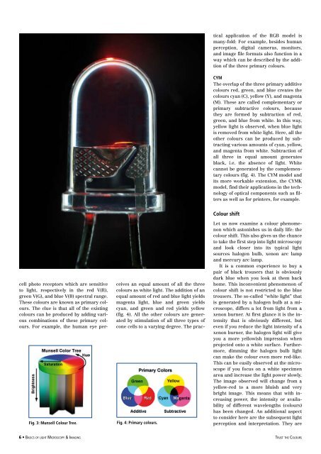
ceives an equal amount <strong>of</strong> all the three<br />
colours as white light. The addition <strong>of</strong> an<br />
equal amount <strong>of</strong> red and blue light yields<br />
magenta light, blue and green yields<br />
cyan, and green and red yields yellow<br />
(fig. 4). All the other colours are generated<br />
by stimulation <strong>of</strong> all three types <strong>of</strong><br />
cone cells to a varying degree. The practical<br />
application <strong>of</strong> the RGB model is<br />
many-fold: For example, besides human<br />
perception, digital cameras, monitors,<br />
and image file formats also function in a<br />
way which can be described by the addition<br />
<strong>of</strong> the three primary colours.<br />
CYM<br />
The overlap <strong>of</strong> the three primary additive<br />
colours red, green, and blue creates the<br />
colours cyan (C), yellow (Y), and magenta<br />
(M). These are called complementary or<br />
primary subtractive colours, because<br />
they are formed by subtraction <strong>of</strong> red,<br />
green, and blue from white. In this way,<br />
yellow light is observed, when blue light<br />
is removed from white light. Here, all the<br />
other colours can be produced by subtracting<br />
various amounts <strong>of</strong> cyan, yellow,<br />
and magenta from white. Subtraction <strong>of</strong><br />
all three in equal amount generates<br />
black, i.e. the absence <strong>of</strong> light. White<br />
cannot be generated by the complementary<br />
colours (fig. 4). The CYM model and<br />
its more workable extension, the CYMK<br />
model, find their applications in the technology<br />
<strong>of</strong> optical components such as filters<br />
as well as for printers, for example.<br />
Fig. 3: Munsell Colour Tree.<br />
cell photo receptors which are sensitive<br />
to light, respectively in the red V(R),<br />
green V(G), and blue V(B) spectral range.<br />
These colours are known as primary colours.<br />
The clue is that all <strong>of</strong> the existing<br />
colours can be produced by adding various<br />
combinations <strong>of</strong> these primary colours.<br />
For example, the human eye per-<br />
Fig. 4: Primary colours.<br />
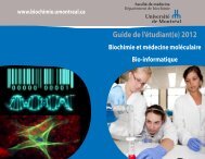
Colour shift<br />
Let us now examine a colour phenomenon<br />
which astonishes us in daily life: the<br />
colour shift. This also gives us the chance<br />
to take the first step into light microscopy<br />
and look closer into its typical light<br />
sources halogen bulb, xenon arc lamp<br />
and mercury arc lamp.<br />
It is a common experience to buy a<br />
pair <strong>of</strong> black trousers that is obviously<br />
dark blue when you look at them back<br />
home. This inconvenient phenomenon <strong>of</strong><br />
colour shift is not restricted to the blue<br />
trousers. The so-called “white light” that<br />
is generated by a halogen bulb at a microscope,<br />
differs a lot from light from a<br />
xenon burner. At first glance it is the intensity<br />
that is obviously different, but<br />
even if you reduce the light intensity <strong>of</strong> a<br />
xenon burner, the halogen light will give<br />
you a more yellowish impression when<br />
projected onto a white surface. Furthermore,<br />
dimming the halogen bulb light<br />
can make the colour even more red-like.<br />
This can be easily observed at the microscope<br />
if you focus on a white specimen<br />
area and increase the light power slowly.<br />
The image observed will change from a<br />
yellow-red to a more bluish and very<br />
bright image. This means that with increasing<br />
power, the intensity or availability<br />
<strong>of</strong> different wavelengths (colours)<br />
has been changed. An additional aspect<br />
to consider here are the subsequent light<br />
perception and interpretation. They are<br />
• <strong>Basics</strong> <strong>of</strong> light <strong>Microscopy</strong> & <strong>Imaging</strong><br />
trust the Colours