Basics of Light Microscopy Imaging - AOMF
Basics of Light Microscopy Imaging - AOMF
Basics of Light Microscopy Imaging - AOMF
You also want an ePaper? Increase the reach of your titles
YUMPU automatically turns print PDFs into web optimized ePapers that Google loves.
Dear Reader<br />
“A Huge Number <strong>of</strong><br />
Techniques in a Relatively<br />
Short Space”<br />
Although microscopes are becoming more and more easy to use, it still remains important to have an<br />
appreciation <strong>of</strong> the fundamental principles that determine the resolution and contrast seen in microscope<br />
images. This series <strong>of</strong> articles, by authors from Olympus, aims to provide an overview <strong>of</strong> the<br />
physical principles involved in microscope image formation and the various commonly used contrast<br />
mechanisms together with much practically oriented advice. Inevitably it is impossible to cover any <strong>of</strong><br />
the many aspects in great depth. However, their approach, which is to discuss applications and provide<br />
practical advice for many aspects <strong>of</strong> modern day microscopy, will prove attractive to many.<br />
The articles begin by setting the scene with a discussion <strong>of</strong> the factors that determine the resolving<br />
power <strong>of</strong> the conventional microscope. This permits the introduction <strong>of</strong> important concepts such as<br />
numerical aperture, Airy disc, point spread function, the difference between depth <strong>of</strong> focus and depth<br />
<strong>of</strong> field and the concept <strong>of</strong> parfocality. Common contrast mechanisms such as darkfield and phase<br />
contrast are then introduced, followed by differential interference contrast and polarisation contrast<br />
imaging. Throughout the discussion, the importance <strong>of</strong> digital image processing is emphasised and<br />
simple examples such as histogram equalisation and the use <strong>of</strong> various filters are discussed.<br />
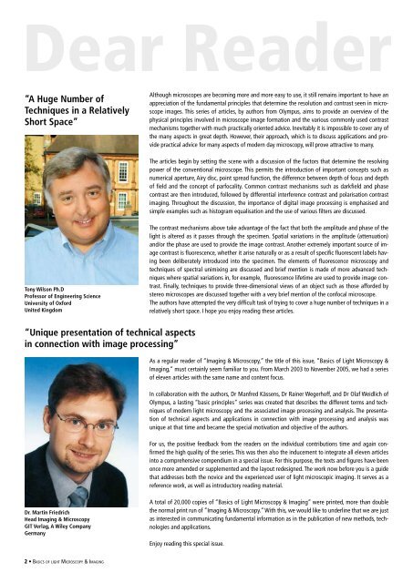
Tony Wilson Ph.D<br />
Pr<strong>of</strong>essor <strong>of</strong> Engineering Science<br />
University <strong>of</strong> Oxford<br />
United Kingdom<br />
The contrast mechanisms above take advantage <strong>of</strong> the fact that both the amplitude and phase <strong>of</strong> the<br />
light is altered as it passes through the specimen. Spatial variations in the amplitude (attenuation)<br />
and/or the phase are used to provide the image contrast. Another extremely important source <strong>of</strong> image<br />
contrast is fluorescence, whether it arise naturally or as a result <strong>of</strong> specific fluorescent labels having<br />
been deliberately introduced into the specimen. The elements <strong>of</strong> fluorescence microscopy and<br />
techniques <strong>of</strong> spectral unimixing are discussed and brief mention is made <strong>of</strong> more advanced techniques<br />
where spatial variations in, for example, fluorescence lifetime are used to provide image contrast.<br />
Finally, techniques to provide three-dimensional views <strong>of</strong> an object such as those afforded by<br />
stereo microscopes are discussed together with a very brief mention <strong>of</strong> the confocal microscope.<br />
The authors have attempted the very difficult task <strong>of</strong> trying to cover a huge number <strong>of</strong> techniques in a<br />
relatively short space. I hope you enjoy reading these articles.<br />
“Unique presentation <strong>of</strong> technical aspects<br />
in connection with image processing”<br />
As a regular reader <strong>of</strong> “<strong>Imaging</strong> & <strong>Microscopy</strong>,” the title <strong>of</strong> this issue, “<strong>Basics</strong> <strong>of</strong> <strong>Light</strong> <strong>Microscopy</strong> &<br />
<strong>Imaging</strong>,” must certainly seem familiar to you. From March 2003 to November 2005, we had a series<br />
<strong>of</strong> eleven articles with the same name and content focus.<br />
In collaboration with the authors, Dr Manfred Kässens, Dr Rainer Wegerh<strong>of</strong>f, and Dr Olaf Weidlich <strong>of</strong><br />
Olympus, a lasting “basic principles” series was created that describes the different terms and techniques<br />
<strong>of</strong> modern light microscopy and the associated image processing and analysis. The presentation<br />
<strong>of</strong> technical aspects and applications in connection with image processing and analysis was<br />
unique at that time and became the special motivation and objective <strong>of</strong> the authors.<br />
For us, the positive feedback from the readers on the individual contributions time and again confirmed<br />
the high quality <strong>of</strong> the series. This was then also the inducement to integrate all eleven articles<br />
into a comprehensive compendium in a special issue. For this purpose, the texts and figures have been<br />
once more amended or supplemented and the layout redesigned. The work now before you is a guide<br />
that addresses both the novice and the experienced user <strong>of</strong> light microscopic imaging. It serves as a<br />
reference work, as well as introductory reading material.<br />
Dr. Martin Friedrich<br />
Head <strong>Imaging</strong> & <strong>Microscopy</strong><br />
GIT Verlag, A Wiley Company<br />
Germany<br />
A total <strong>of</strong> 20,000 copies <strong>of</strong> “<strong>Basics</strong> <strong>of</strong> <strong>Light</strong> <strong>Microscopy</strong> & <strong>Imaging</strong>” were printed, more than double<br />
the normal print run <strong>of</strong> ”<strong>Imaging</strong> & <strong>Microscopy</strong>.” With this, we would like to underline that we are just<br />
as interested in communicating fundamental information as in the publication <strong>of</strong> new methods, technologies<br />
and applications.<br />
Enjoy reading this special issue.<br />
• <strong>Basics</strong> <strong>of</strong> light <strong>Microscopy</strong> & <strong>Imaging</strong>


